Antigens of
Coccidioides posadasii
as an Important Tool
for the Immunodiagnosis of Coccidioidomycosis
Rossana de Aguiar Cordeiro• Kharla Rabelo Nobre Patoilo•Silviane Bandeira Praciano • Delia Jessica Astete Medrano •Francisca Jakelyne de Farias Marques•
Liline Maria Soares Martins•Kelsen Dantas Eulalio•Antoˆnio de Deus Filho• Maria do Amparo Salmito Cavalvanti•Maria Auxiliadora Bezerra Fechine•
Raimunda Samia Nogueira Brilhante•Zoilo Pires de Camargo•Marcos Fa´bio Gadelha Rocha• Jose´ Ju´lio Costa Sidrim
Received: 24 September 2012 / Accepted: 21 November 2012 / Published online: 14 December 2012 ÓSpringer Science+Business Media Dordrecht 2012
Abstract Serologic diagnosis has been presented as a safe alternative for coccidioidomycosis. However, commercial kits based on coccidioidal antibodies available in the USA are considered too expensive for laboratories outside that country. In this study, we describe the preparation of antigens for detection of human coccidioidal antibodies by the immunodif-fusion test (ID) and enzyme immunoassay (EIA).
Antigens were tested against serum samples from patients with coccidioidomycosis, histoplasmosis and paracoccidioidomycosis, as well as healthy individu-als. The highest reactivity in the ID tests was seen in the F0-90 antigen. In the EIAs, the best results were obtained with the F60-90 antigen. None of the serum samples from healthy individuals were recognized by any of the antigen extracts tested by ID or EIA. In conclusion, the F0-90 and F60-90 antigens have the potential to be commercially employed in presumptive diagnosis of coccidioidomycosis by ID or EIA, respectively. The tests could improve serological diagnosis of coccidioidomycosis in South America.
Keywords CoccidioidomycosisDiagnosis
AntigenImmunodiffusionEnzyme immunoassay
Introduction
Coccidioidomycosis (CM) is a deep infection caused by Coccidioides immitis and C. posadasii, found exclusively in the Americas [1]. Both species are soil-inhabitant dimorphic fungi and infect humans and animals after inhalation of their asexual arthroconidia [2]. Approximately 60 % of infected individuals are asymptomatic and develop a protective cell-mediated response [3]. The remaining 40 % can show any of the following clinical manifestations: acute pneumonia,
R. de Aguiar Cordeiro (&)K. R. N. Patoilo S. B. PracianoD. J. A. MedranoF. J. de Farias MarquesR. S. N. BrilhanteJ. J. C. Sidrim Postgraduate Program in Medical Microbiology, Department of Pathology and Legal Medicine, Federal University of Ceara´, Fortaleza, Ceara, Brazil
e-mail: rossanacordeiro@ufc.br
L. M. S. MartinsA. de Deus Filho
Federal University of Piauı´, Teresina, Piauı´, Brazil
K. D. EulalioM. d. A. S. Cavalvanti
Instituto de Doenc¸as Tropicais Natan Portella, Teresina, Piauı´, Brazil
M. A. B. Fechine
Universidade da Integrac¸a˜o Internacional da Lusofonia Afro-Brasileira, Fortaleza, CE, Brazil
Z. P. de Camargo
Department of Microbiology, Immunology, and Parasitology, Federal University of Sa˜o Paulo, Sa˜o Paulo, SP, Brazil
M. F. G. Rocha
chronic progressive pneumonia, pulmonary nodules and cavities and disseminated disease [3, 4]. Since clinical findings are unspecific, CM can mimic several other community-acquired pneumonias [5,6], tuber-culosis [7] and malignancy [8]. Therefore, the impor-tance of correct diagnosis is unquestionable.
Many approaches are currently available for labo-ratory diagnosis of CM: recovery ofCoccidioidesspp. by culture [9, 10]; detection of fungal spherules on clinical specimens by microscopy [11, 12]; histopa-thology [11,12]; molecular-based techniques directly with clinical specimens [13,14] or cultures [15,16]; serology [10, 12]; and skin testing [17]. Although a positive culture of Coccidioides spp. remains the ‘‘gold standard’’ for a conclusive diagnosis [9], clinical laboratories must have BL3 facilities for secure handling of fungal cultures, which is an important hindrance to many institutions [18]. There-fore, strategies for improvement of CM diagnosis should be encouraged [10,18,19].
Diagnosis based on serology has been considered a useful alternative in CM [20]. Although antibodies (Ab) have virtually no effect on protection against the disease, Ab titers are considered a good predictor of the infection status [11]. Several approaches have been used to measure the serologic response for diagnosis and follow-up of patients with CM: complement fixation, immunodiffusion (ID), latex agglutination and enzyme immunoassay (EIA). In Brazil, serolog-ical diagnosis of CM is mainly performed by ID and EIA techniques [18, 21,22]. Although both ID and EIA commercial kits are currently available for detection of coccidioidal antibodies, they are too expensive for many laboratories outside the USA. In this paper, we describe the preparation of different in-house antigens for detection of human coccidioidal IgG by ID and EIA that can be an alternative to commercial kits.
Materials and Methods
Fungal Strain
A C. posadasii strain from the Specialized Medical Mycology Center’s fungal collection (CEMM, Federal University of Ceara´, Fortaleza, Brazil) was evaluated in this study. The strain (CEMM 01-06-092) was previ-ously identified by mycological analysis and specific
PCR, as described by Cordeiro et al. [23] and Umeyama et al. [16], respectively. Strain manipulations were performed within a class II biological safety cabinet in a biosafety level 3 laboratory.
Culture Medium and Growth Conditions
The fungal culture was initially grown in potato dextrose agar for 10 days at 30°C. After this period,
approximately 2 ml of sterile saline solution was added to the agar slant, and the culture was gently scraped with a cotton swab. The suspension was transferred to a 500-ml flask containing 100 ml of 2 % glucose and 1 % yeast extract broth (GYE) and incubated for 10 days at 30 °C, being considered a
pre-inoculum. After this period, the pre-inoculum was transferred to a 3-l flask containing 1,500 ml of GYE and further incubated as described above for 30 days.
Fungal Antigens
Antigen extraction was based in the protocol described by Brilhante et al. [21], with modifications. The fungal culture was treated with 0.2 g l-1 of thimerosal (ethylmercurithiosalicylic acid sodium salt; Synth, Brazil), and the mixture was incubated at 30 °C for
24 h or 30 days in order to obtain different exoanti-gens or cellular antiexoanti-gens, respectively. The superna-tant was collected by paper filtration, and the proteins were salted out with the following concentrations of ammonium sulfate (Sigma, USA): 0–90, 0–30, 30–60 and 60–90 %. The mixtures were kept at 4°C for
24 h, and then the precipitated proteins were recov-ered by centrifugation (10,0009g, 30 min, 4°C) and
dialyzed exhaustively against a 10X volume of distilled water in a dialysis membrane with a 10 kDa molecular mass cutoff. The samples were stored at
-20°C until used for serological tests. Five
differ-ent antigenic samples were obtained: exoantigen from 24-hour thimerosal treatment (Exo) and cellular antigens from 30-day thimerosal treatment (F0-30, F30-60, F60-90 and F0-90).
Antigen Characterization
of each extract was determined by SDS–PAGE, according to Laemmli [25], with modifications. Sam-ples were mixed with loading buffer (0.25 M Tris–HCl pH 8.0, 10 % SDS, 10 % b-mercaptoethanol, 20 % glycerol, 0.25 mg ml-1bromophenol blue), heated at 100 °C for 15 min and then centrifuged (13,0009g, 5 min). Volumes of 20ll of the supernatant were electrophoresed in 12.5 % polyacrylamide gels under denaturation conditions. Gels were run at room tem-perature (18°C) for 5 h at 80 volts in buffer containing
25 mM Tris base, 200 mM, glycine and 0.1 % SDS pH 8.0. After electrophoresis, the gels were stained with Coomassie brilliant blue R250 (Sigma, USA). The molecular weights were determined through E-Capt software (version 12.7 for Windows), in comparison with standard protein markers (Bio-Rad Laboratories, USA).
The presence of charged carbohydrate was evalu-ated according to Rocha et al. [26] (2008). In brief, each antigenic extract was electrophoresed in a 6 % (wt/vol) polyacrylamide gel at room temperature for 4 h at 80 volts in diaminopropane acetate buffer (50 mM; pH 9.0). Gels were stained with 0.1 % (wt/ vol) toluidine blue solution. For comparison, high- and low-molecular-weight standards (Bio-Rad, USA) as well as standard chondroitin 4-sulfate and chondroitin 6-sulfate (Sigma, USA) were subjected to the same protocols, as indicated.
Serum Samples
A total of 26 serum samples from patients with acute pulmonary or disseminated CM were included in this study. All of them were diagnosed by culture and ID test with a commercial kit (IDCF antigen, Immy Immunomycologics, USA). Patients were negative for HIV and tuberculosis, by way of serologic and Mantoux test, respectively. Heterologous serum sam-ples from patients with culture-based diagnosis for histoplasmosis (HM; n=10) and
paracoccidioido-mycosis (PM;n=7) were also included. In addition, serum samples from clinically healthy individuals without reactivity after commercial ID (n=100)
were included as negative controls. None of the healthy individuals have clinical evidence of tubercu-losis or another respiratory disease. The investigation was approved by the ethics committee of Federal University of Ceara´ (Process number 35/04).
ID Assays
ID assays were performed as described by Camargo et al. [27]. Briefly, the tests were performed with 1 % agarose in PBS solution. Slides were prepared with a central well surrounded by six wells (diameter 3 mm), each one placed 6 mm away. Each well was filled with 20ll of antigen preparation or serum sample, and the slides were incubated for 24 h in a moist chamber at 28°C. Serum samples from patients with CM, HM
and PM were also tested after heating at 63°C for
13 min for IgM inactivation [28]. The slides were washed with 5 % sodium citrate for 1 h and then for 24 in PBS buffer. Then the slides were stained with 0.15 % Coomassie brilliant blue in ethanol/acetic acid/deionized water (4:2:4) and destained with the solvent mixture whenever necessary. Precipitin bands between antigen and antibody wells were then recorded.
EIAs
In brief, microtiter plates (Corning Costar, USA) were coated with 50 ll of each antigen preparation (1.0, 2.5 and 5.0 lg of protein per well) and incubated overnight at 4 °C. After blockage with 1 % BSA
(Calbiochem, USA) for 90 min at 37°C, the plates
were washed four times with PBS-Tween 20 (0.05 %), and 50 ll of each serum sample (1:400–1:51200 dilution) was added to each well. The plates were incubated at 37°C for 1 h and then washed again
three times with PBS-Tween. The plates were incu-bated with 100ll peroxidase-labeled goat anti-human IgG (1:2000 dilution; Sigma, USA) for 1 h at 37°C.
After four washes with PBS-Tween, the reaction was developed by adding 100ll OPD (orthophenylenedi-amine) solution (0.4 mg ml-1; Sigma, USA) and 0.01 % (v/v) H2O2in 0.1 M citrate/phosphate buffer,
pH 5.0. After incubation for 30 min in the dark, the reaction was stopped by adding 25ll 1.25 M H2SO4.
The absorbance was measured on a microplate reader at 492 nm. The EIAs were performed using serum samples from patients with pulmonary or dissemi-nated CM (n=26) and heterologous serum samples
from patients with HM (n =10) and PM (n=7). A
Data Analysis
To calculate the sensitivity, specificity, positive pre-dictive value (PPV) and negative prepre-dictive value (NPV) for the experimental ID tests, the follow-ing formulas were used: Sensitivity was equal to A/(A?C); specificity was equal to D/(B?D); PPV
was equal to A/(A?B); and NPV was equal to D/ (C?D), where A is a specimen with a positive result by both experimental ID and the traditional tests (culture and/or commercial ID test); B is a specimen with a positive result by experimental ID but negative results by the traditional tests; C is a specimen with a negative result by experimental ID but positive results by the traditional test; and D is a specimen with negative results by both types of assays (experimental ID and traditional tests).
Sensitivity, specificity and optimal cutoff values of indirect EIAs were determined for each antigen preparation by using receiver operating characteristic (ROC) curve analysis. The area under the ROC curve (AUC) was calculated and compared.
Results
Antigenic Extracts
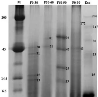
Five antigenic extracts were obtained: Exo, F0-30, F30-60, F60-90 and F0-90. The Bradford assay revealed the following amounts of total proteins in the antigenic extracts: Exo, 21.57 lg ml-1; F30-60, 9.78 lg ml-1; F60-90, 303.0lg ml-1; and F0-90, 567.0lg ml-1. The protein content of the F0-30 antigen was undetectable (\0.1lg ml-1). The SDS–
PAGE profile of each antigenic extract revealed proteins with different molecular weights (Fig.1). Preliminary analysis did not reveal the presence of charged carbohydrates in any of the antigenic extracts.
ID
By way of ID reactions, differences in reactivity of each antigenic extract were seen. Poor reactivity against sera of patients with CM (n =26) was seen in
Exo (n=9, 34.61 %) and in F0-30 (n=4, 15.38 %). The remaining antigenic preparations presented the following reactivity results: 11 (42.30 %), 15 (57.69 %) and 23 (88.46 %) for F30-60, F60-90 and
F0-90, respectively. Similar pattern results were seen with both heated and unheated serum samples. None of the heterologous serum samples or the negative control samples (healthy individuals) presented posi-tive reaction by ID. The data concerning sensitivity, specificity, positive predictive value and negative predictive value of each antigenic extract are shown in Table 1.
EIA
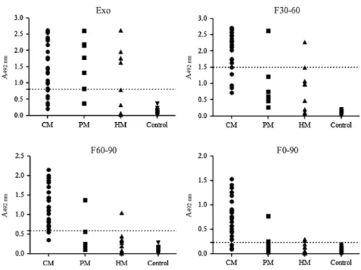
Indirect EIAs were performed with only the following protein-rich extracts: Exo, F30-60, F60-90 and F0-90 (Fig.2). Reproducible results were obtained when the antigenic extracts were tested at 2.5 lg protein ml-1.
Fig. 1 Protein profile of antigenic extracts in 12.5 % poly-acrylamide gel under denaturation conditions. Numbers corre-spond to the molecular weight of the main proteins in each antigenic preparation. M. Molecular weight marker
Table 1 Reactivity parameters regarding immunodiffusion (ID) tests with five antigenic extracts
Antigen Sensitivity (%)
Specificity (%)
PPV (%)
NPV (%)
Exo 34.61 100 100 87.31
F0–30 15.38 100 100 84.17
F30–60 42.30 100 100 88.63
F60–90 57.69 100 100 91.40
F0–90 88.46 100 100 97.50
Serum samples from 26 patients with CM and 75 individuals without CM were tested for IgG reactivity by indirect EIA. The number of positive serum samples detected was 20 (76.92 %) for Exo; 22 (84.61 %) for F30-60; 24 (92.30 %) for F60-90 and 23 (88.46 %) for F0-90. Cross-reactivity was seen in Exo (6 PM; 4 HM), F30-60 (1 PB; 2 HM), F 60-90 (1 PM; 1 HM) and F0-90 (1 PM; 0 HM). None of the serum samples from healthy individuals were recog-nized by any of antigen extracts. After analysis of the ROC curve for each antigen (Fig.3), the cutoff parameters were determined as follows: 0.8280, 1.497, 0.5895 and 0.3270 for Exo, F30-60, F60-90 and F0-90, respectively. The parameters regarding specificity, sensitivity, confidence intervals and AUC for each antigen are displayed in Table2.
Discussion
Coccidioidesserology has a long and well-documented history. Pioneering studies developed by Cooke [29] with grinded cultures ofC. immitisas antigenic extract, and the outstanding research conducted by Smith et al. [30,31] with more than 39,000 serological tests have assisted to set up the currently serological protocols for the diagnosis and prognosis of CM.
In the recent years, several studies have highlighted the importance of serological tests for the diagnosis and follow-up of patients with CM [10,20,32–34]. Presumptive diagnosis of CM can be achieved by ID tests, which can detect IgM or IgG antibodies raised against ‘‘tube precipitin’’ or ‘‘complement fixation’’ antigens and thus provide information on the status of the disease [35,36]. ID testing is very popular among routine laboratories because of its simplicity and reliability [37]. Despite these advantages, ID tests can present great variability regarding sensitivity [37] and cross-reactions with histoplasmosis (HM) sera may occur [36,37], even against purified antigens [38].
In the present study, it was shown that all of the antigenic extracts tested by ID were able to recognize serum samples from patients with CM. However, the highest sensitivity and specificity values were obtained with antigen F0-90. ID performed against F0-90 also showed higher values of PPV and NPV, which indicates that the test with this antigen is most likely correct in its assessment. The absence of cross-reactions suggests the potential of this test for presumptive diagnosis of CM. Despite the low protein content of the F0-30 antigen, positive reactions among CM serum samples were seen. We suppose that trace amounts of structural carbohydrates may have con-tributed to the observed reactivity.
Fig. 2 Distribution of optical density values in EIA assays withCoccidioides posadasiiantigens for patients with
EIAs have been widely employed for CM diagnosis in endemic regions of the USA [10,33,35,39–41]. However, differences regarding sensitivity, specificity and predictive values have been observed among EIAs. Martins et al. [40] showed that combined IgM and IgG EIA tests had specificity of 98.5 % and sensitivity of 94.8 %. By using complement fixation tests as reference, Zartarian et al. [41] presented a commercial EIA that had specificity of 98 % and sensitivity of 100 %. In Latin America, Tiraboschi et al. [42] evaluated the reactivity of an experimental EIA and found sensitivity of 72 % and specificity of 85 %. In our study, we observed that both F0-90 and F60-90 antigens were suitable for detection of coc-cidioidal IgG by indirect EIA. However, the F60-90 antigen showed the highest levels of sensitivity, specificity, PPV and NPV. The reactivity parameters obtained with F60-90 EIA were similar to those found in other studies [33,35,43,44].
EIAs for detection of coccidioidal antibody have shown a considerable degree of cross-reactions with sera from patients with blastomycosis or HM. In a seminal study performed by Yang et al. [38], a recombinant protein with chitinase activity was found to express epitopes that reacted with sera from patients with blastomycosis or HM. In addition, cross-reactiv-ity has been described in all available serological methods, including complement fixation, ID and EIA directed at antigen detection [39], as well as in sera from patients with non-mycotic diseases tested by ID and complement fixation tests [36].
In the present study, cross-reactions with HM and paracoccidioidomycosis (PM) serum samples were seen in the EIAs with all of the antigens. However, fewer cross-reactions occurred with F60-90 (PM=1; HM=1) and F0-90 (PM=1) antigens. Although
one may argue that cross-reactions can compromise the usefulness of our EIA, it is important to note that
Fig. 3 ROC curves graphing sensitivity % (true-positive results) versus 100 % specificity (false-positive results) for the antigens evaluated in this study:aExo,bF30-60, c60–90 anddF0-90. The cutoff values decrease from high to low values as the curves move from left to right
Table 2 Reactivity parameters EIA assays with four antigenic extracts of Coccidioides posadasii
Standard error
PPVpositive predictive value, NPVnegative predictive value
Antigen Cutoff AUC±SE PPV (%) NPV (%) Sensitivity (%) Specificity (%)
Exo 0.8280 0.904±0.029 76.92 91.3 76.92 88.16
F30-60 1.390 0.973±0.014 84.61 95.89 84.61 97.33
F60-90 0.5895 0.985±0.009 92.31 97.26 92.30 97.33
undesired false-positive reactions occur even with commercial kits for coccidioidal antibody detection [39,45]. Cross-reactions against unrelated pathogens in a commercial EIA kit have also been described [46]. Besides that, patients with CM, PM and HM can present peculiar clinical characteristics. In addition, these mycoses occur in distinct endemic areas in South America [47]. Taken together, this information may help to interpret the F60-90 EIA results. Therefore, we believe that F60-90 EIA has potential to be used as a screening tool for CM diagnosis in routine laboratories in South America.
One of the major drawbacks of tests with in-house antigens is that their quality can vary from batch to batch. Although this question has not been extensively investigated in our study, we have also prepared antigenic extracts from two otherC. posadasiicultures (clinical strain CEMM 01-06-085 and environmental stain CEMM 01-06-101) and run the same tests regarding antigen characterization described in Mate-rials and Methods section. Antigenic extracts showed similar reactivity by way of ID and EIA against a random set of sera samples (data not shown). Taken together, these data suggest that little variability is expected to happen with these antigens. AlthoughC. posadasii is considered a dangerous pathogen, it is possible to apply standardized and safe techniques to guarantee the production of both antigens in a commercial scale.
In conclusion, the present study showed that the F0-90 and F60-F0-90 antigens have a potential to be employed in presumptive diagnosis of CM by way of ID or indirect EIA, respectively. The tests are inexpensive, easy to perform and rely on reagents and equipment achievable to small laboratories in South America.
Acknowledgments This work was supported by the National Council for Scientific and Technological Development (CNPq; Brazil; Process: 306637/2010-3 and PRONEX Process 2155-6).
References
1. DiCaudo DJ. Coccidioidomycosis: a review and update. J Am Acad Dermatol. 2006;55:929–42.
2. Laniado-Laborin R. Expanding understanding of epidemi-ology of coccidioidomycosis in the Western hemisphere. Ann NY Acad Sci. 2007;1111:19–34.
3. Borchers AT, Gershwin ME. The immune response in coccidioidomycosis. Autoimmun Rev. 2010;10:94–102.
4. Galgiani JN, Ampel NM, Blair JE, Catanzaro A, Johnson RH, Stevens DA, Williams PL. Infectious Diseases Society of America. Coccidioidomycosis. Clin Infect Dis. 2005;41: 1217–23.
5. Parish JM, Blair JE. Coccidioidomycosis. Mayo Clin Proc. 2008;83:343–8.
6. Valdivia L, Nix D, Wright M, Lindberg E, Fagan T, Lie-berman D, Stoffer T, Ampel NM, Galgiani JN. Coccidioi-domycosis as a common cause of community-acquired pneumonia. Emerg Infect Dis. 2006;12:958–62.
7. Cadena J, Hartzler A, Hsue G, Longfield RN. Coccidioi-domycosis and tuberculosis coinfection at a tuberculosis hospital: clinical features and literature review. Med (Bal-timore). 2009;88:66–76.
8. Chung CR, Lee YC, Rhee YK, Chung MJ, Hong YK, Kweon EY, Park SJ. Pulmonary coccidioidomycosis with peritoneal involvement mimicking lung cancer with peri-toneal carcinomatosis. Am J Respir Crit Care Med. 2011; 183:135–6.
9. Sutton DA. Diagnosis of coccidioidomycosis by culture: safety considerations, traditional methods, and susceptibil-ity testing. Ann NY Acad Sci. 2007;1111:315–25. 10. Ampel NM. New perspectives on coccidioidomycosis. Proc
Am Thorac Soc. 2010;7:181–5.
11. Saubolle MA. Laboratory aspects in the diagnosis of coc-cidioidomycosis. Ann NY Acad Sci. 2007;1111:301–14. 12. Saubolle MA, McKellar PP, Sussland D. Epidemiologic,
clinical, and diagnostic aspects of coccidioidomycosis. J Clin Microbiol. 2007;45:26–30.
13. Binnicker MJ, Buckwalter SP, Eisberner JJ, Stewart RA, McCullough AE, Wohlfiel SL, Wengenack NL. Detection ofCoccidioidesspecies in clinical specimens by real-time PCR. J Clin Microbiol. 2007;45:173–8.
14. de Aguiar Cordeiro R, Nogueira Brilhante RS, Gadelha Rocha MF, Arau´jo Moura FE, de Pires Camargo Z, Costa Sidrim JJ. Rapid diagnosis of coccidioidomycosis by nested PCR assay of sputum. Clin Microbiol Infect. 2007;13: 449–51.
15. Bialek R, Kern J, Herrmann T, Tijerina R, Cecen˜as L, Re-ischl U, Gonza´lez GM. PCR assays for identification of Coccidioides posadasiibased on the nucleotide sequence of the antigen 2/proline-rich antigen. J Clin Microbiol. 2004;42:778–83.
16. Umeyama T, Sano A, Kamei K, Niimi M, Nishimura K, Uehara Y. Novel approach to designing primers for identi-fication and distinction of the human pathogenic fungi and Coccidioides posadasii by PCR amplification. J Clin Microbiol. 2006;44:1859–62.
17. Castan˜o´n-Olivares LR, Laniado-Laborı´n R, Concepcion T, Mun˜oz-Herna´ndez B, Aroch-Caldero´n A, Aranda-Uribe IS, lores-Sa´nchez MA, del Rocı´o Gonza´lez-Martı´nez M, Her-na´ndez-Navarez A, Manjarrez-Zavala ME, Miranda-Mau-ricio S, Palma G, Pe´rez-Mejı´a A. Clinical comparison of two Mexican coccidioidins. Mycopathologia. 2010;169: 427–30.
18. Cordeiro, RA, Brilhante, RSN, Rocha, MFG, Sidrim, JJC. Coccidioidomycosis In: Don Liu (Ed). Molecular detection of human fungal pathogens. Taylor & Francis Group, USA, p. 217–230.
coccidioidomycosis in Ceara´ State, Northeast Brazil: epi-demiologic and diagnostic aspects. Diagn Microbiol Infect Dis. 2010;66:65–72.
20. Pappagianis D. Serologic studies in coccidioidomycosis. Semin Respir Infect. 2001;16:242–50.
21. Brilhante RS, Cordeiro RA, Rocha MF, Fechine MA, Fur-tado FM, Nagao-Dias AT, Camargo ZP, Sidrim JJ. Coc-cidioidal pericarditis: a rapid presumptive diagnosis by an in-house antigen confirmed by mycological and molecular methods. J Med Microbiol. 2008;57:1288–92.
22. Togashi RH, Aguiar FM, Ferreira DB, Moura CM, Sales MT, Rios NX. Pulmonary and extrapulmonary coccidioi-domycosis: three cases in an endemic area in the state of Ceara´. Brazil J Bras Pneumol. 2009;35:275–9.
23. Cordeiro RA, Brilhante RS, Rocha MF, Fechine MA, Camara LM, Camargo ZP, Sidrim JJ. Phenotypic charac-terization and ecological features ofCoccidioidesspp. from Northeast Brazil. Med Mycol. 2006;44:631–9.
24. Bradford M. A rapid and sensitive method for the quanti-tation of microgram quantities of protein utilizing the principle of protein-dye binding. Anal Biochem. 1976;72: 248–54.
25. Laemmli UK. Cleavage of structural proteins during the assembly of the head of bacteriophage T4. Nature. 1970;227:680–5.
26. Rocha FA, Leite AK, Pompeu MM, Cunha TM, Verri WA Jr, Soares FM, Castro RR, Cunha FQ. Protective effect of an extract fromAscaris suumin experimental arthritis models. Infection Immun. 2008;76:2736–45.
27. Camargo Z, Unterkircher C, Campoy SP, Travassos LR. Production ofParacoccidioides brasiliensisexoantigens for immunodiffusion tests. J Clin Microbiol. 1988;26:2147–51. 28. Thorne N, Klingman LL, Teresi GA, Cook DJ. Effects of heat inactivation and DTT treatment of serum on immu-noglobulin binding. Human Immunol. 1995;37(Suppl 1):123.
29. Cooke JV. Immunity tests in coccidioidal granuloma. Proc Soc Exp Biol Med. 1914;12:35.
30. Smith CE, Saito MT, Beard RR, Kepp RM, Clark RW, Eddie BU. Serological tests in the diagnosis and prognosis of coccidioidomycosis. Am J Hyg. 1950;52:1–20. 31. Smith CE, Saito MT, Campbell CC, Hill GB, Saslaw S,
Salvin SB, Fenton JE, Krupp MA. Comparison of comple-ment fixation tests for coccidioidomycosis. Public Health Rep. 1957;72:888–94.
32. de Cordeiro RA, Fechine MA, Brilhante RS, Rocha MF, da Costa AK, Nagao MA, de Camargo ZP, Sidrim JJ. Serologic detection of coccidioidomycosis antibodies in northeast Brazil. Mycopathologia. 2009;167:187–90.
33. Blair JE, Coakley B, Santelli AC, Hentz JG, Wengenack NL. Serologic testing for symptomatic coccidioidomycosis in immunocompetent and immuno suppressed hosts. My-copathologia. 2006;162:317–24.
34. Yeo SF, Wong B. Current status of nonculture methods for diagnosis of invasive fungal infections. Clin Microbiol Rev. 2012;15:465–84.
35. Blair JE, Currier JT. Significance of isolated positive IgM serologic results by enzyme immunoassay for coccidioido-mycosis. Mycopathologia. 2008;166:77–82.
36. Pappagianis D, Zimmer BL. Serology of coccidioidomy-cosis. Clin Microbiol Rev. 1990;3:247–68.
37. Johnson JE, Jeffery B, Huppert M. Evaluation of five commercially available immunodiffusion kits for detection ofCoccidioides immitisandHistoplasma capsulatum anti-bodies. J Clin Microbiol. 1984;20:530–2.
38. Yang MC, Magee DM, Kaufman L, Zhu Y, Cox RA. Recombinant Coccidioides immitis complement-fixing antigen: detection of an epitope shared byC. immitis, His-toplasma capsulatum, andBlastomyces dermatitidis. Clin Diagn Lab Immunol. 1997;4:19–22.
39. Durkin M, Connolly P, Kuberski T, Myers R, Kubak BM, Bruckner D, Pegues D, Wheat LJ. Diagnosis of coccidioi-domycosis with use of the Coccidioides antigen enzyme immunoassay. Clin Infect Dis. 2008;47:e69–73.
40. Martins TB, Jaskowski TD, Mouritsen CL, Hill HR. Com-parison of commercially available enzyme immunoassay with traditional serological tests for detection of antibodies toCoccidioides immitis. J Clin Microbiol. 1995;33:940–3. 41. Zartarian M, Peterson EM, de la Maza LM. Detection of
antibodies toCoccidioides immitisby enzyme immunoas-say. Am J Clin Pathol. 1997;107:148–53.
42. Tiraboschi IN, Marticorena B, Negroni R. ELISA in human coccidioidomycosis. Rev Inst Med Trop Sao Paulo. 1991;33:281–5.
43. Wieden MA, Lundergan LL, Blum J, Delgado KL, Coolb-augh R, Howard R, Peng T, Pugh E, Reis N, Theis J, Gal-giani JN. Detection of coccidioidal antibodies by 33-kDa spherule antigen,CoccidioidesEIA, and standard serologic tests in sera from patients evaluated for coccidioidomycosis. J Infect Dis. 1996;173:1273–7.
44. Kaufman L, Sekhon AS, Moledina N, Jalbert M, Pappagi-anis D. Comparative evaluation of commercial premier EIA and micro immuno diffusion and complement fixation tests for Coccidioides immitis antibodies. J Clin Microbiol. 1995;33:618–9.
45. Kuberski T, Herrig J, Pappagianis D. False-positive IgM serology in coccidioidomycosis. J Clin Microbiol. 2010; 48:2047–9.
46. Wheat LJ. Nonculture diagnostic methods for invasive fungal infections. Curr Infect Dis Rep. 2007;9:465–71. 47. Bonifaz A, Va´zquez-Gonza´lez D, Perusquı´a-Ortiz AM.


