ORIGINAL
ARTICLE
ENGLISH VERSION
spectra of tibial muscles of rats were taken in vivo and in vitro. The diffusion of the lactic acid through the muscle, in vitro, was also monitored. The results suggest that near infrared Raman spectroscopy, in the future, could be an alternative technique for physical evaluation, allowing measurements of the lactic acid concentration in skeletal muscle, through a non-invasive method.
Key words: Lactic acid. Raman spectroscopy. Skeletal rat muscle.
INTRODUCTION
For an individual to perform physical activities, his/her organic functions should be fully adjusted, to better en-dure the intense demands from the exercises1. Thus, from a guided training one may adjust one’s physiological sys-tems to endure physical exertion1.
As voluntary muscle contractions depend on a number of factors, such as the central nervous system, peripheral neurons, neuromuscular junction, and striated skeletal muscles, it is hard to establish a strict definition for muscle fatigue. According to Edwards2, muscle fatigue is the in-ability to sustain the required or expected muscle contrac-tion strength. In principle, any of such factors may be in-volved in the muscle fatigue process; thus the significant physiological factor for the development of fatigue may be of mechanic, metabolic or electrophysiological nature.
In spite of muscle fatigue being a complex phenomenon related to multiple causes, some authors classify it wheth-er from pwheth-eriphwheth-eral or central origin3,4. Accumulation of metabolites such as lactic acid takes place during intense physical activity, and is followed by a fall in tissue pH. There are, however, evidences that the increase of hydro-gen ions concentration may have an inhibitory effect on contractile filaments, including reduction of calcium tropo-nin sensitivity5.
A number of studies have investigated biological factors that influence fitness of athletes, seeking for reference
pat-Intramuscular lactic acid assessment through Raman
spectrography: new perspectives in sports medicine
*
Fabiano de Barros Souza
1, Marcos Tadeu T. Pacheco
2, Antônio B. VilaVerde
2,
Landulfo Silveira Jr.
2, Rodrigo L. Marcos
1and Rodrigo Alvaro B. Lopes-Martins
1* Instituto de Pesquisa e Desenvolvimento – IP&D. Universidade do Vale do Paraíba – UNIVAP.
1. Laboratório de Fisiologia e Farmacodinâmica. 2. Laboratório de Terapia Fotodinâmica. Received in 10/6/03
2nd version received in 7/10/03
Approved in 4/11/03
Correspondence to:
Rodrigo Alvaro B. Lopes Martins, Ph.D. Laboratório de Fisiologia e Farmacodinâmica Instituto de Pesquisa & Desenvolvimento – IP&D Universidade do Vale do Paraíba – UNIVAP Av. Shishima Hifumi, 2.911 – Urbanova 12244-000 – São José dos Campos, SP E-mail: [email protected]
ABSTRACT
terns to prescribe training. Another important aspect is to physiologically characterize a metabolic zone from which an unbalance occurs between metabolite production and clearance, which may cause muscle fatigue. Among such metabolites, lactic acid has been mentioned as an excel-lent indicator of the energy system chiefly used during ex-ercise1,6. Lactic acid is constantly produced in the body, and its concentration increases in muscle areas during a high intensity physical activity7.
Since Fletcher and Hopkins8 showed how lactic acid is formed during muscle contraction, much heed has been paid to the probable mechanisms that control lactic acid production and clearance during exercise. In the late 50 s and early 60 s, Hollmann et al.9 developed the idea of “the onset of anaerobic metabolism to measure cardiorespira-tory performance”. In their studies, they observed that dur-ing exercises with load increments at every three minutes, one would reach a point where pulmonary ventilation (PV) had a higher increase than the oxygen uptake (VO2). As PV and plasma lactic acid changes were similar, Hollmann de-fined this moment of the exercise as “the point of optimal ventilatory efficiency”. Later on, Wasserman and McLlory10, based on a study with cardiac patients, presented the ex-pression “anaerobic threshold”, proposing that ventilatory parameters could be used to estimate the inflexion point of the plasma lactic acid curve.
The “anaerobic threshold” was the focus of a number of investigations over the past decade, and is one of the most polemic and controversial subjects in the recent history of Exercise Physiology. Even though investigators disagree on its basic mechanisms10, the anaerobic threshold (AnT) has been broadly used by investigators, physiologists, phys-ical trainers and physicians. Practphys-ical applications for de-termining AnT include exercise prescription at an adequate intensity13, performance prediction14, and longitudinal as-sessment of aerobic training effects15.
In spite of the significant number of expressions and ref-erences used to establish thresholds, these can be divided into two major categories: OPLA (onset of plasma lactate accumulation) as the exercise intensity prior to an expo-nential plasma lactic acid increase. Even though some au-thors use the same basics of the previous study, for them exercise intensity is defined as lactate threshold (LT)16. There are authors, who use the same expression (LT), define it differently, as LT being the exercise intensity that causes a 1 mM increase in plasma lactic acid above base line values (∆1mM). Coyle17 supports his methodology from finding in LT exercise intensities 5% higher than OPLA’s, and very close to the speed athletes use at a marathon. Furthermore, for cyclists, employing an LT-matching intensity results in a frequency similar to muscle glycogenolysis, leading to a
similar time for fatigue among subjects due to glycogen depletion (three hours)18.
A number of studies investigated the relationship among lactic acid and noradrenaline and adrenaline concentrations, suggesting a strong causative relationship among them19. Mazzeo and Marshall observed that the behavior of cate-cholamines during progressive load exercises is similar to lactic acid behavior, and the adrenaline concentration infle-xion point (adrenaline threshold) may be used to predict LT.
It is well known that there are some difficulties and hur-dles to directly assess the level of lactic acid during exer-cise. The first is that the lactic acid measured is in the blood, and not actually in the muscles used for performing a phys-ical activity. As the exercise-generated lactic acid is pro-duced and cleared particularly by the active muscles, and may be cleared by active and inactive muscles, heart and liver, lactic acid concentrations may be different for exer-cises of similar intensities, depending on the site from where blood is drawn. Thus, comparison of results from different studies should take into account the site blood sample was taken from, and the handling (plasma or total blood) of the samples. Secondly, these are invasive methods, and, on a higher or lower degree, there is a risk of contamination of both, the athlete and the evaluator. A third important rea-son is that typically it is not possible to take samples in real time, during the physical activity, but only before or after the exercise. Considering these factors, from a fitness assessment perspective, further studies of new technolo-gies that provide risk reduction and reliability enhance-ment in lactic acid measureenhance-ments in the muscle during ex-ercise are rather interesting.
Spectroscopy techniques have been used for assessment of biologic tissues, with quite reliable results, as long as one manages to adjust the spectroscopy technique to the necessary information for an accurate assessment of the sample20.
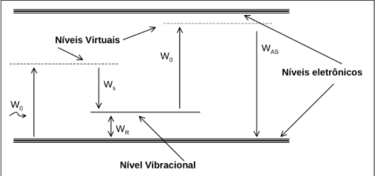
emission. Most of the scattered light may have the same frequency that the incident light (Rayleigh scattering – elas-tic). However, a small fraction of the incident light (hυi) might have its power reduced (h(υi-υR) stokes) or increased (h(υi+υR) anti-stokes) (Raman scattering – inelastic) (fig-ure 1). Considering that the power of light is proportional to the frequency, the change in frequency of the light in-elastically scattered is equal to the vibrational frequency of the scatter molecule. Such energy-exchange process among the molecule, the scatter and the incident light is known as Raman effect. From the energy perspective, the Raman scattering process may be considered as the transi-tion of a molecule from a fundamental stage to a charged vibrational stage, followed by a simultaneous absorption of an incident photon, and emission of a scattered photon (Raman). The scattered Raman light may be assessed by a spectrometer, and its intensity is shown as a function of its changed frequency (Raman displacement). As each mole-cule sample has its own molecular vibrational set, the Ra-man spectrum of a particular sample would be a series of peaks, each one displaced by the characteristic vibrational frequency of that molecule, thus allowing identification of the molecule under investigation. Raman displacement is typically measured in wavelength (cm-1), a convenient unit to relate the frequency change of the scattered light to the frequency of the incident light.
Cells and tissues are made of proteins, nucleic acids, polysaccharides, lipids, vitamins and other components that form the molecular compound, with a highly complex struc-ture. All diseases cause changes in cell and/or tissue bio-chemistry. The current challenge of modern medicine is to find non-invasive and non-destructive analytical techniques to investigate such changes. Few analytical methods meet these requirements and are sensitive enough to reveal com-position and structural details. The most outstanding tech-niques in this field are the NMR, infrared (IR), and Raman spectroscopy. Among them the Raman and the infrared spectroscopy are currently emerging as powerful methods for medical diagnosis, considering that microscopic chang-es characteristic of the disease may be detected by optical spectroscopy, thus allowing a non-invasive in vivo diagno-sis.
Raman and IR spectroscopy may provide detailed bio-chemical information that may be used to detect diseases in their early stages. As the vibrational spectroscopy tech-nique may be employed with the use of optical fibers, sam-ple collection is not necessary, which is a major advantage in face of conventional biopsy techniques and further his-topathological testing21. Once optical diagnosis is made, the assailed organ may be effectively treated before it re-achs an advanced stage. Vibrational spectroscopy
tech-niques, such as Raman and IR, have been recently used to investigate human and animal skin cancer22,23, and for ath-erosclerosis diagnosis24,25. Raman spectroscopy has also been used to study skin inflammatory diseases, and to as-sess the effect of drugs on the skin25. According to the vi-bration nature, which is determined by the molecule sym-metry, infrared or Raman vibrations may be allowed or not. The purpose of this investigation paper is to assess in-frared Raman spectroscopy as a possible method to detect musculoskeletal lactic acid.
MATERIAL AND METHODS]
For the experiments, male Wistar rats with weight rang-ing from 250 to 350 g, from UNIVAP’s Research and Devel-opment Institute were used. The rats were kept at an aver-age temperature of 25oC, light/darkness cycle of 12 h, with water and ad libitum food until the time of experiment.
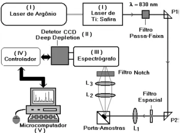
At the time of the experiment, the animals were sedated with sodium pentobarbital (40 mg kg-1 i.v.) via intraperi-toneum, and fastened on a surgical bed. Anterior tibial muscle and tibial nerve were dissected and isolated. The animal was placed on the Raman spectroscopy equipment (figure 2), to acquire the characteristic spectra.
The Raman system
Raman spectroscopy (RS) is a vibrational spectroscopy technique, used to determine molecular structure and ma-terial identification and appraisal. The main application of this technique in the chemical-pharmaceutical are is the non-destructive assessment of solid, liquid and gas prod-ucts, and a swift identification of the sample.
The Raman system block diagram is shown in figure 2. In short, a 5 W argon laser is used to pump a solid Ti:saphire laser. Argon laser was installed and aligned as to provide maximum power. The Ti:saphire laser was installed an aligned as to provide maximum laser power in wave length
Nível Vibracional
Níveis eletrônicos Níveis Virtuais
W0
Ws
WR
W0
WAS
Fig. 1 – Sketch of electron transition for obtaining stoke and anti-stoke
the maximum distance the length of the rat tibial muscle allowed.
Handling of the spectra
The spectra from the acquisitions in the Raman system were filtered by a digital filter, and implemented with use of Matlab software. A good portion of the spectra noises was deleted, for a better observation of the desired peak, and for measurement of peak intensity, in order to com-pare diffusion at different moments after lactic acid injec-tion.
RESULTS
Raman spectrum of rat tibial muscle
Raman spectra of the control rats tibial muscle (i.e., with no lactic acid injection) were performed. Figure 3 shows a characteristic spectrum of rat tibial muscle without lactic acid injected.
Raman lactic acid spectrum in quartz bowl
The Raman lactic acid spectrum presented several and well-resolved peaks within the frequencies of our system, which reaches 1,500 cm-1. These frequencies relate to the tune ranging between 750 and 950 nm. Raman signal
scat-tered on the sample is collected at 90 degrees, with notch-type filters, and focused at the entrance of the spectrograph breach. Notch filters eliminate the scattered Rayleigh light and transmit the Raman signal to the spectrograph, to be dispersed. The light scattered by the spectrograph is de-tected by a liquid nitrogen-refrigerated Deep Depletion CCD. The Ti:saphire laser wavelength passes through a ho-lographic Kaiser filter to eliminate unwanted light and trans-mitting the desired wavelength only. The charging laser is then focused on the sample, after passing the dispersion optic device.
After preparation of the whole system, a fifty-second acquisition procedure was performed to obtain the Raman spectrum of the muscle without lactic acid (basal). Then 50 µl of lactic acid at 86% in a 9.4 mol/l concentration was injected with a 1 µl syringe in the proximal muscle area. After the injection, one waited for three minutes and then the acquisitions in the Raman system were made, each one of them for 50 seconds.
Injection of lactic acid and spectrum acquisition in the in vitro experiment
The muscle was prepared as previously described and fastened to the sample display carrier, for continuous 200 s acquisitions at a 4-minute interval.
To assess acid diffusion in the muscle, a 50 µl lactic acid injection at a concentration of 9.4 mol/l was given. After the injection, seven acquisitions at 4-minute were further made, in a total of 11 acquisitions.
This allowed the design of a chart showing diffusion of lactic acid in the muscle. The distance from the injection site to the laser incidence site on the muscle was of 1.5 cm,
Fig. 2 – Block diagram showing the bench Raman spectroscopy system
-2 0 0 0 2 0 0 4 0 0 6 0 0 8 0 0 10 0 0 12 0 0
7 0 0 80 0 9 0 0 10 00 1 10 0 12 0 0 1 30 0 14 0 0 1 5 00
M úsculo sem ácido láctico injetado
Deslocam ento Ram an (cm-1)
In
ten
si
d
ad
e (u
.a)
Fig. 3 – Spectrum characteristic of the rat tibial muscle without lactic
acid. The plotting represents the six animals used.
1454 830
0 5000 10000 15000 20000 25000 30000 35000 40000
600 700 800 900 1000 1100 1200 1300 1400 1500 1600
Ácido láctico 86%
Deslocam ento Ram an (cm-1)
In
ten
si
d
ad
e (u
.a)
Fig. 4 – Raman spectrum of lactic acid at 86%. One can see two
different lactic acid molecule vibration mode, and their main characteristic peaks were 830 and 1,457 cm-1.
Figure 4 shows the spectrum of lactic acid at 86% (Sig-ma Chemical Co., St. Louis, USA), evidencing the several sample peaks in quartz bowl.
Raman lactic acid spectrum in the tibial muscle of rats after lactic acid injection
Figure 5 shows Raman spectrum of rats tibial muscle after lactic acid injection. We can clearly see the presence of characteristic lactic acid peaks at 830 and 1,457 cm-1.
Lactic acid diffusion seen through Raman spectra
Table 1 shows the values of serial acquisition for the assessment of lactic acid diffusion in the tibial muscle of rats. The result of the linear regression analysis shows a 0.95 positive correlation between time of acquisition and intensity of Raman signal, shown in the table.
DISCUSSION
Raman and infrared spectroscopy techniques are emerg-ing as powerful methods for clinical diagnosis, as micro-scopic changes characteristic of a disease can be detected by optical spectroscopy, thus allowing a non-invasive in vivo diagnosis.
As previously mentioned, Raman and IR spectroscopy may provide detailed biochemical information that can be used to detect a disease at an early stage. As the vibrational spectroscopy technique can be used through optic fibers, excision of a sample is not necessary, being an advantage over conventional biopsy techniques for histopathological testing. Depending on the nature of vibration, which is determined by the symmetry of the molecule, they may or may not be done in infrared or Raman.
In some materials, the Raman signal may be neutralized by fluorescence of impurities if the samples are charged
with visible light. However, with the recent development of IR and Raman with Fourier transformation spectrome-ters operating close to the infrared region (NIR-FT-Raman), this scenario has been totally changed. It was shown that most organic samples may be charged by a Nd:YAG laser with λ = 1,064 nm. Considering that many of the biologi-cal material components do not present absorption bands in this range, there is a very low probability of fluores-cence charging. From this perspective, Raman spectrosco-py has been constantly used in medicine and biology for clinical diagnosis, studies with plants, and assessment of environmental pollutants.
In our investigation, we assessed the use of Raman spec-troscopy as a possible tool to identify metabolites such as lactic acid, based in the fact that each biochemical compo-nent in the tissues has a characteristic vibrational spectrum, with narrow and well-resolved bands. As the particular fo-cus of our investigation was to identify intramuscular lac-tic acid, we showed that this technique allowed a signifi-cant resolution of vibrational bands, providing information on the presence of such metabolite when its concentration increased in the observed muscle.
In the protocols where exogenous lactic acid adminis-tration was done through intramuscular injection, it was possible to clearly identify this substance and separate out from other muscle components. However, Pilotto et al.26, in their study, mention that the Raman system was able to detect physiologic levels of lactic acid in the blood, where the signal had higher intensity and lower noise, facilitating the computer reading of the obtained Raman spectrum. In
14 5 4 83 0
0 50 0 1 00 0 1 50 0
60 0 8 00 10 0 0 1 20 0 1 40 0 16 00
M úsculo com ácido láctico
Deslocam ento Ram an (cm-1)
In
ten
si
d
ad
e (u
.a)
Fig. 5 – Characteristic spectrum of the rat tibial muscle with injected
lactic acid. One can observe lactic acid peaks at 830 e 1,454 cm-1. The plotting represents the six animals used.
TABLE 1
Intensity values in arbitrary units (a.u), in relation to the time of their acquisition after lactic acid injection.
The outcome of the linear regression analysis is a positive 0.95 correlation betw een Raman signal time
for acquisition and intensity Raman (p < 0.0001)
Acquisitions Time (min) Intensity (a.u)
01 04 09,433
02 08 10,566
03 12 12,830
04 16 17,358
05 20 20,377
06 24 15,700
07 28 20,507
08 32 22,962
09 36 25,185
10 40 33,750
the muscle, the signal/noise relationship is changed, show-ing a significant increase of noise and a decrease of the studied substance signal, making hard the physiologic de-tection of lactic acid.
Using the Raman system to detect lactic acid, the need for more specific protocols became clear, where the sys-tem instability is lessened by the type of laser used and the type of system alignment before each experiment. There is no question that the type of system used in our investiga-tion is a powerful tool for a qualitative assessment of bio-logic samples. However, its high complexity makes it hard to be used by exercise physiology experts outside the ex-perimental lab. This technology is still under development, and one sees the need of a more robust and closed system, of higher specificity and lower cost, to be used in the prac-tice by professionals of this field.
On the other hand, Pilotto et al.21 described the difficul-ties in the use of the optic fiber Raman system, particularly regarding the intensity of the noise caused by the strong Raman signal from the optic fiber, which directly inter-feres in the lactic acid signal. For this reason, to reach our results we used the bench Raman system.
As to the Raman displacement of the peaks, a small ex-perimental variability was seen, described by Silveira Jr.20 as signal capture by the spectrograph, and the displace-ment variation is due to the position of the cut-off grade in search of the desired signal.
As for the intensity of the peaks, the system showed a more intense peak close to 830 cm-1, as mentioned by Pi-lotto et al.21, and a less intense one close to 1,456 cm-1, being characteristic of the substance. Even though the sec-ond peak was not observed in the in vitro protocol results, because of signal impairment that required a high number of accumulations during acquisitions, such discrepancy was likely due, according to Silveira Jr.20, to the low power from the Ti:saphire laser alignment.
In the in vitro protocol, the increase of peak intensity during lactic acid diffusion was relevant, i.e., it was possi-ble to show the spreading of the substance from the site of administration until the site of signal retrieval, in an at-tempt to monitor lactic acid diffusion in skeletal muscles (table 1). On the other hand, there was a decrease in inten-sity of the Raman signal in two points, which contradicts Silveira Jr.20, who says that even during constant accumu-lation and acquisitions, there may be small differences in the signal intensity retrieval by the spectrograph, chiefly caused by instability of laser power or geometry problems in the incidence of light on the sample. As the different composition of tissues accounts for the observed spectral
difference, the diagnosis of a disease or tissue classifica-tion may be established by identifying such spectral dif-ferences, with the use of the entire spectrum, or the most important transitions and vibrations20.
Raman spectroscopy is a quite interesting technique to be used in the medical field due to its molecular specifici-ty. However, the intensity of the back-scattered Raman sig-nal intensity is extremely weak compared to other radia-tion decay processes, particularly fluorescence. Typically, the intensity of the Raman signal is 1,000 times less in-tense than fluorescent emission. It is thus necessary the use of appropriate instruments to retrieve such weak signal and sometimes the experiment and results assessment are spoiled due signal intensity variation.
An interesting and very important aspect of applying Raman spectroscopy in biologic samples is the proper se-lection of the charging wavelength. Most biologic mole-cules present electronic level transitions in ultraviolet and visible wavelengths. With the use of visible lasers for Ra-man charging, such an argon ion laser, for instance, the fluorescence of these biomolecules prevails over the Ra-man signal. As these molecules do not present electronic transitions to be charged at the infrared, fluorescence is almost non-existent, the lasers with 700-900 nm wavelength should be preferred.
The Raman spectroscopy showed high potential to de-tect lactic acid in tibial muscle of rats, even though the protocols should be better adjusted to the type of system used, so that, in future investigations, there are no instabil-ity problems in the equipment, as in this investigation. The difficulties found in standardizing the experiments are nat-ural, due to the stage of development of this new technolo-gy, showing that further studies are necessary for the tech-nique to be fully applied.
However, the initial results from our investigation clear-ly show that the system is able to identify lactic acid in the skeletal muscles, in physiologic concentrations, as the vi-brational definition power is of high resolution. Moreover, the system was able to assess lactic acid diffusion over time, even with interference from a low Raman signal.
On the other hand, this system must be adjusted to be used in actual physical exertion in humans, as obstacles are present, such as movement and human skin, which is a barrier to capture the signal.
REFERENCES
1. Fox EL, Foss ML. The physiological basis of physical education and athletics, Chapter 2, Wm. C. Brown Publishers, 1989.
2. Edwards RHT. Hypotheses of peripheral and central mechanisms un-derlying occupational muscle pain and injury. Eur J Appl Physiol 1988; 57:275- 81.
3. Bigland B, Furbush F, Woods J. Fatigue of intermittent submaximal vol-untary contractions: central and peripheral factors. J Appl Physiol 1986; 61:421- 9.
4. Fitts R. Muscle fatigue: the cellular aspects. Am J Sports Med 1996; 24:9-14.
5. Sejersted OM, Vollestad NK. Physiology of muscle fatigue and associ-ated pain. Amsterdam: Elsevier Science Publishers BV, 1993.
6. Billat LV, Koralsztein JP. Significance of the velocity at VO2max and
time to exhaustion at this velocity. Sports Med 1996;22:90-8.
7. Yaznek P. Condicionamento físico do atleta transplantado. São Paulo: Sarvier APM, 1994.
8. Fletcher WM, Hopkins FG. Lactic acid in amphibian muscle. J Physiol 1997;35:242-9.
9. Hollmann W. Historical remarks on the development of the aerobic-anaer-obic threshold up to 1966. Int J Sports Med 1985;6:109-16.
10. Wasserman K, McLlory MB. Detecting the threshold of anaerobic me-tabolism in cardiac patients during exercise. Am J Cardiol 1964;14:844-52.
11. Hagberg J. Exercise hyperventilation in patients with McArdle’s dis-ease. J Appl Physiol 1982;52:991-4.
12. Gaesser O, Poole DC. Lactate and ventilatory threshold: disparity in time course at adaptation to training. J Appl Physiol 1986;6:999-1004.
13. Dwyer J, Bybeer R. Heart rate indices of the anaerobic threshold. Med Sci Sports Exerc 1983;15:72-6.
14. Farrel PA, Wilmore JH, Coyle EF, Billing JE, Costill DL. Plasma lactate accumulation and distance running performance. Med Sci Sports 1979; 11:338-44.
15. Kohrt WM. Longitudinal assessment of responses by triathletes in swim-ming, cycling, and running. Med Sci Sports Exerc 1989;21:569-75. 16. Ivy JL. Alteration in the lactate threshold with changes in substrate
avail-ability. Int J Sports Med 1980;2:139-42.
17. Coyle E, Martin WH, Ehsani AA, Hagberg JM, Bloomfield SA, Sina-core DR, et al. Blood lactate threshold in some well-trained ischemic heart disease patients. J Appl Physiol 1983;54:18-23.
18. Coyle EF. Integration of the physiological factors determining endur-ance performendur-ance ability. Exerc Sport Sci Rev 1995;23:25-63. 19. Stainsby WN, Brooks GA. Control of lactic acid metabolism in
contract-ing muscles and durcontract-ing exercise. Exerc Sport Sci Rev 1990;18:29-63. 20. Silveira JR. Correlação entre a técnica de espectroscopia Raman e a
análise histopatológica das placas ateromatosas em artérias coronárias humanas [Tese de Doutorado]. São Paulo: Universidade de São Paulo, 2001.
21. Kendall C, Stone N, Shepherd N, Geboes K, Warren B, Bennett R, et al. Raman spectroscopy, a potential tool for the objective identification and classification of neoplasia in Barrett’s oesophagus. J Pathol 2003;200: 602-9.
22. Crow P, Stone N, Kendall CA, Uff JS, Farmer JA, Barr H, et al. The use of Raman spectroscopy to identify and grade prostatic adenocarcinoma in vitro. Br J Cancer 2003;89:106-8.
23. Wong Kee Song LM, Marcon NE. Fluorescence and Raman spectrosco-py. Gastrointest Endosc Clin N Am 2003;13:279-96.
24. Moreno PR, Muller JE. Identification of high-risk atherosclerotic plaques: a survey of spectroscopic methods. Curr Opin Cardiol 2002;17:638-47. 25. Van de Poll SW, Romer TJ, Puppels GJ, van der Laarse A. Imaging of atherosclerosis. Raman spectroscopy of atherosclerosis. J Cardiovasc Risk 2002;9:255-61.