research, have been successfully used in the analysis of body segment movements in functional or diagnostic activities in other medical specialties,(2,5) principally in orthopedics and neurology. In pulmonology, such systems provide biomechanical,(6) morphometric(7) and kinematic evidence,(8-11) involved in respiratory diseases or not, and can be applied in different scenarios while maintaining quality and reproducibility.(11) The type of photogrammetry used in study(5,12,13) represents a promising trend, whose operational differentiation from traditional photogrammetry is in the methodological improvement for the analysis of respiratory movement.
This communication presents the performance of photogrammetry in the identification of quantitative alter-In pulmonology, although impairment of the respiratory
mechanics is not restricted to individuals with respiratory diseases,(1) it can produce results of relevant proportions in such individuals. To study the kinetic aspects of respira-tory pathology, one must have knowledge of respirarespira-tory mechanics and kinematics in order to identify and quantify imbalances of the respiratory muscles, as well as seeking out new technologies,(2) as observed in other medical specialties. Equipment for objective monitoring(1,3) is scarce, and those developed in laboratories are occasionally incompatible with the Brazilian public health care system scenario.
In this panorama, movement analysis systems using imaging, whose adaptation(4) as a noninvasive moni-toring technique has gained ground in state-of-the-art
Impacto de fatores externos sobre a mecânica respiratória avaliada por um modelo fotogramétrico específico: biofotogrametria
Denise da Vinha Ricieri1, Nelson Augusto Rosário Filho2
Abstract
This is a report on a methodological adaptation of the photogrammetric technique, which is used in other medical specialties, for use in analyzing respiratory movements. Photogrammetry and a model of photogrammetry designated biofotogrametria para análise da mecânica respiratória (BAMER, photogrammetric analysis of respiratory mechanics) were tested under previously described pathophysiological conditions: post-exercise dynamic hyperinflation using positive end-expiratory pressure. The BAMER model identified an increase in the thoraco-abdominal area following exercise using positive end-expiratory pressure. These results are comparable to those obtained with more robust systems of respiratory kinematics. The use of photogrammetry has value in many areas, since it produces quantitative data, being particularly relevant in pediatrics, in which monitoring resources are scarce.
Keywords: Biomechanics; Photogrammetry; Respiratory mechanics; Thoracic wall.
Resumo
Este é um relato sobre a adaptação metodológica da técnica fotogramétrica, utilizada em outras especialidades, para análise do movimento respiratório. A biofotogrametria e o modelo denominado biofotogrametria para análise da mecânica respiratória (BAMER), foram testados em uma condição fisiopatológica previamente descrita: a presença de hiperinsuflação dinâmica após exercício com uso de pressão expiratória final positiva. O modelo BAMER identificou um aumento da área tóraco-abdominal após exercício com uso de pressão expiratória final positiva, resultado este comparável aos obtidos por sistemas mais robustos em cinemática respiratória. A pesquisa em biofotogrametria possui valor relevante para muitas áreas, posto que agrega dados quantitativos, sendo particularmente relevante na pediatria, onde o moni-toramento é escasso.
Descritores: Biomecânica; Fotogrametria; Mecânica respiratória; Parede torácica.
* Study carried out in the Pediatric Allergy, Immunology and Pulmonology Section of the Postgraduate Program in Child-Adolescent Health of the Pediatrics Department. Universidade Federal do Paraná – UFPR, Federal University of Paraná – Curitiba, Brazil.
1. Assistant Professor in the School of Physical Therapy. Universidade Federal do Paraná – UFPR, Federal University of Paraná – Matinhos, Brazil. 2. Full Professor in the Pediatrics Department. Universidade Federal do Paraná – UFPR, Federal University of Paraná – Curitiba, Brazil.
Correspondence to: Denise da Vinha Ricieri. Hospital de Clínicas da UFPR, Pronto Atendimento Pediátrico Serviço de Alergia, Imunologia e Pneumopediatria, Rua General Carneiro, 181, CEP 80060-900, Curitiba, PR, Brasil.
Tel 55 41 3452-2919. E-mail: [email protected] Financial support: None.
for cardiorespiratory reactions to load. In the second test, performed at the same time of day one week following the first test, the same conditions and load were applied, in addition to using an oronasal mask attached to a unidirectional valve for expir-atory delay set at 5 cmH2O PEEP. The PEEP was introduced 5 min prior to the beginning of the exer-cise in order to adapt the subject to the equipment. It is of interest that, even during the introduction of the mask, without any valve/load, modifications were observed in the thoraco-abdominal behavior.
A Sony TRV-140 camcorder with a shutter speed of 1/60 s, optical axis perpendicular to the sagittal plane of the evaluated individual, and elevated 1.50 m above the floor on a tripod was used to record five respiratory maneuvers at a time, of which only the three intermediate maneuvers were analyzed for each volunteer. The maneuvers were performed from residual volume to total lung capacity and had been previously practiced. The camcorder remained in the same position throughout the experiment, being activated by remote control, for compara-tive records among tests in the images referring to the time points: time point 1 (pre-exercise for the first test); time point 2 (post-exercise for the first test); time point 3 (pre-exercise for the second test, with mask and without the load valve); time point 4 (after 3 min of quiet ventilation, for adaptation to ations in the lateral area of the trunk, induced by
the use of positive end-expiratory pressure (PEEP), using a geometric model, following exercise under controlled conditions. We worked with three male volunteers who were between 40 and 45 years of age, with no history of smoking and with normal spirometric test results. The study was approved by the Ethics in Human Research Committee of the Federal University of Paraná Hospital das Clínicas.
In a model consisting of irregular quadrilaterals over anatomic reference points of the trunk, traced over recorded video images of maximum respiratory maneuvers, we used photogrammetry to calculate the differences between the area of each constituent quadrilateral measured prior to controlled exercise in a stationary cycle ergometer and that measured after the exercise. The exercise was the landmark for the question problem: would the areas of the model, at the end of the test with PEEP, be larger and prone to be detected by photogrammetry when compared to the areas prior to exercise? In order to answer this question, two 30 min exercise tests were performed, with a one-week interval between the two. The tracing of the thoraco-abdominal area of the first test result became the control for compar-ison with the second test, using PEEP.
The tests were performed at 60% of the heart rate reserve, the first test being considered a control
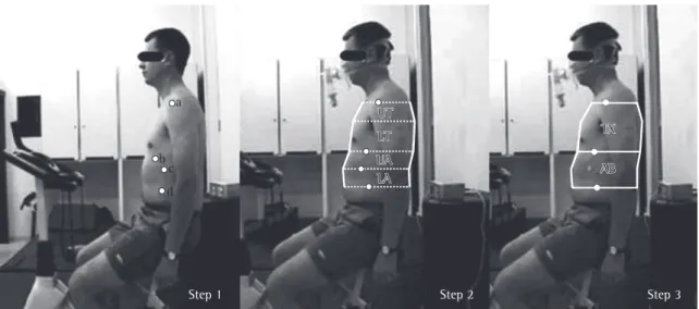
Step 1 Step 2 Step 3
a
b c
d
UT UT
LT LT
UA UA LA LA
TX TX
AB AB
Figure 1 - Photogrammetric analysis of respiratory mechanics model. Step 1: Surface markers delineating the chest wall: (a) acromioclavicular joint, (b) level of xiphoid process; (c) lower angle of the cartilage of the 10th rib (d) umbilicus
sub-compartments. The limits were traced by lines on the silhouette of the body surfaces, anteriorly and posteriorly. The final set was designated biofo-togrametria para análise da mecânica respiratória (BAMER, photogrammetric analysis of respiratory mechanics).
The images captured at each proposed time point were extracted form the footage using the software Corel-R.A.V.E.® 3 (Corel Corporation, Ottawa, Canada). Over those, the BAMER model was traced using Corel-Draw® version 12 (Corel Corporation) and, subsequently, the areas were measured using AutoCAD® 2005 (Autodesk Inc., San Rafael, CA, USA), having the area of one of the surface markers, with an area corresponding to 1.69 cm2, as a calibrator for the conversion. This sequence of procedures equaled the framing of the images recorded on different days, that is, it established a line of identity among the results, which were submitted to statistical treatment in the Statistical Package for the Social Sciences, version 13.0 (SPSS Inc., Chicago, IL, USA), and values of p ≤ 0.05 were considered statistically significant.
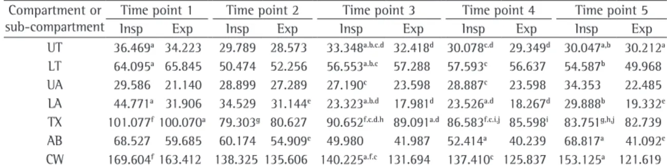
Table 1 gathers the median values obtained from the nine inspiratory and expiratory maneuvers of the three studied subjects, at each time point analyzed, for each division of the model. However, it is important to observe that even in a small trial, the performance of the model confirmed the suspi-cion of the central argument: there was evidence the equipment with PEEP set at 5 cmH2O expiratory
load at the pre-exercise for the second test level); and time point 5 (post-exercise with PEEP).
In order to facilitate identification in the film, the delimitation of the chest wall was highlighted by the distribution of white flat spherical markers, 13 mm in diameter, by the same examiner. The markers remained in place throughout the experiment at the following locations: a) the acromioclavicular joint; b) the xiphoid process; c) the costal arch of the 10th rib; and d) the level of the umbilicus.
Studies which established references for studies on respiratory kinematics(7,14-18) laid the foundation for the model of irregular quadrilaterals. Planes transverse to the references were drawn, and the extent of the chest wall was delineated from the level of the acromioclavicular joint in the upper portion to the level of the umbilicus in the lower portion (Figure 1, Step 1). This line was divided at the level of the xiphoid process(15) (Figure 1, Step 3) into thoracic and abdominal compartments. Other studies(16,18) permitted advancement, with addi-tional divisions, so that each compartment became two new sub-compartments (Figure 1, Step 2). Therefore, a plane at the level of the anterior axillary fold divided the thoracic compartment into upper and lower thoracic sub-compartments; the same occurred for the abdominal compartment, where a plane at the level of the lower angle of the 10th rib cartilage delineated the upper and lower abdominal
Table 1 - Median of the inspiratory and expiratory values in cm2 for the chest wall areas in each compartment and
sub-compartment of the photogrammetric analysis of respiratory mechanics model.* Compartment or
sub-compartment
Time point 1 Time point 2 Time point 3 Time point 4 Time point 5
Insp Exp Insp Exp Insp Exp Insp Exp Insp Exp
UT 36.469a 34.223 29.789 28.573 33.348a.b.c.d 32.418d 30.078c.d 29.349d 30.047a,b 30.212a LT 64.095a 65.845 50.474 52.256 56.553a.b.c 57.288 57.593c 56.637 54.587b 49.968 UA 29.586 21.140 28.899 27.289 27.190c 23.598 28.887c 23.598 34.353 22.485 LA 44.771a 31.906 34.529 31.144e 23.323a.b.d 17.981d 23.526a.d 18.267d 29.888b 19.332e TX 101.077f100.070a 79.303g 80.627 90.652f.c.d.h 89.091a.d 86.583f.c.i.j 85.598i 83.751g,h,j 82.739 AB 68.527 59.685 60.174 54.909e 49.980 41.987 52.414a 40.239 68.817a 41.092e CW 169.604f163.412 138.325 135.606 140.225a.f.c 131.694 137.410c 125.837 153.125a 121.619
The areas measured using BAMER corroborated the following statements: the thoracic inspiratory areas were larger at the post-exercise with PEEP time point (p < 0.05) than at the time points without expiratory load, and this was accompanied by a reduction in the expiratory area of the abdominal compartment (p < 0.05), explained by the reduction in that of the lower abdominal sub-compartment. Healthy subjects who exercised on a stationary cycle ergometer with PEEP(19) had increased pulmonary end-inspiratory volume, mostly due to the thoracic expansion, accompanied by a reduction in abdominal end-inspiratory volume. Similarly, when compared with the inspiratory areas prior to and following the same tests, there was difference (p < 0.05) for the thoracic compartment in the test with PEEP, that is, between the time point 3 and time point 5, as well as between time point 4 and time point 5.
Even though we still need ample scientific inves-tigation, research into the type of photogrammetry used in this study(20) has contributed to pulmon-ology, since it is capable of aggregating quantitative data, giving objectivity to the analysis of respiration patterns and effectiveness in the identification of the kinetic and pathological variations associated with respiratory diseases. This group of contribu-tions is particularly relevant in pediatrics, where resources for respiratory monitoring are scarce.
Therefore, although much remains to be explored, with due caution and rigor, until the definite validation of respiratory photogrammetry as used in this study becomes a reality, we believe that the pathway opened by the use of the BAMER model is promising. It is a relatively simple, low-cost resource, capable of identifying a known patho-physiological phenomenon, such as the increase in the thoraco-abdominal area following exercise using PEEP, as have the more robust systems in respira-tory kinematics.
References
1. Kotani T, Minami S, Takahashi K, Isobe K, Nakata Y, Takaso M, et al. An analysis of chest wall and diaphragm motions in patients with idiopathic scoliosis using dynamic breathing MRI. Spine. 2004;29(3):298-302.
2. Cliquet Jr A, Magalhães da Franca JE, Sônego D, Grana T, Leite FI, Paolillo AR et al. Avanços tecnológicos na prática ortopédica: análises de membros superiores e inferiores. Acta Ortop Bras. 2004;12(1):57-61.
3. Richards JG. The measurement of human motion: A comparison of commercially available systems. Human Movement Science. 1999;18(5):589-602.
of active hyperinflation following the exercise performed using PEEP, detectable by the BAMER model.
Studies with PEEP, applied at rest or during exercise, have shown an increase in the recruit-ment of respiratory muscle activity in different forms: the PEEP modified the level of functional residual capacity and the subsequent inspiratory dynamics.(14) The present study explored the hypoth-esis that the effect could be detectable through the use of the BAMER model.
The pre-exercise inspiratory areas differed for the thoracic compartment and the chest wall. At the pre-exercise time point, the thoracic area in the free test was larger than when PEEP was used, with or without load (p < 0.01); this difference was signifi-cant for the chest wall with mask and without load (p < 0.05). Considering the differentiated nature of the BAMER subdivision, the difference was detected in the upper and lower thoracic sub-compartments and in the lower abdominal sub-compartment, whose areas were larger (p < 0.05) at the pre-exer-cise time points without mask than those obtained with the mask and with load.
A 1996 study indicated an increase in the func-tional residual capacity due to cumulative effect of the expiratory delay at rest, using a 5 cmH2O load. In the present study, the time of use of PEEP at rest was shorter and, although using the same load, no modifications in the thoraco-abdominal area were observed, or at any other BAMER division level.(14) However, at time points 3 and 4, the impact of the mask without load for the patient was identified by the reaction of the respiratory mechanics in significant alterations (p < 0.05) for the thoracic inspiratory areas and the chest wall, for which the BAMER presented variation of the measurements in the upper and lower thoracic sub-compartments and in the lower abdominal sub-compartments, and for the amplitude of the expiratory maneuvers, when the thoracic compartment was different for the modification of the upper thoracic sub-com-partment as well as for alterations in the lower abdominal sub-compartment.
positiva. In: Rev Bras Fisioter. 2006;1(1supl):72-3. Anais do XIII Simpósio Internacional de Fisioterapia Respiratória 2006 Set 5-9; Curitiba.
13. Ricieri DV, Lodovico A, Trentini D, Baraúna MA. Angular photogrammetric analysis of respiratory movements of the chest wall and its correlation with respiratory and anthropometric variables. In: Digital Annals of 14th World Congress for Physical Therapists; 2003 Jun 6-10; Barcelona, Spain.
14. Spahija JA, Grassino A. Effects of pursed-lips breathing and expiratory resistive loading in healthy subjects. J Appl Physiol. 1996;80(5):1772-84.
15. Ferrigno G, Carnevali P, Aliverti A, Molteni F, Beulcke G, Pedotti A. Three-dimensional optical analysis of chest wall motion. J Appl Physiol. 1994;77(3):1224-31.
16. Kenyon CM, Cala SJ, Yan S, Aliverti A, Scano G, Duranti R, et al. Rib cage mechanics during quiet breathing and exercise in humans. J Appl Physiol. 1997;83(4):1242-55.
17. Kondo T, Uhlig T, Pemberton P, Sly PD. Laser monitoring of chest wall displacement. Eur Respir J. 1997;10(8):1865-9. 18. Sanna A, Bertoli F, Misuri G, Gigliotti F, Iandelli I,
Mancini M, et al. Chest wall kinematics and respiratory muscle action in walking healthy humans. J Appl Physiol. 1999;87(3):938-46.
19. Aliverti A, Cala SJ, Duranti R, Ferrigno G, Kenyon CM, Pedotti A, et al. Human respiratory muscle actions and control during exercise. J Appl Physiol. 1997;83(4):1256-69.
20. Ricieri DV. Biofotogrametria: a ciência e seus segredos. 2a. ed. Curitiba: In1spirar; 2005. 90p.
4. Efstathopoulos EP, Costaridou L, Kocsis O, Panayiotakis G. A protocol-based evaluation of medical image digitizers. Br J Radiol. 2001;74(885):841-6.
5. Baraúna MA Duarte F, Sanchez HM, Canto RS, Malusá S, Campelo-Silva CD, et al. Avaliação do equilíbrio estático em indivíduos amputados de membros inferiores através da Biofotogrametria Computadorizada. Rev Bras Fisioter 2006;10(1):83-90.
6. American Thoracic Society/European Respiratory Society. ATS/ERS Statement on respiratory muscle testing. Am J Respir Crit Care Med. 2002;166(4):518-624.
7. Hochman B, Castilho HT, Ferreira LM. Padronização fotográfica e morfométrica na fotogrametria computadorizada do nariz. Acta Cir Bras. 2002;17(4):258-66.
8. Keshner EA. Head-trunk coordination during linear anterior-posterior translations. J Neurophysiol. 2003;89(4):1891-901.
9. Kondo T, Uhlig T, Pemberton P, Sly PD. Laser monitoring of chest wall displacement. Eur Respir J. 1997;10(8):1865-9. 10. Cervera E, del Pobil AP, Martinet P. Improving Image-Based
Visual Servoing with Three-Dimensional Features. Int J Robotics Res. 2003;22(10-11):821-39.
11. Ribeiro AP T-SF, Iunes DH, e Monte-Raso VV. Confiabilidade inter e intra-examinador da fotopodometria e intra-examinador da fotopodoscopia. Rev Bras Fisioter. 2006;10(4):435-9.