Emphysema index in a cohort of patients with no
recognizable lung disease: influence of age*
,**
Índice de enfisema pulmonar em coorte de pacientes sem doença pulmonar conhecida: influência da idade
Bruno Hochhegger, Giordano Rafael Tronco Alves, Klaus Loureiro Irion, José da Silva Moreira, Edson dos Santos Marchiori
Abstract
Objective: To investigate the effects of age on pulmonary emphysema, based on the values of the emphysema index (EI) in a cohort of patients who had never smoked and who had no recognizable lung disease. Methods:
We reviewed the CT scans, reported as normal, of 315 patients. Exclusion criteria were a history of smoking, cardiorespiratory disease, and exposure to drugs that could cause lung disease. From this cohort, we selected 32 patients (16 men and 16 women), matched for gender and body mass index, who were divided equally into two groups by age (< 50 years and ≥ 50 years). We quantified emphysema using a computer program specific to that task. The EI was calculated with a threshold of −950 HU. We also evaluated total lung volume (TLV) and mean lung density (MLD). Results: The overall means for TLV, MLD, and EI were 5,027 mL, −827 HU, and 2.54%, respectively. Mean values in the older and younger groups, respectively, were as follows: for TLV, 5,229 mL vs. 4,824 mL (p > 0.05); for MLD, −846 HU vs. −813 HU (p < 0.04); and for EI, 3.30% vs. 1.28% (p < 0.001). Significant correlations were found between EI and age (r = 0.66; p = 0.001), EI and TLV (r = 0.58; p = 0.001), and EI and MLD (r = −0.67; p < 0.001). The predicted EI per age was defined by the regression equation (r2 = 0.43): p50(EI) = 0.049 × age − 0.5353. Conclusions: It is important to consider the influence of age when quantifying emphysema in patients over 50 years of age. Based on the regression analysis, EI values of 2.6%, 3.5%, and 4.5% can be considered normal for patients 30, 50, and 70 years of age, respectively. Keywords: Pulmonary emphysema; Tomography, spiral computed; Aging; Pulmonary disease, chronic obstructive.
Resumo
Objetivo: Investigar os efeitos da idade no enfisema pulmonar, com base nos valores do índice de enfisema (IE) em uma coorte de pacientes que nunca fumou e que não possuía doença pulmonar conhecida. Métodos: Foram revisados exames de TC, considerados normais, de 315 pacientes. Tabagismo, doenças cardiorrespiratórias e exposição a drogas que poderiam causar doença pulmonar foram critérios de exclusão. Dessa coorte, selecionamos 32 pacientes (16 homens e 16 mulheres), igualmente divididos em dois grupos (idade < 50 anos e idade ≥ 50 anos), que foram pareados por gênero e índice de massa corpórea. Realizou-se a quantificação do enfisema utilizando um programa específico. O IE foi calculado com um limiar de −950 UH. O volume pulmonar total (VPT) e a densidade pulmonar média (DPM) também foram avaliados. Resultados: As médias gerais de VPT, DPM e IE foram 5.027 mL, −827 UH e 2,54%, respectivamente. A comparação entre os mais velhos e os mais novos mostrou as seguintes médias: VPT (5.229 mL vs. 4.824 mL; p > 0,05); DPM (−846 UH vs. −813 UH; p < 0,04) e IE (3,30% vs. 1,28%; p < 0,001). Houve correlações significativas entre IE e idade (r = 0,66; p = 0,001), IE e VPT (r = 0,58; p = 0,001) e IE e DPM (r = −0,67; p < 0,001). O IE previsto por idade foi definido através da equação de regressão (r2 = 0,43): p50(IE) = 0,049 × idade − 0,5353. Conclusões: É importante considerar a influência da idade na quantificação de enfisema em pacientes com mais de 50 anos. Baseado na análise de regressão, valores de IE de 2,6%, 3,5% e 4,5% podem ser considerados normais para pacientes com 30, 50 e 70 anos, respectivamente.
Descritores: Enfisema pulmonar; Tomografia computadorizada espiral; Envelhecimento; Doença pulmonar obstrutiva crônica.
* Study carried out at the Santa Casa Sisters of Mercy Hospital Complex in Porto Alegre, Porto Alegre, Brazil.
Correspondence to: Giordano Rafael Tronco Alves. Rua Prof. Annes Dias, 295, Centro Histórico, CEP 90020-090, Porto Alegre,
RS, Brasil.
Tel. 55 55 9915-9009. E-mail: [email protected] Financial support: None.
Submitted: 5 March 2012. Accepted, after review: 10 April 2012.
Emphysema index in a cohort of patients with no recognizable lung disease: influence of age
J Bras Pneumol. 2012;38(4):494-502
495
1.6 m or more than 1.85 m and those whose weight was below 55 kg or above 90 kg were excluded, given that extreme constitutional differences might have interfered with the final outcomes. Patients in whom CT screening revealed pulmonary, pleural, or cardiac abnormalities were also excluded. The presence of significant respiratory artifacts also constituted an exclusion criterion. The medical records of all patients were reviewed for data analysis. For precise determination of height and weight, a routine questionnaire was administered to all of the patients prior to CT scanning. When available and convenient, information gathered during subsequent medical visits, as well as ancillary test results, was also reviewed. Because all CT scans were retrospectively analyzed and because the patients were to remain anonymous, no written informed consent was required, and the study was approved by the local research ethics committee. After applying all of the exclusion criteria, we selected a cohort of 32 patients. The non-enhanced CT images of the chest of those patients (16 men
and 16 women in the 23-78 year age bracket) were post-processed with the syngo InSpace 4D
software (Siemens Medical Systems, Forchheim, Germany) for emphysema quantification. The cohort was divided into two groups, by age (< 50 years and ≥ 50 years). The younger group comprised 8 males and 8 females, as did the older group. The patients in the two groups were matched for gender and body mass index in order to highlight the influence of age. Total
lung volume (TLV) and mean lung density (MLD) were calculated for values ranging from −1,024 HU to −400 HU, the latter being the standard threshold for the software. A threshold of −950 HU was selected for “emphysema” quantification.
Finally, two experienced thoracic radiologists reviewed the images.
The CT scans were obtained with a CT scanner with 64 rows of detectors (SOMATOM Sensation 64 Systems; Siemens Medical Systems), CT parameters being as follows: collimation, 32 × 0.6 mm (with z-flying focal spot producing 64 overlapping 0.6-mm slices per rotation); rotation time, 0.33 s; and pitch, 1.3. Radiation dose was
set at 120 kV and 200 mAs (dose modulation
was allowed for optimization according to patient size and anatomical shape). Images were reconstructed for contiguous 1.00-mm axial images with a medium sharp reconstruction kernel
Introduction
Pulmonary emphysema is defined as an abnormal permanent enlargement of the air spaces distal to the terminal bronchioles, accompanied by destruction of the alveolar walls and without obvious fibrosis.(1) Pulmonary emphysema is a major public health problem; it is currently ranked 12th as a cause of disease burden worldwide and is projected to rank 5th by 2020 as a cause of life-years lost and lost quality of life.(2)
Degeneration of elastic fibers in the respiratory
bronchioles, alveolar ducts, and alveoli occurs as part of the natural aging process, usually in individuals over 50 years of age.(3,4) As a consequence, the density of lung parenchyma diminishes, because the alveolar ducts become enlarged and the alveoli become shallower.(4) These changes have been designated “senile emphysema”(3,4) and correlate with stage I COPD, which is found in approximately 35% of “healthy”
elderly nonsmokers.(5)
Because pulmonary emphysema is defined on an anatomical basis, CT is currently the modality of choice for an accurate and noninvasive assessment of in vivo pathological changes.(6) Additionally, HRCT and helical CT can detect and quantify pulmonary emphysema, HRCT
and helical CT findings correlating well with histopathological findings.(7-14) Finally, modern
CT scanners with multiple rows of detectors—
multidetector CT (MDCT)—allow the acquisition
of thin (< 1-mm) slices of the whole chest in a few seconds, improving spatial resolution and avoiding respiratory artifacts.
The objective of the present study was to investigate the effects of age on pulmonary emphysema, based on the values of the emphysema index (EI) in a cohort of patients who had never smoked and who had no recognizable lung disease.
Methods
(in HU). Various thresholds have been suggested
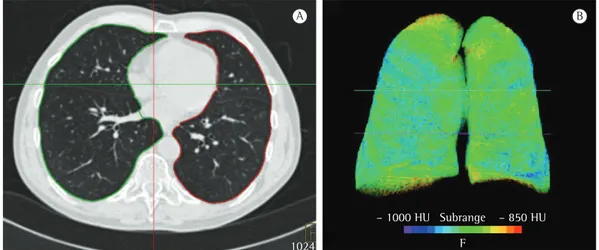
to differentiate between normal and abnormal lungs.(11-13) Based on the acquisition parameters used, we selected a threshold of −950 HU.(11,12) The EI was then calculated by dividing the TLV by the lung volume with densities below −950 HU. The software provides a 3D image showing the
distribution of the areas of emphysema (Figure 1). The normal distribution of the CT densitometry
parameters (TLV, MLD, and EI) was tested by a
normal probability plot with the MedCalc software, version 8.1.1 (MedCalc Software, Mariakerke, Belgium). We accepted a type I error of 5% for patient selection, therefore excluding those above the 95th percentile, which was based on a Student’s t-distribution with 30 degrees of freedom and calculated by the following formula: mean + 1.70 × SEyx
where SEyx is the standard error of the predicted x for each y.
Correlations of TLV, MLD, and EI with age were
calculated by Pearson’s correlation coefficient and tested by the Student’s t-test. The influence of
age on EI and MLD was evaluated by regression
analysis, and the distribution was graphically demonstrated by XY scatter plots. The 50th percentile (p50) of EI was calculated by the following equation:
f(x) = bx + a
where a and b were calculated on the basis of the trend line of the distribution of EI per age. The (B40; Siemens). The patients were scanned from
cranial to caudal, holding their breath at the end
of a maximal inspiratory effort. During the study
period, the CT scanner was periodically calibrated in accordance with the recommendations of the manufacturer. The raw data were entered into
a scale with values ranging from −1,024 HU to 3,072 HU. We chose not to use spirometry
for controlling lung volumes, given that the technique can increase the radiation dose without a significant improvement in precision.(15) All examinations were performed without the injection of intravenous contrast medium. A data matrix of 512 × 512 was selected.
Pulmonary emphysema was quantified by CT densitometry and volumetry, an imaging post-processing technique for calculating the volume of an organ (or of part of an organ). The technique uses a whole set of volumetric CT images and attenuation coefficient values (or
density, expressed in HU) in order to segment the
organ. In addition, the technique can measure
absolute TLC (which includes air, blood, and
lung tissue) and calculate the volume of a lung portion whose density is above or below a selected
threshold. We used the syngo InSpace 4D software
(Siemens Medical Systems), which automatically recognizes the lungs and eliminates any structures with an attenuation coefficient higher than
−400 HU. After automatic segmentation, the software calculates TLV, emphysema volumes, and MLD. The operator can choose a threshold
between normal lung and emphysematous lung
Emphysema index in a cohort of patients with no recognizable lung disease: influence of age
J Bras Pneumol. 2012;38(4):494-502 497
parameters were as follows: TLV (r = 0.07; p = 0.71; 95% CI, −0.29 to 0.41); MLD (r = −0.33; p = 0.07; 95% CI, −0.61 to 0.02); and EI (r = 0.66; p = 0.001;
95% CI, 0.38-0.83). Significant correlations were
found between EI and TLV (r = 0.58; p = 0.001; 95% CI, 0.26-0.78) and between EI and MLD (r = −0.67; p < 0.01; 95% CI, −0.83 to −0.39).
No significant correlations were found between
MLD and age or between EI and age when the
younger group was analyzed separately (r = 0.14 and p = 0.6133; and r = 0.34 and p = 0.1921, respectively).
The SEs of the CT parameters for age (SEyx) were as follows: SETLV,age = 1,278 mL;
SEMLD,age = 39.04 HU; and SEEI,age = 1.70%.
Therefore, the p95 values were as follows:
TLV = 7,199 mL; MLD = −894 HU; EI = 5.43%;
and SEyx for EI and TLV = 1.79%. The best
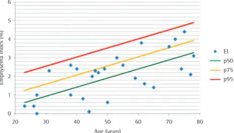
regression equation for the predicted EI per age (r2 =0.43) was as follows:
p50 = 0.049 × age − 0.5353
The SEEI,age for p50 was 0.95%. As shown in
Figure 2, p75 and p95 were calculated by the
following equations:
p75 = p50 + 0.683 × 0.952
p95 = p50 + 1.70 × 0.952
The best regression equation for the predicted
EI per MLD (r2 =0.63) was as follows: p50 = 5EI − 18e − 0.049 × MLD
where e is the constant for EI. best adjustment of the regression equation tested
was measured by determining the r2. The 75th percentile (p75) and the 95th percentile (p95)
were then calculated on the basis of a Student’s t-distribution with 30 degrees of freedom, by the following equations:
p75 = p50 + 0.683 × SEyx
p95 = p50 + 1.70 × SEyx
Finally, the normal distribution was confirmed
for TLV, MLD, and EI, which were plotted as near-straight lines and tested with the
Kolmogorov-Smirnov test.
Results
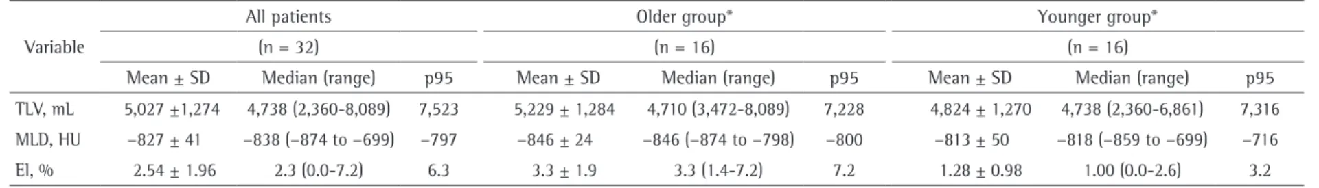
The study population (n = 32) was divided into two groups, by age (< 50 years and ≥ 50 years). The mean age of the individuals in the younger group was 32.8 ± 9.0 years, whereas that of those in the older group was 63.5 ± 8.6 years. Each group comprised 8 men and 8 women, matched for age and body mass index. As shown in Table 1, the overall means for the pulmonary emphysema parameters were
as follows: TLV = 5,027 mL; MLD = −827 HU;
and EI = 2.54%. Mean values in the older and younger groups, respectively, were as follows:
for TLV, 5,229 mL vs. 4,824 mL (p > 0.05); for MLD, −846 HU vs. −813 HU (p < 0.04); and for
EI, 3.30% vs. 1.28% (p < 0.001).
After the exclusion of values above p95, the correlations between age and each of the
Hochhegger B, Alv
es GRT
, Irion KL, Moreira JS, Marchiori E
Table 1 - Emphysema parameters in the groups studied.
Variable
All patients Older group* Younger group*
(n = 32) (n = 16) (n = 16)
Mean ± SD Median (range) p95 Mean ± SD Median (range) p95 Mean ± SD Median (range) p95
TLV, mL 5,027 ±1,274 4,738 (2,360-8,089) 7,523 5,229 ± 1,284 4,710 (3,472-8,089) 7,228 4,824 ± 1,270 4,738 (2,360-6,861) 7,316
MLD, HU −827 ± 41 −838 (−874 to −699) −797 −846 ± 24 −846 (−874 to −798) −800 −813 ± 50 −818 (−859 to −699) −716
Emphysema index in a cohort of patients with no recognizable lung disease: influence of age
J Bras Pneumol. 2012;38(4):494-502
499
of −950 HU as the cut-off point to distinguish
between normal lungs and emphysematous lungs. Ideally, the CT densitometry software should use the same threshold. To our knowledge, our study is the first to address the effects of age on pulmonary emphysema in nonsmokers with
no recognizable lung disease using a 64-MDCT
scanner and volumetric acquisition.
Studies have reported an EI > 0 in healthy
individuals.(22-26) In addition, the EI has been shown to increase with age.(24,26) One group of authors(22) investigated this issue in a cohort of healthy individuals younger than 40 years of age, showing that EI values ≤ 0.35% should be considered normal for volumetric measurements performed with 10-mm collimation, 50 mAs, and a standard reconstruction algorithm; those authors found that the EI was not significantly influenced by age in that age group, a finding that is consistent with those of the present study.
However, other studies,(24,26) particularly those involving older cohorts, have found significant evidence that EI increases with age, as observed in our older group.
The cut-off point of 50 years of age was chosen on the basis of previous studies reporting that the age of 50 years marks the onset of age-related degeneration of elastic fibers in respiratory bronchioles, as well as the onset of enlargement and flattening of the alveoli. (3,5) Interestingly, the age-related changes are remarkably homogeneous, as opposed to the irregular distribution of airspace enlargement in emphysema.(5)
The EI values observed in our cohort of patients were higher than were those reported in a study involving single-slice CT(22) and lower than were those reported in a study involving HRCT.(24) Factors that might have influenced the results include the reconstruction algorithm, radiation
dose, collimation, CT scan manufacturer, and HU
range selected for lung segmentation.(18,20,21,27,28) The
software used in the present study segments the
lungs within a range of −1,024 HU to −400 HU, which results in a TLV that is lower than is that
obtained with lung segmentation within a range
of −1,024 HU to −250 HU.(22) Therefore, although lung volumes can be similar at densities below
−950 HU, proportional differences among TLV
values can be observed at higher densities. Our study has some limitations. The main
limitation was the small sample size. However,
Based on the regression analysis, EI values of 2.6%, 3.5%, and 4.5% can be considered
normal for patients 30, 50, and 70 years of
age, respectively.
Discussion
It has been shown that CT quantification of emphysema correlates well with histopathological findings and pulmonary function test results.
(7-15) The method has been recommended for
use in longitudinal studies of emphysema and is currently considered to be better than functional tests for disease assessment.(6,16) In addition, previous studies have reported that the correlation between CT densitometry and macroscopic morphometry is higher than is that between macroscopic morphometry and subjective visual grading of emphysema.(14)
Emphysema has a long and silent asymptomatic evolution, manifesting clinically only at an advanced stage.(17) Reference EI values establishing
normality are required in order to distinguish between patients with no emphysema and those with mild emphysema or early disease. In order to select a reference value for comparing the EI values in a given patient, we should take into consideration the radiation dose,(18,19) the slice thickness,(18) the reconstruction algorithm,(20) the type of scanner,(21) the HU range selected for lung segmentation (usually −1,024 HU to −400 HU or −1,024 HU to −250 HU),(19,22) and the HU threshold selected in order to distinguish
between normal and emphysematous lung (usually
−970 HU, −950 HU, or −910 HU).(11-13,22) Various HU thresholds have been proposed in
order to distinguish between normal and abnormal lungs.(10-13) The initial suggestion was a threshold of −910 HU for axial scanners, with thicker collimation
(i.e., 10 mm), and for examinations performed with the administration of intravenous contrast medium.(10) For thin-slice collimation (1 mm), Gevenois et al. reported good correlations with pathology specimens when the threshold was set
at −950 HU.(12,13) For examinations using individual axial images (rather than the whole lung volume)
acquired with MDCT scanners, Madani et al.(11) found that the strongest correlation between CT quantification and pathology findings was
obtained with thresholds between −950 HU and −970 HU. However, there is no universally accepted
threshold for volumetric analysis of emphysema by
by CT quantification). Based on our regression analysis, EI values of 2.6%, 3.5%, and 4.5% can be considered normal for patients 30, 50, and
70 years of age, respectively.
Acknowledgements
We would like to thank Dr. Benjamin Pinkey, Dr. Nelson Porto, and Dr. Joe Evans for their
invaluable contribution to the present study.
References
1. The definition of emphysema. Report of a National Heart, Lung, and Blood Institute, Division of Lung Diseases workshop. Am Rev Respir Dis. 1985;132(1):182-5. PMid:4014865.
2. Murray CJ, Lopez AD. Evidence-based health policy--lessons from the Global Burden of Disease Study. Science. 1996;274(5288):740-3. PMid:8966556. http:// dx.doi.org/10.1126/science.274.5288.740
3. Verbeken EK, Cauberghs M, Mertens I, Clement J, Lauweryns JM, Van de Woestijne KP. The senile lung. Comparison with normal and emphysematous lungs. 1. Structural aspects. Chest. 1992;101(3):793-9. PMid:1541148. http:// dx.doi.org/10.1378/chest.101.3.793
4. Gillooly M, Lamb D. Airspace size in lungs of lifelong non-smokers: effect of age and sex. Thorax. 1993;48(1):39-43. PMid:8434351 PMCid:464237. http://dx.doi.org/10.1136/thx.48.1.39
5. Hardie JA, Buist AS, Vollmer WM, Ellingsen I, Bakke PS, Mørkve O. Risk of over-diagnosis of COPD in asymptomatic elderly never-smokers. Eur Respir J. 2002;20(5):1117-22. PMid:12449163. http://dx.doi.org/10.1183/09031936. 02.00023202
6. Newell JD Jr, Hogg JC, Snider GL. Report of a workshop: quantitative computed tomography scanning in longitudinal studies of emphysema. Eur Respir J. 2004;23(5):769-75. PMid:15176695. http://dx.doi.org/10.1183/09031936. 04.00026504
7. Kinsella M, Müller NL, Abboud RT, Morrison NJ, DyBuncio A. Quantitation of emphysema by computed tomography using a “density mask” program and correlation with pulmonary function tests. Chest. 1990;97(2):315-21. PMid:2298057. http://dx.doi.org/10.1378/chest.97.2.315 8. Heussel CP, Herth FJ, Kappes J, Hantusch R, Hartlieb
S, Weinheimer O, et al. Fully automatic quantitative assessment of emphysema in computed tomography: comparison with pulmonary function testing and normal values. Eur Radiol. 2009;19(10):2391-402. PMid:19458953. http://dx.doi.org/10.1007/s00330-009-1437-z 9. Falaschi F, Miniati M, Battolla L, Filippi E, Sostman
HD, Laiolo E, et al. Quantification of pulmonary emphysema with computerized tomography. Comparison with various methods [Article in Italian]. Radiol Med. 1995;90(1-2):16-23.
10. Müller NL, Staples CA, Miller RR, Abboud RT. “Density mask”. An objective method to quantitate emphysema using computed tomography. Chest. 1988;94(4):782-7. PMid:3168574. http://dx.doi.org/10.1378/chest.94.4.782 11. Madani A, Zanen J, de Maertelaer V, Gevenois PA.
Pulmonary emphysema: objective quantification at multi-detector row CT--comparison with macroscopic and it should be recognized that elderly patients
without signs of respiratory disease (also known
as “primary” aging patients) constitute a limited
group, accounting for less than 10% of the total elderly population.(29) Two other important limitations of our study were its retrospective nature and the fact that our patients had been diagnosed with extrathoracic malignancy, which means that they could not be ideally classified
as healthy. However, none of the patients had
been diagnosed with pulmonary emphysema or previous lung disease, as reported in their medical records or as seen on CT scans. Finally, despite our rigorous criteria for selecting and matching the patients, we should state that the equations work better for patients in the same height and weight range and for examinations performed with similar scanners and the same acquisition and software parameters.
One group of authors proposed the use of
the percentile density (PD) rather than the EI
in longitudinal studies of emphysema.(30) The EI is based on the assumption that voxels with densities below a chosen threshold represent emphysema, given that the proportion between lung tissue and air is reduced to a point in which the density of those lung portions is very similar
to the density of air. In contrast, PD (which is usually set at 15%) is defined as the HU value
below which a chosen proportion of the lungs is rated, based on a frequency distribution histogram.
A PD of 15% has been proposed as a parameter
to evaluate emphysema progression.(6) However, Madani et al.(11) found that a PD of 1% correlated best with histopathological findings. We chose to
use EI rather than PD because we do not agree that PD, regardless of the chosen setting, can
actually quantify emphysema. For instance, if
PD is applied to a completely consolidated lung,
the frequency distribution histogram will always
have 1% or 15% of voxels below the HU value of the selected PD (regardless of the percentile
Emphysema index in a cohort of patients with no recognizable lung disease: influence of age
J Bras Pneumol. 2012;38(4):494-502
501
21. Yuan R, Mayo JR, Hogg JC, Paré PD, McWilliams AM, Lam S, et al. The effects of radiation dose and CT manufacturer on measurements of lung densitometry. Chest. 2007;132(2):617-23. PMid:17573501. http:// dx.doi.org/10.1378/chest.06-2325
22. Irion KL, Marchiori E, Hochhegger B, Porto Nda S, Moreira Jda S, Anselmi CE, et al. CT quantification of emphysema in young subjects with no recognizable chest disease. AJR Am J Roentgenol. 2009;192(3):W90-6. PMid:19234245. http://dx.doi.org/10.2214/AJR.07.3502
23. Bnà C, Zompatori M, Ormitti F, Sverzellati N, Verduri A. High resolution CT (HRCT) of the lung in adults. Defining the limits between normal and pathologic findings. Radiol Med. 2005;109(5-6):460-71. 24. Gevenois PA, Scillia P, de Maertelaer V, Michils A, De
Vuyst P, Yernault JC. The effects of age, sex, lung size, and hyperinflation on CT lung densitometry. AJR Am J Roentgenol. 1996;167(5):1169-73.PMid:8911175. 25. Vikgren J, Boijsen M, Andelid K, Ekberg-Jansson A,
Larsson S, Bake B, et al. High-resolution computed tomography in healthy smokers and never-smokers: a 6-year follow-up study of men born in 1933. Acta Radiol. 2004;45(1):44-52. PMid:15164778. http://dx.doi. org/10.1080/02841850310002970
26. Horiuchi N, Fujita J, Suemitsu I, Yamasaki Y, Higa F, Tateyama M. Low-dose multislice CT and high-resolution CT assessment of pulmonary emphysema in public school teachers. Lung. 2007;185(1):25-30.PMid:17294335. http://dx.doi.org/10.1007/s00408-006-0082-4 27. Heussel CP, Kappes J, Hantusch R, Hartlieb S, Weinheimer
O, Kauczor HU, et al. Contrast enhanced CT-scans are not comparable to non-enhanced scans in emphysema quantification. Eur J Radiol. 2010;74(3):473-8. PMid:19376661. http://dx.doi.org/10.1016/j. ejrad.2009.03.023
28. Schilham AM, van Ginneken B, Gietema H, Prokop M. Local noise weighted filtering for emphysema scoring of low-dose CT images. IEEE Trans Med Imaging. 2006;25(4):451-63. PMid:16608060. http:// dx.doi.org/10.1109/TMI.2006.871545
29. Bonomo L, Larici AR, Maggi F, Schiavon F, Berletti R. Aging and the respiratory system. Radiol Clin North Am. 2008;46(4):685-702, v-vi. PMid:18922288. http:// dx.doi.org/10.1016/j.rcl.2008.04.012
30. Stoel BC, Parr DG, Bakker EM, Putter H, Stolk J, Gietema HA, et al. Can the extent of low-attenuation areas on CT scans really demonstrate changes in the severity of emphysema? Radiology. 2008;247(1):293-4; author reply 294. http://dx.doi.org/10.1148/radiol.2471071608 microscopic morphometry. Radiology. 2006;238(3):1036-43.
PMid:16424242. http://dx.doi.org/10.1148/ radiol.2382042196
12. Gevenois PA, de Maertelaer V, De Vuyst P, Zanen J, Yernault JC. Comparison of computed density and macroscopic morphometry in pulmonary emphysema. Am J Respir Crit Care Med. 1995;152(2):653-7. PMid:7633722. 13. Gevenois PA, De Vuyst P, de Maertelaer V, Zanen
J, Jacobovitz D, Cosio MG, et al Comparison of computed density and microscopic morphometry in pulmonary emphysema. Am J Respir Crit Care Med. 1996;154(1):187-92. PMid:8680679.
14. Bankier AA, De Maertelaer V, Keyzer C, Gevenois PA. Pulmonary emphysema: subjective visual grading versus objective quantification with macroscopic morphometry and thin-section CT densitometry. Radiology. 1999;211(3):851-8. PMid:10352615. 15. Gierada DS, Yusen RD, Pilgram TK, Crouch L, Slone RM,
Bae KT, et al. Repeatability of quantitative CT indexes of emphysema in patients evaluated for lung volume reduction surgery. Radiology. 2001;220(2):448-54. PMid:11477250.
16. Stolk J, Putter H, Bakker EM, Shaker SB, Parr DG, Piitulainen E, et al. Progression parameters for emphysema: a clinical investigation. Respir Med. 2007;101(9):1924-30. PMid:17644366. http://dx.doi.org/10.1016/j. rmed.2007.04.016
17. Litmanovich D, Boiselle PM, Bankier AA. CT of pulmonary emphysema--current status, challenges, and future directions. Eur Radiol. 2009;19(3):537-51. PMid:18825385. http://dx.doi.org/10.1007/s00330-008-1186-4 18. Madani A, De Maertelaer V, Zanen J, Gevenois PA.
Pulmonary emphysema: radiation dose and section thickness at multidetector CT quantification--comparison with macroscopic and microscopic morphometry. Radiology. 2007;243(1):250-7. PMid:17392257. http:// dx.doi.org/10.1148/radiol.2431060194
19. Zaporozhan J, Ley S, Weinheimer O, Eberhardt R, Tsakiris I, Noshi Y, et al. Multi-detector CT of the chest: influence of dose onto quantitative evaluation of severe emphysema: a simulation study. J Comput Assist Tomogr. 2006;30(3):460-8. PMid:16778622. http://dx.doi.org/10.1097/00004728-200605000-00018 20. Boedeker KL, McNitt-Gray MF, Rogers SR, Truong DA,
About the authors
Bruno Hochhegger
Radiologist. Moinhos de Vento Hospital and Santa Casa Sisters of Mercy Hospital Complex in Porto Alegre, Porto Alegre, Brazil.
Giordano Rafael Tronco Alves
Medical Student. Federal University of Santa Maria, Santa Maria, Brazil.
Klaus Loureiro Irion
Radiologist. Liverpool Heart and Chest Hospital NHS Foundation Trust, Liverpool, United Kingdom.
José da Silva Moreira
Pulmonologist. Santa Casa Sisters of Mercy Hospital Complex in Porto Alegre, Porto Alegre, Brazil.
Edson dos Santos Marchiori