Meningitis and pneumonia in Guatemalan
children: the importance of
Haemophilus
influenzae
type b and
Streptococcus
pneumoniae
Edwin J. Asturias,
1,2Monica Soto,
3Ricardo Menendez,
4Patricia L. Ramirez,
3Fabio Recinos,
5Remei Gordillo,
4Elizabeth Holt,
1and Neal A. Halsey
1Objective. To determine the epidemiology ofHaemophilus influenzaetype b (Hib) and
Streptococcus pneumoniaeinvasive infections in hospitalized Guatemalan children. This is an important issue since Hib vaccine has not been incorporated into the routine immunization program in Guatemala and information from hospital records in 1995 indicated a low inci-dence of Hib andS. pneumoniaeas causes of meningitis and invasive infections.
Methods. Children who were hospitalized in Guatemala City with clinical signs compati-ble with bacterial infections were evaluated for evidence of Hib orS. pneumoniaeinfection. Normally sterile body fluids were cultured, and antigen detection was performed on cere-brospinal fluid (CSF) and pleural fluid.
Results. Of 1 203 children 1–59 months of age hospitalized over a 28-month period, 725 of them (60.3%) had a primary diagnosis of pneumonia, 357 (29.7%) of meningitis, 60 (5.0%) of cellulitis, and 61 (5.1%) of sepsis and other conditions. Hib was identified in 20.0% of chil-dren with meningitis andS. pneumoniae in 12.9%. The average annual incidence of Hib meningitis was 13.8 cases per 100 000 children under 5 years of age, and 32.4% of meningi-tides caused by Hib and 58.7% ofS. pneumoniaemeningitides occurred prior to 6 months of age. Case fatality rates were 14.1%, 37.0%, and 18.0%, respectively, for children with Hib,
S. pneumoniae,and culture-negative and antigen-negative meningitis. Prior antibiotic ther-apy was common and was associated with significant reductions in CSF–culture–positive re-sults for children with other evidence of Hib orS. pneumoniaemeningitis.
Conclusions. Improvements in case detection, culture methods, and latex agglutination for antigen detection in CSF resulted in identification of Hib andS. pneumoniaeas important causes of severe disease in Guatemalan children. Using a cutoff of > 10 white blood cells per cubic millimeter in CSF would improve the sensitivity for detection of bacterial meningitis and help estimate the burden of bacterial meningitis in Guatemala and other developing countries.
Haemophilus influenzaetype b; meningitis,Haemophilus;Streptococcus pneumoniae; vac-cination; Guatemala.
ABSTRACT
Key words
1 Johns Hopkins Bloomberg School of Public Health,
Department of International Health, Baltimore, Maryland, United States of America. Send corre-spondence to: Neal A. Halsey, Johns Hopkins Bloomberg School of Public Health, 615 N. Wolfe St., W5401, Baltimore, Maryland 21205, United States of America; telephone: 410-955-6964; fax:
410-502-6733; e-mail: [email protected] (no reprints available).
2 The Center for Disease Studies and Control,
Guatemala City, Guatemala.
3 Hospital General San Juan de Dios, Department of
Pathology, Guatemala City, Guatemala.
4 Hospital Roosevelt, Department of Pathology,
Guatemala City, Guatemala.
5 Hospital General del Instituto Guatemalteco de
In many industrialized countries the incidence of invasive disease caused byHaemophilus influenzaetype b (Hib) decreased dramatically following widespread use of Hib conjugate vac-cines (1–5). Before these vacvac-cines were available, an estimated one in 200 chil-dren in the United States of America developed invasive Hib disease before 5 years of age (1). Information from a limited number of developing coun-tries in Africa, Asia, and Latin Amer-ica indAmer-icates that Hib is an important cause of childhood morbidity and mortality, responsible for 25%–65% of bacterial meningitis as well as 15%– 25% of severe pneumonia in some areas (4–9). Case fatality rates for meningitis in developing countries are often higher than reported in devel-oped countries because of delays in di-agnosis and suboptimal antimicrobial therapy (4, 10–13). Conjugate vaccines offer the potential for preventing these important causes of morbidity and mortality, but the importance of Hib and Streptococcus pneumoniae is often underappreciated due to failure to ob-tain cultures from sick children and suboptimal microbiologic methods. Introduction of Hib vaccines in devel-oping countries like Guatemala has progressed slowly, despite evidence of its effectiveness. Decision makers in developing countries need data before committing limited resources for the purchase of relatively expensive vac-cines. Since Hib vaccine had not been incorporated into the public routine immunization program in Guatemala, we initiated a surveillance program to detect invasive diseases caused by Hib andS. pneumoniaein order to estimate the burden of severe disease in chil-dren in Guatemala City.
METHODS
Study population
Between 1 October 1996 and 31 Jan-uary 1999 we evaluated children 1 to 59 months of age admitted with clini-cal signs compatible with bacterial infections to the three major referral
hospitals in Guatemala City. We pro-spectively determined the proportion of infections caused by Hib and S. pneumoniae. The three hospitals—Hos-pital General San Juan de Dios, Hospi-tal Roosevelt, and HospiHospi-tal General del Instituto Guatemalteco de Seguri-dad Social (IGSS) (Spanish name )—cap-ture approximately 85% of Guatemala City children hospitalized with menin-gitis. Private hospitals and sanatori-ums serve the remaining 15% of chil-dren presenting with these illnesses. These major referral hospitals also serve adjacent municipalities sur-rounding Guatemala City, which to-gether constitute the Guatemalan met-ropolitan area. The proportion of children who reside in the surround-ing metropolitan area and who de-velop serious infections resulting in re-ferral to these hospitals is unknown. For this reason we calculated inci-dence rates only for children who lived in Guatemala City; the remain-ing analyses include all eligible chil-dren, regardless of residence. Based on projections from the 1994 census, the National Statistics Institute estimates that 326 779 children less than 5 years of age were living in the Guatemalan metropolitan area in 1997; of these, 121 003 were living in Guatemala City.
Case definitions
Children with possible invasive bac-terial disease were identified by daily reviews of admission logbooks and laboratory results at each institution. Meningitis was defined as either a cerebrospinal fluid (CSF) white blood cell count (WBC)≥10 cells/mm3or a
positive bacterial culture or latex ag-glutination antigen test for Hib or S. pneumoniae in the CSF. Data were also analyzed using the World Health Organization (WHO) definition of probable meningitis: CSF protein > 100 mg/dL or glucose < 40 mg/dL or WBC > 100/mm3 with > 80%
neu-trophils. Bacterial meningitis was con-firmed by a positive bacterial CSF cul-ture or latex agglutination antigen test for Hib or S. pneumoniae(14). A child
was diagnosed as having pneumonia if he or she had fever, cough, tachyp-nea, and/or a chest radiograph show-ing infiltrates. Children with wheezshow-ing were excluded unless the chest radi-ograph revealed lobar consolidation; we did that because the primary pur-pose of this study was to identify chil-dren with bacterial disease. Confirmed bacterial pneumonia was defined as detection of any bacteria by blood or pleural fluid culture or a positive pleural fluid latex agglutination test for Hib or S. pneumoniae. The admit-ting physician diagnosed sepsis based on fever and signs of severe illness (e.g., toxic appearance and hypoten-sion). A child with meningitis, pneu-monia, and/or sepsis was classified as having a primary diagnosis of menin-gitis. A child with sepsis and nia was classified as having pneumo-nia. Clinical signs of septic arthritis, epiglottitis, pericarditis, abscess, or cellulitis were used to define these other invasive bacterial diseases. For these illnesses, Hib or S. pneumoniae
disease was diagnosed by culture from blood or other normally sterile fluid.
Laboratory procedures
A preliminary review of 365 CSF samples obtained in one of the hospi-tals in 1995 from children suspected of having meningitis revealed only one Hib isolate and oneS. pneumoniae iso-late. The chocolate agar used in the hospitals at that time was made from discarded human blood. This agar was demonstrated to be less sensitive than commercial chocolate agar sup-plemented with IsoVitaleX™ (Becton Dickinson Microbiology Systems, Cockeysville, Maryland, United States), based on serial dilution of a log phase growth of Hib. Prior to initiating sur-veillance, the laboratories began using supplemented commercial chocolate agar.
Subcultures on IsoVitaleX™-enriched chocolate agar and MacConkey agar were performed at 24 hours and, if tur-bidity developed, in the next 7 days. In May 1997, automated blood cultures for detection of bacterial growth (BACTEC, Becton Dickinson Microbi-ology Systems, Lutherville, Maryland, United States) were introduced at two of the hospitals; that was done at the third hospital in January 1998. Cerebrospinal fluid, pleural fluid, and fluid from other normally sterile sites were also cultured in broth and on IsoVitaleX™-enriched chocolate agar and MacConkey agar following stan-dard procedures.H. influenzaeisolates were confirmed as serotype b by de-tection of Hib antigen by latex aggluti-nation (Directigen, Becton Dickinson Microbiology Systems, Lutherville, Maryland, United States) in the same body fluid or supernatant of broth cul-tures. Isolates with colony morphol-ogy consistent with S. pneumoniae
were confirmed by optochin disk sen-sitivity. Aliquots of CSF, serum, pleural fluid, and other usually sterile fluids were stored at 2 °C to 8 °C and tested within 24 hours for Hib andS. pneumoniae antigens by latex aggluti-nation. Prior to being tested, sera, pleural fluid, and joint fluid were heat-treated in accordance with the manu-facturer’s recommendations. Testing of sera was discontinued after the first year because of the limited added value and the high cost of the tests. Antigen testing for all other fluids was conducted throughout the surveil-lance period.
Statistical methods
Data were collected on standard forms, entered into a database using Epi Info version 6.0 software (Centers for Disease Control and Prevention, Atlanta, Georgia, United States), and analyzed using SPSS-PC version 10 software (SPSS, Inc., Chicago, Illinois, United States). Proportions were com-pared using two-tailed chi-square or Fisher’s exact tests. Meningitis inci-dence rates for Guatemala City
resi-dents were estimated based on the city’s projected population of children less than 5 years of age in 1997 and the assumption that 85% of Guatemala City children with meningitis were cared for at the three study hospitals.
Ethical reviews
The Committee on Human Research of the Bloomberg School of Public Health of Johns Hopkins University and the board for research and educa-tion at each hospital approved the study.
RESULTS
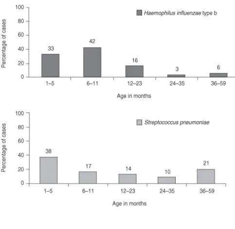
Of the 1 203 children studied, 1 080 (89.8%) had cultures of blood or usu-ally sterile body fluids. Blood culture isolation rates were significantly im-proved (P< 0.001) following the intro-duction of the automated blood cul-ture systems, from 6.3% (24/384) to 14.4% (72/501). Receipt of antibiotics prior to admission was reported for 563 (46.8%) of the children. Meningitis and pneumonia were the diseases most commonly associated with Hib andS. pneumoniaeinfections (Table 1). The highest incidence of Hib and S. pneumoniaewas in the first 12 months
of age, and 33.3% of Hib and 37.7% of S. pneumoniae occurred before 6 months of age (Figure 1).
Meningitis
Of the 357 children with meningitis, 87 of them (24.4%) also had pneumo-nia, 7 (2%) had sepsis, and 4 (1.1%) had both pneumonia and sepsis. Of the 357, 204 of them (57.1%) were male. Bacterial etiology was confirmed in 157 of the 357 children (44.0%), 146 by positive CSF culture or antigen test and 11 by a positive blood culture and inflammatory changes in the CSF. Of the 157 bacteriologically confirmed cases of meningitis, Hib was identified in 71 (45.2%) andS. pneumoniaein 46 (29.3%) (Table 1). Among these cases of bacterial meningitis, Directigen antigen detection identified 26 of the 71 cases of meningitis due to Hib (36.6%) and 10 of the 46 cases of S. pneumoniaemeningitis (21.7%) not de-tected by CSF or blood cultures. Two false-positive CSF Hib latex agglutina-tion tests were observed. In one of those tests a child had S. aureus iso-lated from CSF. In the other false-positive test a child hadS. aureus bac-teremia associated with a peripheral white blood cell count of 51 810 and the CSF culture had no growth; the
TABLE 1. Distribution of invasive diseases by etiology and diagnosis for children 1–59 months of age, Guatemala City, Guatemala, 1 October 1996–31 January 1999
Total cases No with culture
Haemophilus Streptococcus Other organisms or latex
Diagnosis influenzae pneumoniae bacteria found agglutination Total
Meningitis 71 46 40a 200 357 357
Pneumonia 24 30 34b 612 700 725
Sepsis 3 1 0 29 33 34
Cellulitis 6 0 1 49 56 60
Septic arthritis 1 0 2 6 9 9
Epiglottitis 2 0 0 1 3 3
Other 0 0 0 4 4 15
Total 107 77 77 901 1 162 1 203
aThese 40 “other bacteria” were:Neisseria meningitis(7),Salmonellaspp. (8),Staphylococcus aureus(8),Escherichia coli (4),Pseudomonas aeruginosa(5), and other organisms, includingStreptococcusspp. and other enteric gram-negative bac-teria (8).
CSF white blood cell count was 389, with a glucose of 1 mg/dL, and pro-tein of 155 mg/dL.
Antibiotic therapy before hospital admission was reported for 156 of the 357 children with meningitis (43.7%). Among the 37 children with Hib meningitis who received antibiotics before admission, 19 of them (51.4%) had a positive CSF or blood culture, while 26 of the 34 (76.5%) who had not received prior antibiotics had posi-tive CSF cultures (P= 0.05). Similarly, among children with S. pneumoniae
meningitis, 9 of the 16 children (56.3%) with prior antibiotic use had positive CSF cultures, but 27 of the 30 children (90.0%) with no prior antibiotics had positive CSF cultures (P= 0.02).
The average incidence rate for men-ingitis of any cause was 85.4 per 100 000 children/year among children under 5 years of age in Guatemala City; the rate for Hib meningitis was 13.8 per 100 000 and forS. pneumoniaemeningitis, 11.7 per 100 000 children. Fifty-four percent of meningitis occurred prior to 6 months of age and 79.6% before 12 months of age. Of the 71 children with Hib meningitis, 23 of them (32.4%) were younger than 6 months of age, and 27 of the 46 children with
S. pneumoniaemeningitis (58.7%) were also less than 6 months of age. Of the 35 children with meningitis due to
Neisseria meningitidis, Salmonella spp., Escherichia coli, Pseudomonasspp., and other enteric organisms, 22 of them (62.9%) had onset of disease between 1 and 6 months of age. The age distrib-ution for children with no organism identified from CSF was similar to the age distribution for confirmed bacter-ial meningitis. A majority (59.4%) of children with meningitis presented during the dry season (December through May), but no seasonal pattern was observed for meningitis caused by Hib orS. pneumoniae(Figure 2).
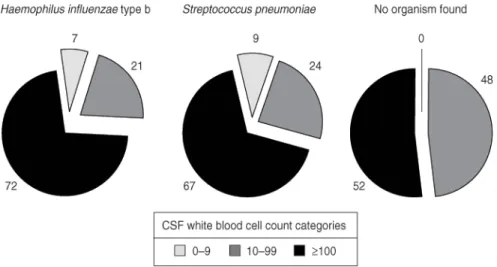
The median CSF white blood cell count for children with bacterial meningitis was 330/mm3. The CSF
contained under 100 WBC/mm3 in
28.6% of children with Hib and 32.6% of children withS. pneumoniae menin-gitis (Figure 3). Of the 71 children with Hib meningitis, 5 of them (7.0%) had FIGURE 1. Age distribution (percentage) of children 1–59 months of age hospitalized with
Haemophilus influenzae type b or with Streptococcus pneumoniae in Guatemala City, Guatemala, 1 October 1996–31 January 1999
under 10 WBC/mm3in the CSF; that
was also true for 4 of the 46 children (8.7%) with S. pneumoniaemeningitis. These 9 children with low CSF white cell counts had positive CSF latex ag-glutination tests for Hib orS. pneumo-niae. Additionally, one of these 5 chil-dren with Hib and all 4 of the chilchil-dren withS. pneumoniaemeningitis had the organism cultured from the CSF.
Of the 357 children with meningitis as defined by CSF > 10 WBC/mm3or
positive culture or latex agglutination test, 280 of them (78.4%) met the WHO criteria for probable bacterial meningi-tis (> 100 WBC/mm3or protein > 100
mg/dL or glucose < 40 mg/dL). Of the 77 children who did not meet the WHO criteria, 9 (11.7%) had positive cultures or latex agglutination tests. Twenty-four additional children who had < 100 WBC/mm3in the CSF met
the WHO criteria for probable bacter-ial meningitis based on CSF protein or glucose. None of these children had a positive blood or CSF culture or latex agglutination test.
The overall case fatality rate (CFR) for meningitis was 23.0% (Table 2). Children withS. pneumoniae meningi-tis had a higher CFR than children with Hib meningitis (37.0% vs. 14.1%,
P= 0.01). Children with no organism identified by culture or antigen detec-tion had a CFR of 18.0%. The CFR for
children with meningitis and pneumo-nia was somewhat higher than the CFR for children with meningitis only (27.5% vs. 20.1%, P= 0.19). The CFR for the 37 children with Hib meningitis who resided outside Guatemala City was higher than the CFR for the chil-dren with Hib meningitis who lived within the city limits (24.3% vs. 3.0% respectively,P< 0.02).
Pneumonia
Seven hundred and twenty-five children had a primary diagnosis of pneumonia; 425 of them (58.6%) were male. Of the 725, 267 of them (36.8%) were under 6 months of age, and 204 of them (28.1%) were between 6 and 12 months of age. The median age for children with primary pneumonia was
8.1 months. A chest radiograph was obtained on 692 of the 725 children with pneumonia (95.4%); lobar or seg-mental consolidation was observed in 463 of the 692 (66.9%) and pleural effu-sions in 65 of the 692 (9.4%).
Just over one half (367 of 725, or 50.6%) of children with pneumonia re-ceived antibiotics before admission. Seven hundred of 725 (96.6%) children with pneumonia had blood or pleural fluid cultures or pleural fluid latex testing. Hib was identified in the blood or pleural fluid in 24 of 700 children (3.4%), andS. pneumoniaein 30 of the 700 (4.3%). An organism was identi-fied by culture or latex agglutination in pleural fluid from 41 of 68 children (60.3%) with pleural effusion. One ad-ditional child with pleural effusion hadStaphylococcus aureusisolated from the blood. Of the 68 children with pneumonia and pleural effusion, S. pneumoniae was identified in 28 of them (41.2%), S. aureus in 8 (11.8%), and Hib in 5 (7.4%).
The overall CFR was 8.0% (58 of 725) (Table 3) for children with a primary diagnosis of pneumonia and 10.1% (7 of 69) for children with pneumonia and pleural effusion.
Sepsis
Presumptive sepsis was diagnosed on admission in 164 children; 7 of these children also had meningitis, 119 also had pneumonia, and 4 also had meningitis and pneumonia. The me-dian age was 5.3 months for children with presumptive sepsis, and 58.5% (96 of 164 were male. Of the 164 chil-dren, 24 children with presumptive FIGURE 3. Distribution (percentage) of cerebrospinal fluid (CSF) white blood cell count
by etiology in children 1–59 months of age with meningitis, Guatemala City, Guatemala, 1 October 1996–31 January 1999
TABLE 2. Case fatality rate by etiology for children with meningitis, Guatemala City, Guatemala, 1 October 1996–31 January 1999
No. of No. of Case fatality Etiology children studied deaths rate (%)
Haemophilus influenzae typeb 71 10 14.1
Streptococcus pneumoniae 46 17 37.0
Other bacteria 40 19 47.5
No organisms found 200 36 18.0
sepsis (14.6%) had an organism iso-lated from blood. Thirty-four of the 164 (20.7%) children with presumptive sepsis were given the final diagnosis of sepsis, as no other focal infection was identified. Hib was identified in 3 of the 33 children (9.1%) with sepsis who had a blood culture, andS. pneu-moniaein 1 child (3.0%).
DISCUSSION
The use of improved culture meth-ods and latex agglutination antigen detection resulted in significantly in-creased rates of identification of Hib andS. pneumoniaeas causes of bacter-ial invasive diseases in Guatemalan children. Other factors contributing to the prior underestimation of Hib and
S. pneumoniae as important causes of severe disease include infrequently obtained blood cultures and high rates of antibiotic use prior to hospitaliza-tion. The estimated annual incidence of Hib and S. pneumoniae meningitis for children under 5 years of age in this population was lower than what has been observed in some other de-veloping countries (4, 5, 15, 16). The true incidence is probably higher than 13.8 per 100 000, given the frequent use of antibiotics prior to hospitaliza-tion and the high CFR (18.0%) in chil-dren with negative CSF cultures and negative latex agglutination antigen detection tests. Other investigators have shown that prior antibiotic
ther-apy was associated with decreased con-centrations of bacteria in the CSF, and many children with culture-negative meningitis have evidence of a bacterial etiology (17, 18). In this study the like-lihood of obtaining a positive culture for children with other evidence of Hib or S. pneumoniae meningitis was significantly lower for children who had reported prior antibiotic therapy than for untreated children.
The high CFRs for Hib meningitis and forS. pneumoniaemeningitis were consistent with observations in other developing countries (4–8). The higher CFR for children with meningitis re-siding outside of Guatemala City was most likely due to delayed therapy and selective referral of children with more severe disease.
Although children with proven bac-terial meningitis generally have high CSF white blood cell counts, using a cutoff point of > 100 WBC/mm3to
de-fine bacterial meningitis would have resulted in missing nine children with Hib or S. pneumoniae meningitis (14). None of the children with fewer than 100 WBC/mm3 in the CSF who met
the WHO criteria for possible menin-gitis based on elevated CSF protein or low glucose had evidence of bacterial etiology based on culture or latex ag-glutination testing of CSF. We believe that a simplified definition of > 10 WBC/mm3is a better screening
crite-rion for suspect meningitis.
In the 1980s, before the introduction of Hib conjugate vaccines in the
United States and other countries where blood cultures were obtained more commonly, meningitis consti-tuted approximately 50% of all in-vasive Hib disease (2, 4). However, meningitis constituted 66.4% of all invasive Hib disease diagnosed in Guatemalan children, most likely due to decreased detection of nonmeningi-tis Hib disease in children referred to hospitals.
The cases of pneumonia due to Hib andS. pneumoniaein this study are un-doubtedly an underestimate of the true burden of disease due to these or-ganisms because of the low use of blood cultures in children presenting with suspect pneumonia and the treat-ment of many children with pneumo-nia as outpatients. During the first year of the study we attempted to in-crease the identification of Hib andS. pneumoniaein children presenting with sepsis or pneumonia by testing sera using latex agglutination. Latex agglu-tination for testing heated sera or pleural fluid resulted in good sensitiv-ity and specificsensitiv-ity for detection of Hib in other studies (19–21). While we were able to identify several cases of Hib disease, the cost-effectiveness of this test is low.
Surveillance for target diseases is important to help understand the po-tential value of vaccines that are under consideration for introduction into im-munization programs. Conjugate Hib andS. pneumoniaevaccines could pvent many of the serious infections re-sulting in hospitalization of Guate-malan children (6, 9, 13, 22–25). An evaluation of the potential impact of Hib conjugate vaccine is under way fol-lowing the introduction of this vaccine in the Hospital General del Instituto Guatemalteco de Seguridad Social.
Acknowledgments. This study
was supported by grants from Glaxo-SmithKline Biologicals and the World Health Organization. Some latex ag-glutination test kits and IsoVitaleX supplements for blood cultures were kindly provided by Becton Dickinson Microbiology Systems. We are particu-larly grateful to the physicians and TABLE 3. Case fatality rate by diagnosis, Guatemala City, Guatemala,
1 October 1996–31 January 1999
No. of No. of Case fatality Diagnosis children studied deaths rate (%)
Meningitis 357 82 23.0
Pneumonia 725 58 8.0
Sepsis 34 6 17.7
Cellulitis 60 0 0.0
Septic arthritis 9 1a 11.1
Epiglottitis 3 1 33.3
Other 15 1 6.7
Total 1 203 149 12.4
nurses of the Hospital General San Juan de Dios, Hospital Roosevelt, and the Hospital General del Instituto Guatemalteco de Seguridad Social for
enrolling and caring for these patients, to James D. Dick for his contributions to the design and improvement of the microbiology methods employed,
to Bonnie King for data management, and to Melissa Romine and Tina Proveaux for their assistance with manuscript preparation.
1. Adams WG, Deaver KA, Cochi SL, Plikaytis BD, Zell ER, Broome CV, et al. Decline of childhoodHaemophilus influen-zaetype b (Hib) disease in the Hib vaccine era. JAMA 1993;269(2):221–226.
2. Broome CV. Epidemiology ofHaemophilus influenzaetype b infections in the United States. Pediatr Infect Dis J 1987;6(8):779– 782.
3. Cochi SL, Broome CV, Hightower AW. Im-munization of US children with Hemo-philus influenzaetype b polysaccharide vac-cine. A cost-effectiveness model of strategy assessment. JAMA 1985;253(4): 521–529. 4. Peltola H. Worldwide Haemophilus
in-fluenzaetype b disease at the beginning of the 21st century: global analysis of the dis-ease burden 25 years after the use of the polysaccharide vaccine and a decade after the advent of conjugates. Clin Microb Rev 2000;13:302–317.
5. Levine OS, Schwartz B, Pierce N, Kane M. Development, evaluation and implemen-tation of Haemophilus influenzae type b vaccines for children in developing coun-tries: current status and priority actions. Pediatr Infect Dis J 1998:17:S95–112. 6. Levine OS, Lagos R, Munoz A, Villaroel J,
Alvarez AM, Abrego P, et al. Defining the burden of pneumonia in children pre-ventable by vaccination against Haemo-philus influenzaetype b. Pediatr Infect Dis J 1999;18(12):1060–1064.
7. Peltola H. Haemophilus influenzaetype b disease and vaccination in Latin America and the Caribbean. Pediatr Infect Dis J 1997;16(8):780–787.
8. Peltola H. Need forHaemophilus influenzae type b vaccination in Asia as evidenced by epidemiology of bacterial meningitis. Pediatr Infect Dis J 1998;17(9 Suppl): S148–151.
9. Mulholland K, Hilton S, Adegbola R, Usen S, Oparaugo A, Omosigho C, et al. Randomised trial ofHaemophilus influen-zaetype-b tetanus protein conjugate vac-cine for prevention of pneumonia and meningitis in Gambian infants. Lancet 1997;349(9060):1191–1197.
10. Salih MA, Khaleefa OH, Bushara M, Taha ZB, Musa ZA, Kamil I, et al. Long term sequelae of childhood acute bacterial
meningitis in a developing country. A study from the Sudan. Scand J Infect Dis 1991;23(2):175–182.
11. World Health Organization. Antimicro-bial and support therapy for bacterial meningitis in children: report of the meet-ing of 18–20 June, 1997, Geneva, Switzer-land. Geneva: WHO; 1998. Available from: http://www.who.intemc-documents/ meningitis/whoemcbac982c.html [Inter-net site]. Accessed 19 February 2001. 12. Molyneux E, Walsh A, Phiri A, Molyneux
M. Acute bacterial meningitis in children admitted to the Queen Elizabeth Central Hospital, Blantyre, Malawi in 1996–97. Trop Med Int Health 1998;3(8):610–618. 13. Mulholland EK, Adegbola RA. The
Gam-bianHaemophilus influenzaetype b vaccine trial: what does it tell us about the burden ofHaemophilus influenzaetype b disease? Pediatr Infect Dis J 1998;17(9 Suppl): S123–125.
14. Levine OS, Schuchat A, Schwartz B, Wenger JD, Elliot J. Generic protocol for population-based surveillance of Haemo-philus influenzae type b. Geneva: World Health Organization, Global Programme for Vaccines and Immunization; 1997. (WHO/VRD/GEN/95.05).
15. Limcangco MR, Salole EG, Armour CL. Epidemiology of Haemophilus influenzae type b meningitis in Manila, Philippines, 1994 to 1996. Pediatr Infect Dis J 2000; 19(1):7–11.
16. Dagan R, Fraser D, Greif Z, Keller N, Kaufstein M, Shazberg G, et al. A nation-wide prospective surveillance study in Is-rael to document pediatric invasive infec-tions, with an emphasis on Haemophilus influenzaetype b infections. Israeli Pedi-atric Bacteremia and Meningitis Group. Pediatr Infect Dis J 1998;17(9 Suppl): S198–203.
17. Shoma S, Rahman M, Yasmin M. Rapid detection ofHaemophilus influenzaetype b in Bangladeshi children with pneumonia and meningitis by PCR and analysis of antimicrobial resistance. J Health Popul Nut 2001;19(4):268–274.
18. Feldman WE. Effect of prior antibiotic therapy on concentrations of bacteria in CSF. Am J Dis Child 1978;132(7):672–674.
19. Ajello GW, Bolan GA, Hayes PS, Leh-mann D, Montgomery J, Feeley JC, et al. Commercial latex agglutination tests for detection ofHaemophilus influenzaetype b andStreptococcus pneumoniae antigens in patients with bacteremic pneumonia. J Clin Microbiol 1987;25(8):1388–1391. 20. Rubin LG, Carmody L. Pneumococcal and
Haemophilus influenzaetype b antigen de-tection in children at risk for occult bac-teremia. Pediatrics 1987;80(1):92–96. 21. Boersma WG, Lowenberg A, Holloway Y,
Kuttschrutter H, Snijder JA, Koeter GH. Rapid detection of pneumococcal antigen in pleural fluid of patients with commu-nity acquired pneumonia. Thorax 1993; 48(2):160–162.
22. Levine OS, Ortiz E, Contreras R, Lagos R, Vial P, Misraji A, et al. Cost-benefit analy-sis for the use of Haemophilus influenzae type b conjugate vaccine in Santiago, Chile. Am J Epidemiol 1993;137(11):1221– 1228.
23. Lagos R, Levine OS, Avendano A, Hor-witz I, Levine MM. The introduction of routineHaemophilus influenzaetype b con-jugate vaccine in Chile: a framework for evaluating new vaccines in newly indus-trializing countries. Pediatr Infect Dis J 1998;17(9 Suppl):S139–148.
24. Wenger JD, DiFabio JL, Landaverde JM, Levine OS, Gaafar T. Introduction of Hib conjugate vaccines in the non-industrial-ized world: experience of four ‘newly adopting’ countries. Vaccine 1999;18: 736– 742.
25. Mulholland K, Levine O, Nohynek H, Greenwood BM. Evaluation of vaccines for the prevention of pneumonia in chil-dren in developing countries. Epidemiol Rev 1999;21(1):43–55.
Manuscript received 27 September 2002. Revised version accepted for publication on 23 July 2003.
Objetivo. Determinar las características epidemiológicas de las infecciones invaso-ras porHaemophilus influenzaetipo b (Hib) yStreptococcus pneumoniaeen niños hospi-talizados en Guatemala. La importancia de este tema radica en que la vacunación con-tra Hib no ha sido incorporada a los programas de inmunización establecidos en Guatemala y en que los registros hospitalarios de 1995 indicaban una baja incidencia de meningitis e infecciones invasoras causadas por Hib yS. pneumoniae.
Métodos. Los niños hospitalizados en la Ciudad de Guatemala con signos clínicos
de infección bacteriana se estudiaron en busca de indicios de infección por Hib o
S. pneumoniae. Se cultivaron líquidos corporales normalmente estériles y se hicieron pruebas de detección de antígenos en líquidos cefalorraquídeo (LCR) y pleural.
Resultados. De los 1 203 niños de 1 a 59 meses de edad hospitalizados en un período de 28 meses, 725 (60,3%) tenían un diagnóstico primario de neumonía, 357 (29,7%) de meningitis, 60 (5,0%) de celulitis y 61 (5,1%) de sepsis u otras afecciones. En 20,0% de los niños con meningitis se detectó Hib y en 12,9% S. pneumoniae. La incidencia media anual de meningitis por Hib fue de 13,8 casos por 100 000 niños menores de 5 años de edad; 32,4% de los casos de meningitis causados por Hib y 58,7% de los cau-sados porS. pneumoniaeocurrieron en niños menores de 6 meses de edad. La tasa de letalidad fue de 14,1%, 37,0% y 18,0%, respectivamente, para los casos de meningitis por Hib, porS. pneumoniaey con resultados negativos tanto en el cultivo como en las pruebas de detección de antígeno. El tratamiento previo con antibióticos fue frecuente y se vio asociado con una reducción significativa de resultados positivos en el cultivo de LCR en los niños que presentaban otros signos de meningitis por Hib oS. pneumo-niae.
Conclusiones. El perfeccionamiento de la detección de casos, los métodos de cultivo y las pruebas de aglutinación con látex para la detección antigénica en LCR permitió identificar a Hib yS. pneumoniaecomo causas importantes de enfermedades graves en niños guatemaltecos. El empleo de un punto de corte de más de 10 leucocitos por milímetro cúbico de LCR mejoraría la sensibilidad de la detección de la meningitis bacteriana y ayudaría a calcular la carga de esta enfermedad en Guatemala y otros países en desarrollo.
RESUMEN