Biomarkers of endothelial function in cardiovascular
diseases: hypertension
Biomarcadores de função endotelial em doenças cardiovasculares: hipertensão
Josynaria Araújo Neves1
*
, Josyanne Araújo Neves1, Rita de Cássia Meneses Oliveira1
Abstract
he incidence of systemic arterial hypertension is increasing worldwide. he foundation of prevention is identiication of people with hypertension. Nowadays, biomarkers are used to diagnose and stratify diseases and estimates prognosis. he objective of this study was to review articles published over the last 5 years on the subject of biomarkers of cardiovascular diseases. he PubMed, SciELO, Science Direct and MEDLINE databases were searched using the keywords: arterial hypertension, cardiovascular biomarkers, nitric oxide, endothelial function and asymmetric dimethylarginine. he studies reviewed show that cardiovascular diseases have complex etiologies. his article describes evidence demonstrating interactions between nitric oxide and asymmetric dimethylarginine that are involved in regulation, in metabolism, and in determination of intracellular levels, and also discusses other biomarkers related to hypertension. Some studies indicate that biomarkers are useful tools for prediction of cardiac events, whereas others state that they have little to contribute to assessments. Careful selection of tests and combinations of tests may be the key to validating use of biomarkers, in view of their low speciicity for diagnosing hypertension.
Keywords: biomarkers; hypertension; endothelial function; ADMA.
Resumo
A incidência de hipertensão arterial sistêmica está aumentando mundialmente. Sua prevenção baseia-se na identiicação dos hipertensos. Atualmente, biomarcadores são utilizados com ins de diagnosticar, estratiicar e prognosticar doenças. Neste estudo, objetivou-se revisar artigos dos últimos cinco anos relacionados a biomarcadores nas doenças cardiovasculares. Pesquisaram-se dados de PubMed, SciELO, Science Direct e MEDLINE, mediante as palavras-chave: hipertensão arterial, biomarcadores cardiovasculares, óxido nítrico, função endotelial e dimetilarginina assimétrica. Os estudos levantados mostram que as doenças cardiovasculares possuem uma etiologia complexa. Neste artigo, evidenciaram-se interações entre o óxido nítrico e a dimetilarginina assimétrica na regulação, no metabolismo e na determinação dos níveis intracelulares, e reviram-se outros biomarcadores relacionados à hipertensão. Alguns estudos indicam os biomarcadores como uma ferramenta útil na predição de eventos cardíacos, e outros reportam que eles contribuem pouco para a avaliação. A seleção e combinação desses pode ser uma alternativa para validar o uso dos biomarcadores devido à pouca especiicidade existente para diagnosticar a hipertensão.
Palavras-chave: biomarcadores; hipertensão; função endotelial; ADMA.
1 Universidade Federal do Piauí – UFPI, Núcleo de Pesquisa em Plantas Medicinais – NPPM, Teresina, PI, Brazil.
Financial support: None.
Conlicts of interest: No conlicts of interest declared concerning the publication of this article. Submitted: March 02, 2016. Accepted: July 28, 2016.
INTRODUCTION
In view of its dimensions, the risks involved,
and the dificulty of controlling it, systemic arterial hypertension (SAH) can be considered a severe public health problem and one that is associated with a high mortality rate, since it predisposes patients to the
development of cardiovascular diseases (CVDs).1
Cardiovascular diseases affect more than 83.6 million North Americans and, in Brazil, the Ministry of health registered 326,000 deaths caused by these diseases during 2010, corresponding to around 1000 deaths
per day.2
According to Georgiopoulou et al.,3 the incidence of
SAH has increased worldwide. Durande and Gutterman4
state that endothelial cells perform a wide range of homeostatic functions. It has been suggested that the combination of vascular endothelial dysfunction and
SAH is related to local and systemic inlammation.5 It is
known that inlammation is a physiological response to protect against harmful and/or pathogenic stimuli and that endothelial dysfunction is a proinlammatory state involving changes to endothelial functions and that it is associated with SAH, which in turn is a multifaceted disease.
Free radicals are among the pathological factors involved, provoking tissue damage and endothelial dysfunction by disturbing the nitric oxide (NO) equilibrium, causing elevated oxidative stress, high proinlammatory cytokine levels (tumor necrosis
factor alpha - TNF-α; interleukins IL-6 and IL-1β)
and excessive production of inlammatory chemokines
[macrophage inlammatory protein alpha-1 (MIP-1α)
and monocyte chemoattractant protein-1 (MCP-1)].6,7
Systemic arterial hypertension is a pathology that constitutes a risk factor that is associated with high rates of morbidity and mortality because it contributes
to exacerbation of other CVDs and kidney diseases.8
As such, SAH is clearly associated with development of vascular lesions and the emergence of dysfunctions in target organs such as the brain, heart, blood vessels,
and kidneys.9
Biomarkers are widely employed in clinical cardiovascular medicine both for diagnosis and stratification of risk and also for estimating the
prognosis of these pathologies.10 One example of an
application of biomarkers is testing osteoprotegerin levels in cases of heart failure, which may be related to
SAH as a cause of hypertrophy of the myocardium.11
The objective of this study is to conduct a review of articles in the scientiic literature related to the subject of SAH and biomarkers. Over the last 10 years, asymmetric dimethylarginine (ADMA) has emerged
as a promising cardiovascular biomarker. Therefore, searches were run for reports relating to plasma ADMA as a new biomarker of putative cardiovascular risk, such as in hypertension, and also for reports describing the contributions made by incorporation of other models of biomarkers, such as endothelial stem cells, troponin T, vitamin D, and uric acid.
A review of the current literature was conducted using bibliographic references published between 2010 and 2015, identiied by searching the PubMed, SciELO, Science Direct, and MEDLINE databases, using the following keywords and combinations of them: arterial hypertension, cardiovascular biomarkers, nitric oxide, endothelial function, and asymmetric dimethylarginine. Searches were run for these terms in both Portuguese and in English.
ARTERIAL BLOOD PRESSURE AND HYPERTENSION
Arterial blood pressure (BP) can be deined as the force exerted by the blood against a given unit of area of the vascular wall. This pressure is generated by the heart and it is the force (potential energy) that enables blood low and tissue perfusion. Blood low through the circulation provides organs and tissues with oxygen according to need and removes the metabolites produced by cell activity. Maintenance of an appropriate blood pressure is therefore of fundamental importance for the circulatory system to function well.
Understanding the pathophysiology of SAH requires an understanding of the mechanisms that control BP, which is regulated by activities that are integrated between the cardiovascular, renal, neural, and endocrine systems, and by physical factors such as the force of the heart’s contractions and the elasticity of the major thoracic arteries. According
to Franceschini et al.,8 maintenance of BP within the
normal range is dependent on variations in cardiac output and peripheral resistance, with countless substances and physiological systems interacting in a complex manner to guarantee adequate BP levels in a diverse range of scenarios.
Systemic arterial hypertension is a circulatory disorder.12
This multifactorial clinical condition is associated with metabolic and functional/structural abnormalities of target organs (heart, kidneys, and blood vessels), characterized by sustained elevated BP levels, generally above the target levels (systolic pressure ≥ 140 mmHg
and/or diastolic pressure ≥ 90 mmHg).13,14 Systemic
and mortality,15,16 even when patients are taking
antihypertensive medications.
The literature lists certain environmental factors that have been linked with progression and complications of cardiovascular pathologies, including smoking, obesity, inactivity, and others. The combinations of these factors in people with hypertension appear to vary with age, physical inactivity, hyperglycemia, and dyslipidemia, while obesity increases the prevalence of associations
with multiple risk factors.14 Lima et al.17 conducted
a study of Brazilian myocardial revascularization patients and concluded that arterial hypertension, obesity, and inactivity were the most common factors and that 79.5% of the patients exhibited at least three risk factors. It has also been observed that the factors that provoke changes to BP are emerging in
ever-younger age groups.18
Nobre et al.19 point out that the genesis of hypertension
encompasses genetic, environmental, vascular, and neural elements. Hyperactivity of the sympathetic nervous system has been proposed as an important
mechanism of hypertension and CVD.20
Social determinants, such as urbanization, income, aging, and education, inluence development of the
pathology.12 As a result, national and international
guidelines for prevention of SAH recommend lifestyle changes, and this approach has gained support over recent years as survival of patients with chronic
diseases has increased.15
The vascular endothelium plays a role in the pathophysiologic mechanisms leading to SAH.
ENDOTHELIUM AND PATHOPHYSIOLOGIC CHANGES
In physiological conditions, endothelial cells are controlled by hemodynamic factors such as BP and blood low, leading to responses that are dependent on production of chemical mediators and that result in
changes to blood low.21 These cells play a fundamental
role in regulation of vascular tone by synthesis and release of relaxation and contraction factors involved
in cardiovascular homeostasis.22
According to Days et al.,23 the most important
endothelium-derived relaxation factors are NO, endothelium-derived hyperpolarizing factor (EDHF), and prostacyclin (PGI2). The principal contractile factors are prostaglandin H2 (PGH2), thromboxane A2, angiotensin II (Ang II), reactive oxygen species (ROS), and endothelin (ET-1).
Endothelium is an organ with multiple functions. Endothelium’s ability to modulate the vascular lumen by regulating vascular tone (controlling local dilation
and contraction) or by releasing vasoactive factors is one of the primary responses to physiological stimuli
generated by blood low and blood pressure.24-27
In hypertension, the complex mechanism that raises BP via NO deiciency involves increase of sympathetic system tone, of the renin-angiotensin
system (RAS), and of oxidative stress.28,29 All cells,
including endothelial cells, have complex enzymatic and non-enzymatic antioxidants systems that act in synergy to defend the organism from damage caused
by free radicals.30
Nitric oxide is an inorganic, gaseous, highly reactive free radical that is produced by oxidation of the amino acid L-arginine, which is converted
into L-citrulline.23 Nitric oxide is generated from
L-arginine by endothelial NO-synthase in the presence of cofactors. Once produced, it diffuses to vascular smooth muscle cells and activates guanylate cyclase (GC), which results in vasodilation mediated by release of the second messenger cyclic guanosine monophosphate (cGMP) and activation of G-dependent protein kinase, resulting in a reduction in intracellular calcium concentration, followed by vasorelaxation. In addition to this activity, NO has many other functions that take part in regulation of transcription of genes, translation of mRNA, and protein modiication of several enzymes involved in mitochondrial respiration,
in mitogenesis, and in growth.31 The NO and cGMP
signaling system is a well-characterized modulator of cardiovascular function in general and of BP in
particular32 (Figure 1).
There are three isoforms of the enzyme responsible for nitric oxide synthesis (NOS). The neuronal form (nNOS), found in neurons and activated by calcium, the inducible form (iNOS), stimulated by inlammatory cytokines, microbial products, and
mechanical disturbance,33 and the enzyme endothelial
nitric oxide synthase (eNOS), located on chromosome 7 (7q35-36) are responsible for synthesis of NO in the circulation, with vasoprotective and vasodilatory
capacities.34 In addition to inhibiting proliferation
of smooth muscle cells, they prevent recruitment, adhesion, and differentiation of inlammatory cells, platelet aggregation, and production of thrombogenic
thromboplastin.21,35
The activity of eNOS and nNOS is dependent on
the calcium/calmodulin complex (Ca2+/CaM). As such,
it is controlled by variations in the concentration of intracellular calcium, which is an important cytoplasmic signaler. In contrast, iNOS is independent of increases
in intracellular Ca2+ concentrations, according
to Forsterman and Sessa,31 and it is expressed in
by cytokines, inlammatory agents, and mechanical
disturbance, resulting in elevated NO low.23,34
BIOMARKERS OF NO/ADMA
Defects of endothelial function and of NO production have been linked with atherosclerosis, hypertension, diabetes, obesity, inactivity, smoking, advanced age, low bioavailability of L-arginine, and presence of
infectious agents.36
Studies with metabolites of NO, such as nitrite/nitrate (NOx), show that they act as monitors of the state of health of patients with CVDs and can be used as
biomarkers in clinical settings.37 Rajendran et al.38
provided further support for this approach, assessing endothelial function in terms of circulating endothelial biomarkers measured in plasma because there are many attractive candidates for endothelial biomarkers. However, they also pointed out that for many pathologies these molecules offer weak selectivity and speciicity and so if they are used individually they have little predictive value.
Considering that production of NO has been
associated with low bioavailability of L-arginine,36
and that this substrate is an analog of ADMA, studies have suggested that the L-arginine/ADMA ratio has
a direct impact on bioavailability of NO.39 According
to Sharma et al.,40 the L-arginine/ADMA ratio in
plasma from healthy people is approximately 100:1. However, in a pathophysiologic situation, ADMA exhibits higher concentrations when compared to L-arginine and so ADMA may act to competitively inhibit eNOS, since it prevents formation of NO and,
consequently, reduces synthesis of this substrate.41
Elevated ADMA levels may therefore inhibit NO synthesis, compromising endothelial function.
High plasma ADMA levels have been reported in patients with vascular risk factors and have a high predictive value for cardiovascular and cerebrovascular
events, while Nishiyama et al.42 identiied ADMA as
a biomarker of arterial hypertension. The results of this bibliographic review lead to the conclusion that there have not been enough studies of the effects of intracellular ADMA levels on hypertension in the last 5 years.
Studies have also revealed that elevated/high ADMA levels reduce the participation of NO in regulation of vascular tone by acting to directly inhibit eNOS and to reduce its bioavailability by increasing the production and release of ROS by activation of the RAS, leading to vascular dysfunction in isolated
arterial vessels in vitro.41
There is evidence to suggest that the imbalance in NO and ROS levels (reduced and increased,
Figure 1. Endothelial signaling and mechanism of relaxation or contraction of smooth muscle. eNOS: endothelial nitric oxide synthase enzyme; NO: nitric oxide; GC: guanylate cyclase; cGMP: cyclic guanosine monophosphate; Ca2+: calcium ions; ECE: endothelin
respectively) activates the sympathetic nervous system, which is a mechanism that appears to be involved in
the neurogenic aspects of hypertension.43 Therefore,
ADMA could be used as a biomarker, indicating a
reduction in the bioavailability of NO.41
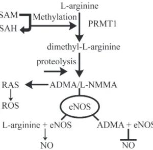
BIOSYNTHESIS OF ADMA
The substrate ADMA is formed after proteolysis of proteins containing methylated residues of arginine. This methylation is facilitated by the enzyme methyltransferase (PRMTs), which uses the S-adenosylmethionine (SAM) protein as the methyl
donor group.44 It is known that there are two types
of PRMTs, differentiated according to their speciic catalytic activity: type 1 catalyzes formation of ADMA and NG-monomethyl-L-arginine (L-NMMA); while type 2 catalyzes formation of symmetrical dimethylarginine (SDMA) and L-NMMA (Figure 2).
Currently, there is interest in degradation of ADMA by dimethylarginine dimethylaminohydrolase
(DDAH),45 which is constituted by two isoforms
(DDAH-1 and DDAH-2). Degradation of ADMA by the enzymatic isoform DDAH-1 takes place in
tissues46 and it has been suggested that modulation
of DDAH-1 activity by agonists of the farnesoid X receptor (FXR), for example -(2,6-dichlorophenyl)-4-(3’-carboxy-2-chlorostilben-4-yl) oxymethyl-5-isopropylisoxazole (GW4064), could be used as a
therapeutic target in treatments for congestive heart
failure and other CVDs.47
According to Caplin et al.,48 80 to 90% of ADMA
is primarily metabolized by DDAHs. There is an alternative route via alanine-aminotransferase
2 glyoxylate (AGXT2) in the kidneys.49 Since ADMA
accumulates in plasma, it could be considered a pathophysiologic cofactor in cardiovascular and kidney diseases.
According to Davids et al.,50 it should be borne
in mind that in some cases the levels of ADMA in circulation do relect intracellular concentrations.
However these levels are not in equilibrium.51
According to Nemeth et al.,45 elevated ADMA
levels in the pericardial liquid of cardiac patients may be indicative of important pathophysiologic mechanisms, such as reduction in bioavailability of NO, contributing to the development of cardiac hypertrophy and remodeling. These authors therefore propose that analysis of this liquid could be used as a diagnostic tool because of interference in the content and effects of pericardial liquid, opening up new treatment options to beneicially modify cardiac function and structure.
It is known that ADMA has been identiied as a risk factor of endothelial dysfunction acting in several different cardiovascular diseases, accelerating their
progression.52 Recent studies in patients with chronic
kidney disease (CKD) suggest that there is a relationship or association between ADMA levels and ibroblast growth factor 23 (FGF-23), and also a relationship
with markers of endothelial cell damage.53,54
Several different studies have assessed conventional and innovative biomarkers for prediction of cardiovascular
events. Reriani et al.55 point out that there is a need
to evaluate the utility of putative biomarkers for evaluation of cardiovascular risk, when compared with endothelial function, because of the small additional value offered by these biomarkers when compared with conventional risk factors. There is speculation that the biomarkers in these studies may only have a minimal role to play in stratiication of cardiovascular risk.
OTHER BIOMARKERS
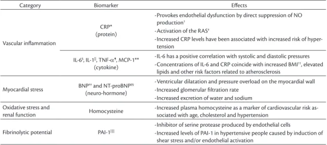
Table 1 shows a list of some well-known biomarkers.
According to Hirata et al.,56 many different markers
have been cited in the literature on vascular endothelial dysfunction, including insulin, adiponectin, vasodilators (nitrite and nitrate), and vasoconstrictors (ROS, endothelin, thromboxane A2). Some of the studies reviewed for this article were focused on endothelial
function and investigation of other possible biomarkers. These are described in the following subsections.
Endothelial Stem Cells (ESCs)
Endothelial stem cells are mononuclear cells that express a combination of endothelial markers (VEGFR2) and progenitors (CD34/CD133) and have a high proliferative capacity. They can be isolated from peripheral blood, bone marrow, and umbilical cord blood. There is no single speciic marker that can be used to identify the cells and currently the most widely accepted method is coexpression of the markers CD133, CD34 and VEGFR2. While these markers are not exclusive to ESCs, their presence in combination is characteristic of a speciic type
of progenitor cell at a speciic stage of maturity.57,58
It has been found that ESCs help in endothelial repair processes and the formation of new vessels, which makes them candidates to be used as markers of cardiovascular health.
Troponin T (hs-cTnT)
McEvoy et al.59 conducted an assessment of the
sensitivity of troponin T (hs-cTnT), which is a marker of subclinical myocardial injury that could possibly identify individuals at risk of hypertension or left ventricular hypertrophy. They concluded that hs-cTnT was associated with the incidence of hypertension and with risk of left ventricular hypertrophy in a clinical population with no history of CVD. However, they emphasized the need for further research to determine
whether hs-cTnT could identify, and beneit clinical screening of, BP or hypertension.
Authors such as Bugnicourt et al.60 and Tu et al.61
have demonstrated that Troponin concentrations could predict a new onset of atrial ibrillation, since they are independently associated with worse clinical outcomes due to severe adverse cardiac events. The exact mechanism by which atrial ibrillation leads to elevated hs-TN is not fully understood and further studies are needed. However, biomarkers such as hs-TNI or hs-TNT are easy to handle and impose lower inancial costs.
Use of hs-TN could identify patients at very high risk and, consequently, allow them to be allocated special treatment with a greater degree of control and deinition of stricter limits of possible risk factors,
such as BP, high cholesterol, and diabetes mellitus.62
It is known that hs-TNI levels are very sensitive for identiication of damage to tissues in the heart.
Vitamin D
The effects of vitamin D on calcium homeostasis and bone metabolism have received a great deal of emphasis in the literature. However, more recent studies have reported that it is associated with several different health problems, including effects on cardiovascular health, since vitamin D can interact with many different mechanisms.
According to the authors Brondum-Jacobsen et al.,63
Grandi et al.,64 Sokol et al.,65 and Wang et al.,66 vitamin
D levels can be used as biomarkers in patients with CVDs.
Table 1. Some of the markers assayed exhibited relationships with cardiac risk.
Category Biomarker Efects
Vascular inlammation
CRP* (protein)
-Provokes endothelial dysfunction by direct suppression of NO
production†
-Activation of the RAS‡
-Increased CRP levels have been associated with increased risk of
hyper-tension
IL-6§, IL-1||, TNF-α¶, MCP-1**
(cytokine)
-IL-6 has a positive correlation with systolic and diastolic pressures
-Concentrations of IL-6 and CRP coincide with increased BMI††, elevated
lipids and other risk factors related to atherosclerosis
Myocardial stress BNP(neuro-hormone)‡‡ and NT-proBNP§§
-Ventricular dilatation and pressure overload on the myocardial wall
-Increased glomerular iltration rate
-Increased excretion of water and sodium
Oxidative stress and
renal function Homocysteine -Increased plasma homocysteine as a marker of cardiovascular risk as-sociated with age, cholesterol and hypertension
Fibrinolytic potential PAI-1||||
-Inhibitor of serine protease produced by endothelial cells
-Increased levels of PAI-1 in hypertensive people caused by induction of
shear stress and/or endothelial activation
The principal vitamin D in circulation is in the form of 25-hydroxyvitamin D. This circulating form binds directly to the vitamin D receptor (VDR) to exert its effect, or it can be converted in the kidneys
by 1 α-hydroxylase to 1.25-dihydroxyvitamin D,
also known as the hormone calcitriol.67,68 It should
be remembered that calcitriol acts by binding with VDR, which is found in the vascular endothelium, vascular smooth muscle, and the myocardium, which raises the possibility of a direct biological effect of vitamin D in the cardiovascular system. The majority of studies report that low levels of 25-hydroxyvitamin D are associated with cardiovascular risk factors such as hypertension, diabetes, and inlammation.
Uric Acid (UA)
Uric acid is currently one of the areas of greatest
interest in research related to SAH. Yanik and Feig69
have demonstrated an association between hyperuricemia and arterial hypertension, providing a basis for using serum levels as a biomarker for diagnosis.
Uric acid is formed by breakdown of adenosine and guanine via hypoxanthine, which is converted into
xanthine and UA by xanthine oxidase.70 The by-products
of reactions catalyzed by xanthine oxidase include ROS such as the superoxide anion, which reacts with protons and NO to generate new ROS. These, in turn, provoke damage to the cardiovascular endothelium
and microvasculature.71
Mamas et al.72 point out that the majority of
biomarkers are not metabolites, like the troponins, which are proteins. Analysis of the metabolites in bodily luids has become an important part of diagnosis and of estimation of prognosis and assessment of therapeutic interventions in clinical applications, since the metabolome is the inal product, downstream of transcription and translation, which means that is more closely related to phenotype.
Currently, the search for biomarkers in the form of speciic metabolites in tissues and/or bodily luids is very intense. One such, ADMA, is naturally a product of human metabolism that is found in circulation and is therefore one of the metabolites found in bodily luids. Biomarkers that can be detected in blood serum or plasma have gained importance because of their eficacy for diagnosis of pathologies. However, despite
all of the advances that have been made, Zhao et al.73
point out that there are still technological limitations, including the lack of a single method for extensive analysis of the entire metabolome, limited spectral libraries and databases, and certain disadvantages of the software available for processing data and extracting biomarkers. The same authors also point
to the need to ind a reasonable method for analysis of metabolites that could substitute or complement the traditional method of diagnosis.
According to Kessler et al.,74 recent advances in
molecular biology have highlighted the possibilities for prevention and treatment, since the high yields and the statistics of genotyping will, in the future, enable identiication of, and intervention in, speciic genomic loci with the potential to change the risk of CVD and, consequently, increase prevention and treatment options for individual people. This is in line
with indings published by O’Donnell and Nabel,75
who conducted genome-wide association studies (GWAS), discovering genetic loci associated with CVDs, including biomarkers in the blood.
According to Zhao et al.,76 the majority of clinical
tests use methods that include tests with a single biomarker, histopathology, and immunohistochemistry. The same authors also state that recent assay methods are generally of low speciicity and sensitivity for a given disease in particular and that the traditional biomarkers only change signiicantly once the disease has caused substantial damage or dysfunction.
Clinical use of biomarkers is compromised in adults with congenital cardiac disease and diagnosis and follow-up of treatment are commonly based on
tests of cardiopulmonary exercise.77 Additionally, the
risk categories employed are very rudimentary and
were identiied more than 50 years ago.78
In general, the ideal biomarker should offer certain characteristics (reliability, sensitivity, and speciicity for the disease), should exhibit minimal variation and should have a low baseline level in healthy people and a high level in the presence of diseases. Additionally, a useful biomarker should be quantiiable using simple and relatively inexpensive methods with results that
are reproducible in many different laboratories.79
FINAL COMMENTS
It should be stressed that CVDs have complex etiology, involving many different factors, interactions, and interrelations, that in combination provoke the physiological mechanisms. Efforts to develop diagnostic methods are concentrating on identiication of biomarkers, since there is a growing need for noninvasive methods to evaluate, monitor, and adjust endothelial function and hypertension.
that other potential markers, such as ESCs, troponin T, vitamin D, and UA, also play roles in these processes. It should be stressed that while some of the studies reviewed indicate that biomarkers have potential for prediction of cardiac events, others state that they contribute relatively little to assessments, especially in low-risk populations. Another issue identiied is related to the speciicity of biomarkers: it is essential to select and combine them because of the correlations between mechanisms of action, and this has attracted the interest of that section of the scientiic community that is focused on reduction of cardiovascular risk, since it could be a viable option for validating these tests, in view of their low speciicity for diagnosis of hypertension.
REFERENCES
1. Gama H, Damasceno A, Silva-Matos C, Diogo D, Azevedo A, Lunet N. Low prevalence of hypertension with pharmacological treatments and associated factors. Rev Saude Publica. 2013;47(2):301-8. PMid:24037357. http://dx.doi.org/10.1590/S0034-910.2013047004328. 2. Simão AF, Précoma DB, Andrade JP, et al. I Diretriz Brasileira de prevenção cardiovascular. Arq Bras Cardiol. 2013;101(6, Supl. 2):1-63. PMid:24554026. http://dx.doi.org/10.5935/abc.2013S012. 3. Georgiopoulou VV, Kalogeropoulos AP, Butler J. Heart failure in hypertension: prevention and treatment. Drugs. 2012;72(10):1373-98. PMid:22747449. http://dx.doi.org/10.2165/11631100-000000000-00000. 4. Durand ML, Gutterman DD. Diversity in mechanisms of endothelium-dependent vasodilation in health and disease. Microcirculação. 2013;20(3):239-47. PMid:23311975. http://dx.doi.org/10.1111/ micc.12040.
5. Dharmashankar K, Widlansky ME. Vascular endothelial function and hypertension: insights and directions. Curr Hypertens Rep. 2010;12(6):448-55. PMid:20857237. http://dx.doi.org/10.1007/ s11906-010-0150-2.
6. Collier P, Watson CJ, Voon V, et al. Can emerging biomarkers of myocardial remodelling identify asymptomatic hypertensive patients at risk for diastolic dysfunction and diastolic heart failure? Eur J Heart Fail. 2011;13(10):1087-95. PMid:21719449. http://dx.doi. org/10.1093/eurjhf/hfr079.
7. Krebs C, Fraune C, Schmidt-Haupt R, et al. CCR5 deficiency does not reduce hypertensive end-organ damage in mice. Am J Hypertens. 2012;25(4):479-86. PMid:22258337. http://dx.doi. org/10.1038/ajh.2011.243.
8. Franceschini N, Reiner AP, Heiss G. Recent findings in the genetics of blood pressure and hypertension traits. Am J Hypertens. 2011;24(4):392-400. PMid:20948529. http://dx.doi.org/10.1038/ ajh.2010.218.
9. Nascimento LR, Molina MCB, Faria CP, et al. Reproducibility of arterial pressure measured in the ELSA-Brasil with 24-hour pressure monitoring. Rev Saude Publica. 2013;47(Suppl 2):113-21. PMid:24346728. http://dx.doi.org/10.1590/S0034-8910.2013047003825. 10. Mamas M, Dunn WB, Neyses L, Goodacre R. The role of metabolites and metabolomics in clinically applicable biomarkers of disease. Arch Toxicol. 2011;85(1):5-17. PMid:20953584. http://dx.doi. org/10.1007/s00204-010-0609-6.
11. Lopez B, Gonzalez A, Lindner D, et al. Osteopontin-mediated myocardial fibrosis in heart failure: a role for lysyl oxidase?
Cardiovasc Res. 2013;99(1):111-20. PMid:23619422. http://dx.doi. org/10.1093/cvr/cvt100.
12. World Health Organization. Global atlas on cardiovascular disease prevention and control. Geneva: WHO; 2011.
13. Sociedade Brasileira de Hipertensão. VI Diretrizes Brasileiras de Hipertensão. Arq Bras Cardiol. 2010 [citado em 2016 mar 02];95(1):1-41. http://publicacoes.cardiol.br/consenso/2010/ Diretriz_hipertensao_associados.pdf
14. Sociedade Brasileira de Cardiologia; Sociedade Brasileira de Hipertensão; Sociedade Brasileira de Nefrologia. VI Diretrizes Brasileiras de hipertensão. Arq Bras Cardiol. 2010;5(Supl.1):1-51. 15. World Health Organization. A global brief on hypertension: silent
killer, global public health crisis. Geneva: WHO; 2013.
16. Martins LC, Figueiredo VN, Quinaglia T, et al. Characteristics of resistant hypertension: ageing, body mass index, hyperaldosteronism, cardiac hypertrophy and vascular stiffness. J Hum Hypertens. 2011;25(9):532-8. PMid:20927128. http://dx.doi.org/10.1038/ jhh.2010.95.
17. Lima FAT, Araújo TL, Lopes MVO, Silva LF, Monteiros ARM, Oliveira SKP. Fatores de risco da doença coronariana em pacientes que realizaram revascularização miocárdica. Rev Rene. 2012 [citado em 2016 mar 02];13:853-60. http://www.revistarene.ufc.br/revista/ index.php/revista/article/viewFile/1080/pdf
18. Anyaegbu EI, Dharnidharka VR. Hypertension in the Teenager. Pediatr Clin North Am. 2014;61(1):131-51. PMid:24267462. http:// dx.doi.org/10.1016/j.pcl.2013.09.011.
19. Nobre F, Coelho EB, Lopes PC, Geleilete TJM. Hipertensão arterial sistêmica primária. Medicina. 2013 [citado em 2016 mar 02];46:256-272. http://revista.fmrp.usp.br/2013/vol46n3/ rev_Hipertens%E3o%20arterial%20sist%EAmica%20prim%E1ria. pdf
20. Grassi G. Sympathetic neural activity in hypertension and related diseases. Am J Hypertens. 2010;23(10):1052-60. PMid:20651696. http://dx.doi.org/10.1038/ajh.2010.154.
21. Prati C, Demougeot C, Guillot X, Godfrin-Valnet M, Wendling D. Endothelial dysfunction in joint disease. Joint Bone Spine. 2014;81(5):386-91. PMid:24565889. http://dx.doi.org/10.1016/j. jbspin.2014.01.014.
22. Pereira AC, Paulo M, Araújo AV, Rodrigues GJ, Bendhack LM. Nitric oxide synthesis and biological functions of nitric oxide released from ruthenium compounds. Braz J Med Biol Res. 2011;44(9):947-57. PMid:21755266. http://dx.doi.org/10.1590/ S0100-879X2011007500084.
23. Dias RG, Negrão CE, Krieger MH. Óxido nítrico y sistema cardiovascular: activación celular, reactividad vascular y variante genética. Arq Bras Cardiol. 2011 [citado em 2016 mar 02];96(1):68-75. http:// www.scielo.br/pdf/abc/v96n1/es_v96n1a12 PMid:21308339. 24. Ando J, Yamamoto K. Effects of shear stress and stretch on
endothelial function. Antioxid Redox Signal. 2011;15(5):1389-403. PMid:20854012. http://dx.doi.org/10.1089/ars.2010.3361. 25. Ferreira LB, Albuquerque AAS, Celotto AC. Fator hiperpolarizante
derivado do endotélio e doenças cardiovasculares. Rev Soc Cardiol. 2011;21:40-5.
26. Melo JB, Figueiredo JAF No, Campos RCA, Meireles MF, Costa ECC, Leal MCM. Estudo da função endotelial no Brasil: prevenção de doenças cardiovasculares. Rev Bras Cardiol. 2014;27:616-23. 27. Menezes IAC, Santos MRV, Cunha CLP. O índice de perfusão
modulation in L-NAME-tread rats. Auton Neurosci. 2013;179(1-2):17. PMid:23810687. http://dx.doi.org/10.1016/j.autneu.2013.06.002. 29. Nakmareong S, Kukongviriyapan U, Pakdeechote P, et al. Tetrahydrocurcumin alleviates hypertension, aortic stiffening and oxidative stress in rats with nitric oxide deficiency. Hypertens Res. 2012;35(4):418-25. PMid:22072109. http://dx.doi.org/10.1038/ hr.2011.180.
30. Lee S, Park Y, Zuidema MY, Hannink M, Zhang C. Effects of interventions on oxidative stress and inflammation of cardiovascular diseases. World J Cardiol. 2011;26(1):18-24. PMid:21286214. http:// dx.doi.org/10.4330/wjc.v3.i1.18.
31. Forstermann U, Sessa WC. Nitric synthases: regulation and function. Eur Heart J. 2012;33(7):829-37, 837a-837d. PMid:21890489. http:// dx.doi.org/10.1093/eurheartj/ehr304.
32. Aroor AR, Demarco VG, Jia G, et al. The role of tissue Renin-Angiotensin-Aldosterone system in the devolopment of endothelial dysfunction and arteial stiffenes. Front Endocrinol. 2013;4:161. PMid:24194732. http://dx.doi.org/10.3389/fendo.2013.00161. 33. Petrella E, Pignatti L, Neri I, Facchinetti F. The L-arginine/nitric
oxide pathway is impaired in overweight/obese pregnant women. Pregnancy Hypertens. 2014;4(2):150-5. PMid:26104420. http:// dx.doi.org/10.1016/j.preghy.2014.01.001.
34. Zhang X, Lynch AI, Davis BR, et al. Pharmacogenetic association of NOS3 variants with cardiovascular disease in patients with hypertension: the genhat study. PLoS One. 2012;7(3):e34217. PMid:22470539. http://dx.doi.org/10.1371/journal.pone.0034217. 35. Teixeira BCA, Lopes AL, Macedo RCO, et al. Marcadores inflamatórios, função endotelial e riscos cardiovasculares. J Vasc Bras. 2014;13:2. http://dx.doi.org/10.1590/jvb.2014.054.
36. Bressler J, Pankow JS, Coresh J, Boerwinkle E. Interaction between the NOS3 Gene and obesity as a determinant of risk of type 2 diabetes: the atheros clerosis risk in communities study. PLoS One. 2013;8(11):e79466. PMid:24278136. http://dx.doi.org/10.1371/ journal.pone.0079466.
37. Ghasemi A, Zahediasl S, Azizi F. Reference values for serum nitric oxide metabolites in an adult population. Clin Biochem. 2010;43(1-2):89-94. PMid:19782059. http://dx.doi.org/10.1016/j. clinbiochem.2009.09.011.
38. Rajendran P, Rengarajan T, Thangavel J, et al. The vascular endothelium and human diseases. Int J Biol Sci. 2013;9(10):1057-69. PMid:24250251. http://dx.doi.org/10.7150/ijbs.7502. 39. Celik M, Iyisoy A, Celik T, Yilmaz MI, Yuksel UC, Yaman H. The
relationship between L-arginine/ADMA ratio and coronary collateral development in patients with low glomerular filtration rate. Cardiol J. 2012;19(1):29-35. PMid:22298165. http://dx.doi. org/10.5603/CJ.2012.0006.
40. Sharma V, Ten-Have GA, Ytrebo L, et al. Nitric oxide and L-arginine metabolism in a devascularized porcine model of acute liver failure. Am J Physiol Gastrointest Liver Physiol. 2012;303(3):G435-41. PMid:22421619. http://dx.doi.org/10.1152/ajpgi.00268.2011. 41. Veresh Z, Debreczeni B, Hamar J, Kaminski PM, Wolin MS, Koller A.
Asymmetric dimethylarginine reduces nitric oxide donor-mediated dilation of arterioles by activating the vascular renin-angiotensin system and reactive oxygen species. J Vasc Res. 2012;49(4):363-72. PMid:22652896. http://dx.doi.org/10.1159/000337485. 42. Nishiyama Y, Ueda M, Katsura KI, et al. Asymmetric dimethylarginine
(ADMA) as a possible risk marker for ischemic stroke. J Neurol Sci. 2010;290(1-2):12-5. PMid:20060545. http://dx.doi.org/10.1016/j. jns.2009.12.020.
43. Hirooka Y, Kishi T, Sakai K, Takeshita U, Sunagawa K. Imbalance of central nitric oxide and reactive oxygen species in the regulation of sympathetic activity and neural mechanisms of hypertension.
Am J Physiol Regul Integr Comp Physiol. 2011;300(4):R818-26. PMid:21289238. http://dx.doi.org/10.1152/ajpregu.00426.2010. 44. Rocha MS, Teerlink T, Janssen MCH, et al. Asymmetric dimethylarginine
in adults with cystathionine β-synthase deficiency. Atheroscler.
2012;222(2):509-11. PMid:22484094. http://dx.doi.org/10.1016/j. atherosclerosis.2012.03.009.
45. Nemeth Z, Cziraki A, Szabados S, Biri B, Keki S, Koller A. Elevated levels of Asymmetric Dimethylarginine (ADMA) in the pericardial fluid of cardiac patients correlate with cardiac hypertrophy. PLoS One. 2015;10(8):e0135498. PMid:26313940. http://dx.doi. org/10.1371/journal.pone.0135498.
46. Hu X, Atzler D, Xu X, et al. Dimethylarginine dimethylaminohydrolase-1 is the critical enzyme for degrading the cardiovascular risk factor asymmetrical dimethylarginine. Arterioscler Thromb Vasc Biol. 2011;31(7):1540-6. PMid:21493890. http://dx.doi.org/10.1161/ ATVBAHA.110.222638.
47. Liu X, Fassett J, Wei Y, Chen Y. Regulation of DDAH1 as a potential therapeutic target for treating cardiovascular diseases. Evid Based Complement Alternat Med. 2013;2013:619207. PMid:23878601. 48. Caplin B, Wang Z, Slaviero A, et al. Alanine-glyoxylate
aminotransferase-2 metabolizes endogenous methylarginines, regulates NO, and controls blood pressure. Arterioscler Thromb Vasc Biol. 2012;32(12):2892-900. PMid:23023372. http://dx.doi. org/10.1161/ATVBAHA.112.254078.
49. Caplin B, Leiper J. Endogenous nitric oxide synthase inhibitors in the biology of disease: markers, mediators, and regulators? Arterioscler Thromb Vasc Biol. 2012;32(6):1343-53. PMid:22460557. http://dx.doi.org/10.1161/ATVBAHA.112.247726.
50. Davids M, van Hell AJ, Visser M, Nijveldt RJ, van Leeuwen PA, Teerlink T. Role of the human erythrocyte in generation and storage of asymmetric dimethylarginine. Am J Physiol Heart Circ Physiol. 2012;302(8):1762-70. PMid:22367507. http://dx.doi.org/10.1152/ ajpheart.01205.2011.
51. Davids M, Teerlink T. Plasma concentrations of arginine and asymmetric dimethylarginine do not reflect their intracellular concentrations in peripheral blood mononuclear cells. Metabolism. 2013;62(10):1455-61. PMid:23890667. http://dx.doi.org/10.1016/j. metabol.2013.05.017.
52. Sibal L, Agarwal SC, Home PD, Boger RH. The Role of Asymmetric Dimethylarginine (ADMA) in Endothelial Dysfunction and Cardiovascular Disease. Curr Cardiol Rev. 2010;6(2):82-90. PMid:21532773. http://dx.doi.org/10.2174/15734031079116265 9.
53. Malyszko J, Koc-Zorawska E, Matuszkiewicz-Rowinska J, Malyszko J. FGF23 and Klotho in relation to markers of endothelial dysfunction in kidney transplant recipients. Transplant Proc. 2014;46(8):2647-50. PMid:25380886. http://dx.doi.org/10.1016/j. transproceed.2014.09.015.
54. Tripepi G, Kollerits B, Leonardis D, et al. Competitive interaction between fibroblast growth factor 23 and asymmetric dimethylarginine in patients with CKD. J Am Soc Nephrol. 2015;26(4):935-44. PMid:25150156. http://dx.doi.org/10.1681/ASN.2013121355. 55. Reriani MK, Lerman LO, Lerman A. Endothelial function as a
functional expression of cardiovascular risk factors. Biomark Med. 2010;4(3):351-60. PMid:20550469. http://dx.doi.org/10.2217/ bmm.10.61.
56. Hirata Y, Nagata D, Suzuki E, Nishimatsu H, Suzuki J, Nagai R. Diagnosis and treatment of endothelial dysfunction in cardiovascular disease. Int Heart J. 2010;51(1):1-6. PMid:20145343. http://dx.doi. org/10.1536/ihj.51.1.
disease. Clin Sci (Lond). 2011;120(7):263-83. PMid:21143202. http:// dx.doi.org/10.1042/CS20100429.
58. Lee PS, Poh KK. Endothelial progenitor cells in cardiovascular diseases. World J Stem Cells. 2014;26(3):355-66. PMid:25126384. 59. McEvoy JW, Chen Y, Nambi V, et al. High-sensitivity cardiac troponin T
and risk of hypertension. Circulation. 2015;132(9):825-33. PMid:26152706. http://dx.doi.org/10.1161/CIRCULATIONAHA.114.014364. 60. Bugnicourt JM, Rogez V, Guillaumont MP, Rogez JC, Canaple
S, Godefroy O. Troponin levels help predict new-onset atrial fibrillation in ischaemic stroke patients: a retrospective study. Eur Neurol. 2010;63(1):24-8. PMid:19923841. http://dx.doi. org/10.1159/000258679.
61. Tu HT, Campbell BC, Churilov L, et al. Frequent early cardiac complications contribute to worse stroke outcome in atrial fibrillation. Cerebrovasc Dis. 2011;32(5):454-60. PMid:22005390. http://dx.doi.org/10.1159/000332028.
62. Lackland DT, Elkind MS, D’Agostino R Sr, et al. Inclusion of stroke in cardiovascular risk prediction instruments: a statement for healthcare professionals from the American Heart Association/ American Stroke Association. Stroke. 2012;43(7):1998-2027. PMid:22627990. http://dx.doi.org/10.1161/STR.0b013e31825bcdac. 63. Brondum-Jacobsen P, Benn M, Jensen GB, Nordestgaard BG. 25-hydroxyvitamin D levels and risk of ischemic heart disease, myocardial infarction, and early death: population-based study and meta-analyses of 18 and 17 studies. Arterioscler Thromb Vasc Biol. 2012;32(11):2794-802. PMid:22936341. http://dx.doi. org/10.1161/ATVBAHA.112.248039.
64. Grandi NC, Breitling LP, Brenner H. Vitamin D and cardiovascular disease: systematic review and meta-analysis of prospective studies. Prev Med. 2010;51(3-4):228-33. PMid:20600257. http://dx.doi. org/10.1016/j.ypmed.2010.06.013.
65. Sokol SI, Tsang P, Aggarwal V, Melamed ML, Srinivas VS. Vitamin d status and risk of cardiovascular events: lessons learned via systematic review and meta-analysis. Cardiol Rev. 2011;19(4):192-201. PMid:21646873. http://dx.doi.org/10.1097/CRD.0b013e31821da9a5. 66. Wang L, Song Y, Manson JE, et al. Circulating 25-hydroxy-vitamin D and risk of cardiovascular disease: a meta-analysis of prospective studies. Circ Cardiovasc Qual Outcomes. 2012;5(6):819-29. PMid:23149428. http://dx.doi.org/10.1161/CIRCOUTCOMES.112.967604. 67. Lou YR, Molnár F, Peräkylä M, et al. 25-Hydroxyvitamin D(3) is an
agonistic vitamin D receptor ligand. J Steroid Biochem Mol Biol. 2010;118(3):162-70. PMid:19944755. http://dx.doi.org/10.1016/j. jsbmb.2009.11.011.
68. Roger VL, Go AS, Lloyd-Jones DM, et al. Heart disease and stroke statistics-2012 update: a report from the American Heart Association. Circulation. 2012;125(1):e2-220. PMid:22179539. http://dx.doi.org/10.1161/CIR.0b013e31823ac046.
69. Yanik M, Feig DI. Serum urate: a biomarker or treatment target in pediatric hypertension? Curr Opin Cardiol. 2013;28(4):433-8. PMid:23736817.
70. Grassi D, Ferri L, Desideri G, et al. Chronic hyperuricemia, uric acid deposit andcardiovascular Risk. Curr Pharm Des. 2013;19(13):2432-8. PMid:23173592. http://dx.doi.org/10.2174/1381612811319130 011.
71. Kaufman M, Guglin M. Uric acid in heart failure: a biomarker or therapeutic target? Heart Fail Rev. 2013;18(2):177-86. PMid:22584465. http://dx.doi.org/10.1007/s10741-012-9322-2.
72. Mamas M, Dunn WB, Neyses L, Goodacre R. The role of metabolites and metabolomics in clinically applicable biomarkers of disease.
Arch Toxicol. 2011;85(1):5-17. PMid:20953584. http://dx.doi. org/10.1007/s00204-010-0609-6.
73. Zhao Y-Y, Cheng X-L, Vaziri ND, Liu S, Lin R-C. UPLC-based metabonomic applications for discovering biomarkers of diseases in clinical chemistry. Clin Biochem. 2014;47(15):16-26. PMid:25087975. http://dx.doi.org/10.1016/j.clinbiochem.2014.07.019.
74. Kessler T, Erdmann J, Dichgans M, Schunkert H. Shared genetic aetiology of coronary artery disease and atherosclerotic stroke-2015. Curr Atheroscler Rep. 2015;17(4):498. PMid:25690589. http:// dx.doi.org/10.1007/s11883-015-0498-5.
75. O’Donnell CJ, Nabel EG. Genomics of cardiovascular disease. N Engl J Med. 2011;365(22):2098-109. PMid:22129254. http://dx.doi. org/10.1056/NEJMra1105239.
76. Zhao YY, Cheng XL, Vaziri ND, Liu S, Lin RC. UPLC-based metabonomic applications for discovering biomarkers of diseases in clinical chemistry. Clin Biochem. 2015;47(15):16-26. PMid:25087975. http://dx.doi.org/10.1016/j.clinbiochem.2014.07.019.
77. Giannakoulas G, Dimopoulos K, Bolger AP, et al. Usefulness of natriuretic Peptide levels to predict mortality in adults with congenital heart disease. Am J Cardiol. 2010;105(6):869-73. PMid:20211335. http://dx.doi.org/10.1016/j.amjcard.2009.11.041. 78. Goff DC Jr, Lloyd-Jones DM, Bennett G, et al. 2013 ACC/AHA guideline on the assessment of cardiovascular risk: a report of the American College of Cardiology/American Heart Association Task Force on practice guidelines. Circulation. 2014;129(25, Suppl2):S49-73. PMid:24222018. http://dx.doi.org/10.1161/01. cir.0000437741.48606.98.
79. Mokra D, Kosutova P. Biomarkers in acute lung injury. Respir Physiol Neurobiol. 2014;209:52-8. PMid:25466727.
*
Correspondence
Josynaria Araújo Neves Universidade Federal do Piauí – UFPI Campus Universitário Ministro Petrônio Portela Avenida Universitária, s/n – Ininga CEP 64049-550 - Teresina (PI), Brazil E-mail: [email protected]
Author information
JAN - Biologist, PhD candidate at Rede Nordeste de Biotecnologia (RENORBIO), Universidade Federal do Piauí (UFPI), Centro de Ciências da Saúde, Núcleo de Pesquisas em Plantas Medicinais. JAN - Biologist, Food technologist and PhD candidate at Rede Nordeste de Biotecnologia (RENORBIO), Universidade Federal do Piauí (UFPI), Centro de Ciências da Saúde, Núcleo de Pesquisas em Plantas Medicinais; Professor at Instituto Federal do Maranhão (IFMA). RCMO - Professor at Universidade Federal do Piauí (UFPI), Centro de Ciências da Saúde, Núcleo de Pesquisas em Plantas Medicinais.
Author contributions
Conception and design: JAN, JAN Analysis and interpretation: JAN, JAN Data collection: JAN, JAN Writing the article: JAN, JAN Critical revision of the article: JAN, JAN, RCMO Final approval of the article*: JAN, JAN, RCMO Statistical analysis: N/A. Overall responsibility: JAN, JAN, RCMO