A comparative analysis of transcranial Doppler parameters
acquired during carotid stenting and semi-eversion
carotid endarterectomy
Uma análise comparativa de parâmetros do Doppler transcraniano adquiridos durante
a colocação de stent carotídeo e endarterectomia carotídea por semieversão
Germano da Paz Olveira1
*
, Ana Terezinha Guillaumon1, Sérgio Clementino Benvindo2, Joana Mayra Teixeira Lima3, Sérgio Ricardo Freire Barreto4, Wagner Mauad Avelar4, Fernando Cendes4
Abstract
Background: Carotid endarterectomy (CEA) and carotid artery stenting (CAS) have both been proposed for treatment of critical atherosclerotic stenosis located at the carotid bifurcation. Monitoring of hyperintense microembolic signals (MES) by transcranial Doppler ultrasound (TCD) is considered a method of quality control, both in CEA and in CAS.
Objective: To analyze temporal distribution of MES throughout both semi-eversion CEA and CAS procedures and to evaluate changes in mean velocity of blood low through the ipsilateral middle cerebral artery (MCA).
Method: hirty-three procedures (17 CEA and 16 CAS) were prospectively monitored using TCD and the data were related to three diferent stages of surgery (pre-cerebral protection, during cerebral protection and post-cerebral protection). Chi-square, Mann-Whitney, ANOVA and contrast tests were used for statistical analysis. Results: he MES were uniformly distributed in the CEA group, but not in the CAS group (p = 0.208). he number of MES was higher in the CAS group in all stages. he average low in the MCA was similarly lower in both groups during the protection stage. Conclusion: CEA provoked a lower incidence of MES per procedure than CAS in all stages. he behavior of the averages of the mean of blood low through the MCA was similar in both groups.
Keywords: carotid stenosis; carotid endarterectomy; transcranial Doppler ultrasonography.
Resumo
Contexto: A endarterectomia carotídea (EC) e a angioplastia carotídea (AC) são propostas para o tratamento de estenoses críticas localizadas na bifurcação carotídea. O monitoramento dos sinais de microembolias (SMs) pela ultrassonograia Doppler transcraniana (UDT) é considerado um método de controle de qualidade para ambas as técnicas. Objetivos: Analisar a distribuição temporal dos SMs ao longo de diferentes estágios da EC por semieversão e da AC, e avaliar o signiicado das mudanças nas médias das velocidades médias do luxo na artéria cerebral média ipsilateral (ACM). Método: Trinta e três procedimentos (17 ECs e 16 ACs) foram monitorados com UDT, e os dados foram coletados prospectivamente para diferenciar os diferentes estágios cirúrgicos (pré, durante e pós-proteção cerebral). Para análise estatística foram usados os testes qui-quadrado, Mann-Whitney, análise de variância (ANOVA) e contraste. Resultados: Em ambos os grupos, os SMs foram distribuídos uniformemente (p = 0,208). Em todos os tempos, o número de SMs foi superior no grupo AC. A média das velocidades médias do luxo na ACM foi menor durante o tempo de proteção em ambos os grupos. Conclusão: A EC teve uma menor incidência de SMs que a AC em todos os estágios. A média das velocidades médias na ACM teve comportamento similar em ambos os grupos.
Palavras-chave: estenose das carótidas; endarterectomia das carótidas; ultrassonograia Doppler transcraniana.
1Universidade Estadual de Campinas – UNICAMP, Departamento de Cirurgia, Campinas, SP, Brazil. 2Centro Universitário Uninovafapi, Teresina, PI, Brazil.
3Universidade Federal do Piauí – UFPI, Teresina, PI, Brazil.
4Universidade Estadual de Campinas – UNICAMP, Departamento de Neurologia, Campinas, SP, Brazil.
Financial support: None.
Conlicts of interest: No conlicts of interest declared concerning the publication of this article. Submitted: May 11, 2016. Accepted: July 01, 2016.
INTRODUCTION
Carotid endarterectomy (CEA) and carotid artery stenting (CAS) have both been proposed for treatment of critical atherosclerotic stenosis located
at the carotid bifurcation. It has been scientiically
accepted that CAS is increasingly an important alternative to CEA.1-3
Transcranial Doppler ultrasound (TCD) is a non-invasive technique that has been used during carotid revascularization with the objective of monitoring hemodynamic changes as well as for identifying hyperintense microembolic signals (MES) resulting from embolic materials.4
Some studies have reported that microemboli can predict cerebrovascular symptoms,5-7 but this has not
been conirmed by others.8,9 There is evidence that
these microemboli can also contribute to dementia.10,11
Monitoring of MES by TCD is considered a method for quality control, both in CEA and in CAS.12,13
This study aims to analyze the temporal distribution of MES throughout three different stages of both CEA (semi-eversion technique) and CAS and to evaluate
changes in mean velocity of blood low in the middle
cerebral artery (MCA).
METHODS
Institutional review board approval was obtained to prospectively analyze patients who would undergo elective carotid revascularization with TCD monitoring. The TCD endpoints were the number of ipsilateral MES generated during the procedure, its temporal distribution, and also the variation of the averages of mean velocity in the ipsilateral MCA. The procedures were divided into three stages: pre-cerebral protection (until the internal carotid artery [ICA] is clamped or
a distal ilter deployed), during cerebral protection (until antegrade low in the ICA is re-established or the ilter removed) and post-cerebral protection (after antegrade blood low in the ICA is re-established or the ilter removed). Cerebral protection time was
measured in seconds and corresponded to the duration of the second stage. Another variable analyzed was
the duration of ischemia, which was classiied as the
period of time during which the average velocity of
blood low within the MCA fell below 30 percent of
the pre-procedural velocity.
Patients
From January 2010 to January 2012, a total of 60 carotid revascularization procedures were performed at the Universidade Estadual de Campinas (UNICAMP) University Hospital. The study was
approved by the UNICAMP ethics committee and all patients signed an informed consent form prior to all procedures. Therefore, this study was performed in accordance with the 1964 Declaration of Helsinki. Treatment for symptomatic disease was offered for stenosis greater than 70%, according to the North American Symptomatic Carotid Endarterectomy Trial (NASCET) criterion,14 while for asymptomatic
patients, the cutoff was 80%, in accordance with Asymptomatic Carotid Atherosclerosis Study (ACAS) recommendations.15 The severity of carotid stenosis
was determined by Duplex ultrasound (DUS) and
conirmed by angiography or angiotomography.
Patients were excluded because of the following: absence of a bone window for TCD monitoring (12 patients); carotid restenosis (1 patient); combined procedures (3 patients); presence of concurrent stenosis in the target artery (5 patients); need for urgent intervention (3 patients); and severe renal
insuficiency (Cr > 2.0) (3 patients). Thirty-three
carotid revascularization procedures (17 CEA and 16 CAS) were conducted with TCD monitoring and the data were prospectively entered into a database.
Tables 1 and 2 summarize baseline patient characteristics broken down by method of carotid revascularization. Overall, 87.8% of the patients were male and their mean age was 71.2 years. Hypertension and smoking history (more than 20 pack-years,
quit ≥ 1 year previously) were prevalent in both
treatment groups and both treatment groups included symptomatic and asymptomatic patients.
Carotid revascularization procedures
Criteria for selection of type of procedure (CEA or CAS) were in accordance with American Heart Association and American Stroke Association guidelines.1
Carotid artery stenting was only considered for patients with: open heart surgery < 6 weeks; myocardial infarction < 4 weeks; angina CCS (Canadian Cardiac Score) class III/IV; chronic heart failure class III/IV; ejection fraction < 30%; abnormal cardiac stress test; chronic oxygen therapy; resting pO2 < 60%; forced expiratory volume in 1 second < 50% predicted; previous ipsilateral CEA; cervical radiation treatment; high cervical lesion (at least C2); lesion below the clavicle; contralateral laryngeal palsy.
Carotid endarterectomy
Patients were treated with 200 mg of aspirin during the perioperative period. Plaques were endarterectomized using a standard semi-eversion technique performed by a vascular surgeon. This technique involves a longitudinal arteriotomy limited to the carotid bulb, removal of the plaque using eversion, and closure of the arteriotomy.16 The semi-eversion technique
permits a smaller arteriotomy and, consequently, a shorter clamping time. A bovine pericardial patch was used in cases in which arteries had diameters smaller than 7 mm, which occurred in one patient, with the intention of reducing the total duration of the procedure and the rate of infections. The criteria for using a shunt in this study was a 70 percent reduction
in baseline mean low velocity in the MCA after
internal carotid clamping, but none of the patients in this cohort required shunting. After intravenous administration of heparin (80-100 IU/kg), the internal, external, and common carotid arteries were occluded, in that order. Heparin was reversed selectively by the anesthesiologist using protamine.
It is worth mentioning the method of unclamping used in the study. The internal and external carotid arteries were unclamped before unclamping of the
common carotid artery, to achieve a backward lush
from the ICA. Next, the ICA was clamped and only then was the common carotid artery unclamped,
allowing blood to low into the external carotid artery.
Finally, the ICA was unclamped.
Carotid artery stenting17
Patients were treated with 200 mg of aspirin and 75 mg of clopidogrel during the perioperative period. Patients were heparinized with 80-100 UI/kg, an arch angiogram was performed and the target carotid was selectively cannulated, in all cases by femoral puncture and using a coaxial system comprising a long catheter (5 French) with Simons 2 or Multipurpose tip, and a
long sheath (6 French) with a straight tip. Distal ilters
used were SpiderFx (EV3 Endovascular Inc, Plymouth, Minn). Seven-mm or 8-mm self-expanding stents were deployed and postdilated to 5 or 6 mm. Stents used included the Protégé RX (EV3 Endovascular Inc), an open-cell stent variety. Atropine was administered
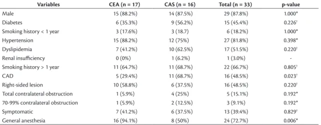
Table 1. Clinical and epidemiologic data (categorical variables).
Variables CEA (n = 17) CAS (n = 16) Total (n = 33) p-value
Male 15 (88.2%) 14 (87.5%) 29 (87.8%) 1.000*
Diabetes 6 (35.3%) 9 (56.2%) 15 (45.4%) 0.226†
Smoking history < 1 year 3 (17.6%) 3 (18.7) 6 (18.2%) 1.000*
Hypertension 15 (88.2%) 12 (75%) 27 (81.8%) 0.398*
Dyslipidemia 7 (41.2%) 10 (62.5%) 17 (51.5%) 0.220†
Renal insuiciency 0 (0%) 1 (6.2%) 1 (3.0%)
-Smoking history > 1 year 11 (64.7%) 11 (68.7%) 22 (66.7%) 0.805†
CAD 5 (29.4%) 11 (68.7%) 16 (48.5%) 0.023†
Right-sided lesion 10 (58.8%) 6 (37.5%) 16 (48.5%) 0.220†
Total contralateral obstruction 1 (5.9%) 4 (25%) 5 (15.1%) 0.192*
70-99% contralateral obstruction 1 (5.9%) 2 (12.5%) 3 (9.1%) 0.192*
Symptomatic 7 (41.2%) 6 (37.5%) 13 (39.4%) 0.829†
General anesthesia 16 (94.1%) 8 (50%) 24 (72.7%) 0.006*
CAD: coronary artery disease; CAS: carotid artery stenting; CEA: carotid endarterectomy. *Fisher test; †chi-square test.
Table 2. Clinical and epidemiologic data (numerical variables).
Groups Variables CEA (n = 17) CAS (n = 16) p-value*
Mean SD Min Med Max Mean SD Min Med Max
Age (years) 71.5 5.6 63 69 81 70.9 7.1 54 72.5 78 0.772
Protection time (s) 1016.5 343.7 540 1020 2160 881 309.2 393 799.5 1360 0.357
Number of MES 89.8 171.4 5 46 745 597.5 343.3 172 610.5 1640 <0.0001
MBP variation (mmHg) 22.6 8 10 23 35 21.1 10.7 5 21 40 0.563
Duration of ischemia (s) 0 0 0 0 0 33.8 83.4 0 0 312
selectively. Residual stenosis of 20% was considered an acceptable result. Contrast was injected after stent deployment to enable control imaging and there were
no indings suggestive of vasospasm.
Transcranial Doppler ultrasound18
Before the procedure, patients were examined by
mapping out the blood low of the MCA to conirm
the acoustic window, using a TCD Sonara-USA, a 2-channel device with a 2 MHz transducer. New measures were taken during the procedures. The TCD
was itted over the temporal bone above the zygomatic
arch on the side of the target carotid. Microembolic
signals were identiied automatically and recorded
on a hard drive, and were collected across a wide insonation gate set at 45 to 55 mm, with a sample size of 5 mm. Each stage of the procedure was timed. The number of MES and measures of the average
velocity of blood low in the MCA were obtained by
analyzing these data after the procedure, excluding signals that were related to interference produced by the electric scalpel or contrast injection. These values were automatically recorded by the machine, which has been validated in previous reports.17,19
Statistical analysis
Descriptive analysis consisted of tables of frequencies for categorical variables and measures of position and dispersion for numerical variables. The chi-squared test or (when necessary) Fisher’s exact test were employed to compare proportions and the Mann-Whitney test was used to compare numerical measures. Analysis of variance (ANOVA) for repeated measures with rank transformation was conducted, followed by contrast tests, in order to compare groups, procedural stages, and the interaction between them. The level of
statistical signiicance used was α = 5%. All statistical
calculations were performed using SAS for Windows (Statistical Analysis System, version 9.2.; SAS Institute Inc, 2002-2008, Cary, NC, USA).
RESULTS
The majority of clinical epidemiological data did not differ statistically between groups, but the groups were different in terms of coronary artery disease (CAD) and type of anesthesia (Tables 1 and 2). One of the 33 patients suffered a stroke, followed by death, 25 days after the procedure. This patient had undergone CAS with local anesthesia. All of the other patients were successfully treated with carotid revascularization with no complications (such as stroke, myocardial infarction, or death). Doppler US of the neck revealed no stenosis at 30 days.
Analysis of the intraoperative TCD data shown in Table 2 reveals important differences in duration of ischemia. However although this could be relevant from a clinical point of view, in terms of statistics this difference cannot be shown because there was no variation in the CEA group. No conceptual ischemia (< 30% of baseline) occurred during the procedure to treat the patient who later died .
There was a statistically signiicant difference
between the groups (p < 0.0001) for the number of MES detected (Table 2). There was a mean of 89.8 (± 171.4) microembolic signals per procedure in the CEA group, while the mean number in the CAS group was 597.5 (± 343.3). A total of 661 microembolic signals were detected during the procedure to treat the patient who eventually died. This patient was in the CAS group, in which the maximum number of signals was 1640.
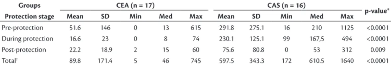
The distribution of MES across the three stages of the procedures can be observed in Table 3 and is also illustrated in Figure 1. In the CEA group, the MES were
uniformly distributed (p = 0.208). However, in the CAS
group, notable differences (p < 0.0001) were detected between the pre-protection and post-protection stages and also between the during protection stage and the post-protection stage. No statistical differences were
found between the irst and second stages. In effect,
the mean number of MES was higher in the CAS group in all three stages.
Table 3. Ipsilateral MES broken down by protection stages and carotid revascularization procedures.
Groups Protection stage
CEA (n = 17) CAS (n = 16)
p-value*
Mean SD Min Med Max Mean SD Min Med Max
Pre-protection 51.6 146 0 13 615 291.8 275.1 16 210 1125 <0.0001
During protection 16.6 23 0 8 74 230.1 125.1 99 167,5 494 <0.0001
Post-protection 22.2 18.9 2 15 60 75.6 80.8 0 53 312 0.009
Total† 89.8 171.4 5 46 745 597.5 343.3 172 610.5 1640 <0.0001
CAS: carotid artery stenting; CEA: carotid endarterectomy; SD: standard deviation. *Comparison between groups using the ANOVA test; †Comparison between the
Another important group of variables analyzed is
the averages for mean blood low velocities in the
ipsilateral MCA (Table 4 and Figure 2). It is important to note that the variations in the average velocities
were similar in both groups (p = 0.152). Furthermore,
in both groups, the mean blood low velocity within
the MCA fell after the transition from pre-protection to during protection, and rose once again during post-protection, attaining values that were higher than both preceding stages (p <0.0001).
Figure 1. Average values with standard deviation of the number of MES in each protection stage.
Table 4. Analysis of the averages of mean velocities of blood low (cm/s) in the ipsilateral MCA, broken down by protection stages
and carotid revascularization procedures. Groups
Protection stage
CEA (n = 17) CAS (n = 16)
p-value*
Mean SD Min Med Max Mean SD Min Med Max
Pre-protection 37.9 8.7 21 36 55 48.4 12.5 31 45.5 78 0.152
During protection 35.7 14.9 11 34 69 39.1 12.2 24.1 38.2 72.5 0.152
Post-protection 48.3 15.3 27 45 85 50 27 0.8 48.5 110 0.152
p-value† <0.0001 <0.0001
-CAS: carotid artery stenting; CEA: carotid endarterectomy. *Comparison between groups using the ANOVA test; †Comparison between protection stages using
the ANOVA test.
DISCUSSION
The correlation between microembolization detected by TCD during CEA and cerebral ischemia was demonstrated in 1994.20 According to one important
study,19 both gaseous and solid emboli can cause
cerebral injuries during carotid revascularization. In the present study, this distinction was not made. Another relevant feature of this study relates to counting microemboli and the manner in which noise related to contrast injection and electrical interference from the electric scalpel was excluded. A previous study has suggested it is necessary to discard these data to avoid methodological errors, but the study authors did not provide details on how to do this.9 These
recommendations were followed in the present study. However, it was noted that the number of MES rose after contrast injection and during use of the electric scalpel, but did not sink back to baseline levels after these phases were over. These extra signals were not discarded in the present study and it is possible that these peculiarities are linked to the observation of a higher number of MES, in comparison to other studies.4,9,17,19
Given the sample size studied, it was not possible to relate microemboli to morbid events. However,
it was demonstrated that MES were signiicantly
more prevalent in patients treated with CAS, and this phenomenon was observed in all three stages of the procedure. Some studies have reported the
same inding.4,9,17,19 The present study also found
that the temporal distribution of microemboli was different in each group. In the CEA group, there was no statistical difference in the distribution of MES across the three stages, but in the CAS group, MES were most prevalent in the pre-protection and during protection stages.
To a certain extent, the results for CEA, contradict
the indings of other authors21 who demonstrated
differences in the temporal distribution of microemboli. They state that distal control of the internal carotid and installation of the shunt, both crucial moments during CEA, would result in more embolization.21
However, there are two aspects that differentiate the present study from that one: 1) in the present study, the technique used for endarterectomy (semi-eversion) results in less distal exposure and manipulation of the internal carotid artery; 2) it was not necessary to use a shunt in any of the cases in the present study.
On the other hand, MES noted during clamping could be explained by possible contralateral microemboli and intracranial atheroemboli. Furthermore, MES were undoubtedly more common when the internal
and external carotid arteries were unclamped, while the common carotid artery was still clamped (see Methods section).
The data observed for the CAS group demonstrated a tendency to greater occurrence of MES in the pre-protection stage, although there was no statistical difference in relation to the second stage. It was clear that it was only once the stent was correctly positioned and arterial manipulation ceased, in the
post-protection stage, that a signiicant drop in the
number of MES occurred. Furthermore, it could be argued that the initial manipulation of the aortic arch and manipulation of the carotid lesion without any protection generate microemboli and that even once
the distal ilter has been installed protection is still incomplete since debris can escape through the ilter or around the ilter, which has been documented.22
Given that many neurological events can occur after removal of the brain-protection device, it can be assumed that a large number of carotid lesions treated with CAS continue to release embolic material after carotid intervention.23,24 These authors suggest that
using closed-cell stents results in a signiicant decrease
in post-procedural neurological events. However, two more recent studies25,26 found no difference between
groups treated using closed and open-cell stents. The results of these more recent studies should avert any criticism of the exclusive use of open-cell stents in the present study.
The increase in velocity of blood low through
the MCA after carotid revascularization has been investigated by several authors.27,28 As was to be
expected, the average blood low velocity in the
ipsilateral middle cerebral artery increased after all procedures.
Another important point is the reduction in average velocities during protection, observed in both groups, notwithstanding that this did not necessarily mean that the patient reached conceptual ischemia (< 30% of baseline). Other authors have observed several cases of this reduction in average blood
low velocity during CEA procedures, but in that
study only 16 out of 49 patients exhibited a drop in blood velocity resulting in ischemia.29 It is relevant
to note that while the CEA group in the present study did not have any cases of conceptual ischemia (< 30% of baseline), there was one case in the CAS group in which ischemia occurred for 312 seconds. However, there is a difference between this study and the one cited above,29 in that it deined ischemia as
In the present study there was a similarity between the groups in terms of how the averages of the mean velocities decreased in the second stage, which was expected for CEA, but went against the initial
hypothesis that CAS with a distal ilter would not disrupt anterograde blood low. In addition to saturation of the ilter, there are other factors that contributed to
this reduction, such as placement of the stent within the residual lumen of the internal carotid and the later balloon expansion of the stent.
In conclusion, in the light of TCD indings, CEA
(semi-eversion technique) resulted in a lower incidence
of MES than CAS with a distal ilter in all protection
stages, with a uniform temporal distribution in the CEA group and greater occurrence of MES in the
irst two stages in the CAS group. The averages of the mean velocity of low within the MCA behaved
similarly in both groups: the averages of the mean
velocities tended to fall from the irst to the second
stage, and then rise from the second to the third,
reaching levels higher than those in the irst stage.
REFERENCES
1. Brott TG, Halperin JL, Abbara S, et al. 2011 ASA/ACCF/AHA/ AANN/AANS/ACR/ASNR/CNS/SAIP/SCAI/SIR/SNIS/SVM/SVS guideline on the management of patients with extracranial carotid and vertebral artery disease: executive summary. A report of the American College of Cardiology Foundation/American Heart Association Task Force on Practice Guidelines, and the American Stroke Association, American Association of Neuroscience Nurses, American Association of Neurological Surgeons, American College of Radiology, American Society of Neuroradiology, Congress of Neurological Surgeons, Society of Atherosclerosis Imaging and Prevention, Society for Cardiovascular Angiography and Interventions, Society of Interventional Radiology, Society of NeuroInterventional Surgery, Society for Vascular Medicine, and Society for Vascular Surgery. Circulation. 2011;124(4):489-532. PMid:21282505. http://dx.doi.org/10.1161/CIR.0b013e31820d8d78. 2. Ringleb PA, Kunze A, Allenberg JR, et al. The stent-supported percutaneous angioplasty of the carotid artery vs. endarterectomy trial. Cerebrovasc Dis. 2004;18(1):66-8. PMid:15178989. http:// dx.doi.org/10.1159/000078752.
3. Mas JL, Trinquart L, Leys D, et al. Endarterectomy Versus Angioplasty in Patients with Symptomatic Severe Carotid Stenosis (EVA-3S) trial: results up to 4 years from a randomised, multicentre trial. Lancet Neurol. 2008;7(10):885-92. PMid:18774745. http://dx.doi. org/10.1016/S1474-4422(08)70195-9.
4. Jordan WD Jr, Voellinger DC, Doblar DD, Plyushcheva NP, Fisher WS, McDowell HA. Microemboli detected by transcranial doppler monitoring in patients during carotid angioplasty versus carotid endarterectomy. Cardiovasc Surg. 1999;7(1):33-8. PMid:10073757. http://dx.doi.org/10.1016/S0967-2109(98)00097-0.
5. Altaf N, Kandiyil N, Hosseini A, Mehta R, MacSweeney S, Auer D. Risk factors associated with cerebrovascular recurrence in symptomatic carotid disease: a comparative study of carotid plaque morphology, microemboli assessment and the European Carotid Surgery Trial risk model. J Am Heart Assoc. 2014;3(3):e000173. PMid:24895159. http://dx.doi.org/10.1161/JAHA.113.000173.
6. Ackerstaff RG, Moons KG, van de Vlasakker CJ, et al. Association of intraoperative transcranial doppler monitoring variables with stroke from carotid endarterectomy. Stroke. 2000;31(8):1817-23. PMid:10926940. http://dx.doi.org/10.1161/01.STR.31.8.1817.
7. Ackerstaff RG, Suttorp MJ, van den Berg JC, et al. Prediction of early cerebral outcome by transcranial doppler monitoring in carotid bifurcation angioplasty and stenting. J Vasc Surg. 2005;41(4):618-24. PMid:15874925. http://dx.doi.org/10.1016/j.jvs.2005.01.034.
8. Bossema ER, Brand N, Moll FL, Ackerstaff RG, van Doornen LJ. Perioperative microembolism is not associated with cognitive outcome three months after carotid endarterectomy. Eur J Vasc Endovasc Surg. 2005;29(3):262-8. PMid:15694799. http://dx.doi. org/10.1016/j.ejvs.2004.11.010.
9. Crawley F, Stygall J, Lunn S, Harrison M, Brown MM, Newman S. Comparison of microembolism detected by transcranial doppler and neuropsychological sequelae of carotid surgery and percutaneous transluminal angioplasty. Stroke. 2000;31(6):1329-34. PMid:10835452. http://dx.doi.org/10.1161/01.STR.31.6.1329. 10. Purandare N, Burns A, Daly KJ, et al. Cerebral emboli as a potential cause of alzheimer’s disease and vascular dementia: Case-control study. BMJ. 2006;332(7550):1119-24. PMid:16648133. http://dx.doi. org/10.1136/bmj.38814.696493.AE.
11. Wapp M, Everts R, Burren Y, et al. Cognitive improvement in patients with carotid stenosis is independent of treatment type. Swiss Med Wkly. 2015;145:w14226. PMid:26700596.
12. Mommertz G, Das M, Langer S, et al. Early control of distal internal carotid artery during carotid endarterectomy: Does it reduce cerebral microemboli? J Cardiovasc Surg. 2010;51(3):369-75. PMid:20523287.
13. Tedesco MM, Dalman RL, Zhou W, Coogan SM, Lane B, Lee JT. Reduction of postprocedure microemboli following retrospective quality assessment and practice improvement measures for carotid angioplasty and stenting. J Vasc Surg. 2009;49(3):607-12. PMid:19135833. http://dx.doi.org/10.1016/j.jvs.2008.10.031. 14. North American Symptomatic Carotid Endarterectomy Trial
Collaborators. Beneficial effect of carotid endarterectomy in symptomatic patients with high-grade carotid stenosis. N Engl J Med. 1991;325(7):445-53. PMid:1852179. http://dx.doi.org/10.1056/ NEJM199108153250701.
15. Walker MD. Executive committee for the asymptomatic carotid atherosclerosis study. Endarterectomy for asymptomatic carotid artery stenosis. JAMA. 1995;273(18):1421-8. PMid:7723155. http:// dx.doi.org/10.1001/jama.1995.03520420037035.
16. Effeney DJ, Stoney RJ. Extracranial cerebrovascular disease. In: Effeney DJ, Stoney RJ, editors. Wylie’s Atlas of Vascular Surgery. Philadelphia: J. B. Lippincott Company; 1992. p. 18-48.
17. Gupta N, Corriere MA, Dodson TF, et al. The incidence of microemboli to the brain is less with endarterectomy than with percutaneous revascularization with distal filters or flow reversal. J Vasc Surg. 2011;53(2):316-22. PMid:21129899. http://dx.doi. org/10.1016/j.jvs.2010.08.063.
18. Ringelstein EB, Droste DW, Babikian VL. Consensus on microembolus detection by tcd. International consensus group on microembolus detection. Stroke. 1998;29(3):725-9. PMid:9506619. http://dx.doi. org/10.1161/01.STR.29.3.725.
19. Skjelland M, Krohg-Sorensen K, Tennoe B, Bakke SJ, Brucher R, Russell D. Cerebral microemboli and brain injury during carotid artery endarterectomy and stenting. Stroke. 2009;40(1):230-4. PMid:18927460. http://dx.doi.org/10.1161/STROKEAHA.107.513341.
study of 100 patients. Br J Surg. 1994;81(10):1435-9. PMid:7820463. http://dx.doi.org/10.1002/bjs.1800811009.
21. Wolf O, Heider P, Heinz M, et al. Microembolic signals detected by transcranial doppler sonography during carotid endarterectomy and correlation with serial diffusion-weighted imaging. Stroke. 2004;35(11):e373-5. PMid:15388901. http://dx.doi.org/10.1161/01. STR.0000143184.69343.ec.
22. Gossetti B, Gattuso R, Irace L, et al. Embolism to the brain during carotid stenting and surgery. Acta Chir Belg. 2007;107(2):151-4. PMid:17515263.
23. Bosiers M, Donato G, Deloose K, et al. Does free cell area influence the outcome in carotid artery stenting? Eur J Vasc Endovasc Surg. 2007;33:135-41. PMID: 17097897. http://dx.doi.org/10.1016/j. ejvs.2006.09.019.
24. Hart JP, Peeters P, Verbist J, Deloose K, Bosiers M. Do device characteristics impact outcome in carotid artery stenting? J Vasc Surg. 2006;44:725-30. PMID: 17011998. http://dx.doi.org/10.1016/j. jvs.2006.06.029.
25. Timaran CH, Rosero EB, Higuera A, Ilarraza A, Modrall JG, Clagett GP. Randomized clinical trial of open-cell vs closed-cell stents for carotid stenting and effects of stent design on cerebral embolization. J Vasc Surg. 2011;54(5):1310-6. PMid:21723064. http://dx.doi. org/10.1016/j.jvs.2011.05.013.
26. Tadros RO, Spyris CT, Vouyouka AG, et al. Comparing the embolic potential of open and closed cell stents during carotid angioplasty and stenting. J Vasc Surg. 2012;56(1):89-95. PMid:22386144. http:// dx.doi.org/10.1016/j.jvs.2011.12.077.
27. Maltezos CK, Papanas N, Papas TT, et al. Changes in blood flow of anterior and middle cerebral arteries following carotid endarterectomy: A transcranial doppler study. Vasc Endovascular Surg. 2007;41(5):389-96. PMid:17942853. http://dx.doi.org/10.1177/1538574407302850. 28. Nowacki P, Zywica A, Podbielski J, Kornacewicz-Jach Z, Drechsler H, Drechsler D. Middle cerebral artery flow after angioplasty and stenting of symptomatic internal carotid artery stenosis. Neurol Neurochir Pol. 2009;43(1):9-15. PMid:19353439.
29. Ali AM, Green D, Zayed H, Halawa M, El-Sakka K, Rashid HI. Cerebral monitoring in patients undergoing carotid endarterectomy using a triple assessment technique. Interact Cardiovasc Thorac Surg. 2011;12(3):454-7. PMid:21098425. http://dx.doi.org/10.1510/ icvts.2010.235598.
*
Correspondence
Germano da Paz Oliveira Rua José Olímpio de Melo, 2436/102 - Ilhotas CEP 64014-063 - Teresina (PI) - Brazil Tel.: +55 (86) 99982-0901 E-mail: [email protected]
Author information
GPO - Vascular and endovascular surgeon from Universidade Estadual de Campinas (UNICAMP); MSc in Sciences from UNICAMP; Primary physician at Serviço de Cirurgia Vascular, Hospital Universitário do Piauí and Hospital Getúlio Vargas. ATG - Vascular and endovascular surgeon; PhD, tenured professor at Departamento de Cirurgia, Universidade Estadual de Campinas (UNICAMP); Chief of the Discipline of Peripheral Vascular Diseases, UNICAMP; Coordinator, Serviço de Alta Complexidade em Cirurgia Vascular e Endovascular, UNICAMP. SCB - Medical student, Centro Universitário Uninovafapi. JMTL - MD from Universidade Federal do Piauí (UFPI), Resident physician of Pediatrics at Universidade de São Paulo (USP). SRFB - General surgeon and critical care physician from Universidade Estadual de Campinas (UNICAMP); Primary physician at the intensive care units of Centro de Apoio à Saúde da Mulher and Hospital de Clínicas da UNICAMP; Collaborating physician at Laboratório de Hemodinâmica Cerebral, UNICAMP. WMA - Neurologist from Universidade Estadual de Campinas (UNICAMP); PhD in Medical Pathophysiology from UNICAMP; Collaborating primary physician at Ambulatório de Neurologia Vascular – Hospital de Clínicas da UNICAMP; head of Laboratório de Hemodinâmica Cerebral, UNICAMP. FC - Neurologist from Universidade Estadual de Campinas (UNICAMP); PhD, tenured professor at Departamento de Neurologia da UNICAMP; Coordinator, Subcomissão de Pós-Graduação, Curso de Fisiopatologia Médica; Member, Comissão de Pesquisa da FCM-UNICAMP.
Author contributions
Conception and design: GPO, ATG, WMA, FC Analysis and interpretation: GPO, ATG, FC Data collection: GPO, ATG, SCB, JMTL, SRFB, WMA, FC Writing the article: GPO, ATG, SRFB, WMA, FC Critical revision of the article: GPO, ATG, SRFB, WMA, FC Final approval of the article*: GPO, ATG, SCB, JMTL, SRFB, WMA, FC Statistical analysis: GPO, ATG, WMA, FC Overall responsibility: GPO, ATG