Semiquantitative analysis of surgical biopsies
of distinct lung lobes of patients with usual
interstitial pneumonia/idiopathic pulmonary fibrosis*
Análise semiquantitativa de biópsias cirúrgicas de diferentes lobos pulmonares de pacientes com pneumonia intersticial usual/fibrose pulmonar idiopática
José Júlio Saraiva Gonçalves, Luiz Eduardo Villaça Leão, Rimarcs Gomes Ferreira, Renato Oliveira, Luiz Hirotoshi Ota, Ricardo Sales dos Santos
Abstract
Objective: To evaluate the differences between surgical biopsies of distinct lung lobes in terms of the histopathological features of usual interstitial pneumonia, using a semiquantitative score. Methods: We selected all of the patients diagnosed with idiopathic pulmonary fibrosis and submitted to surgical biopsy in two distinct lobes between 1995 and 2005 at the Hospital São Paulo and other hospitals operated by the Federal University of São Paulo. In the histological evaluation of the specimens, we used a semiquantitative method based on previous studies, assigning a score to each of the biopsied sites. Results: In this sample of patients, we found no statistically significant differences that would alter the stage of the disease, based on the score used. This finding was independent of the biopsy site (middle lobe or lingular segment). Conclusions: No significant histological differences were found between the lung lobes studied. The definitive histological diagnosis of usual interstitial pneumonia did not alter the stage of the disease.
Keywords: Lung diseases, interstitial; Pulmonary fibrosis; Thoracic surgery; Pathology; Thoracic surgery, video-assisted.
Resumo
Objetivo: Avaliar as diferenças histológicas da pneumonia intersticial usual entre biópsias cirúrgicas de lobos pulmonares distintos, utilizando um escore semiquantitativo. Métodos: Foram selecionados todos os pacientes com o diagnóstico de fibrose pulmonar idiopática e submetidos à biópsia cirúrgica em dois lobos distintos no Hospital São Paulo e em hospitais afiliados da Universidade Federal de São Paulo, no período entre 1995 e 2005. Foi utilizado um método semiquantitativo na avaliação histológica dos espécimes, com base em estudos prévios, aplicando-se um escore para cada local submetido à biópsia. Resultados: Nenhuma diferença estatisticamente significante foi encontrada nesta amostra de pacientes que viesse alterar o estágio da doença, com base no escore utilizado. Este achado foi independente do local da biópsia (lobo médio ou segmento lingular). Conclusões: Não foram observadas diferenças histológicas significantes entre os lobos pulmonares estudados. O diagnóstico histo-lógico definitivo de pneumonia intersticial usual não alterou o estágio da doença.
Descritores: Doenças pulmonares intersticiais; Fibrose pulmonar; Cirurgia torácica; Patologia; Cirurgia torácica vídeo-assistida.
* Study carried out in the Department of Thoracic Surgery, Universidade Federal de São Paulo/Escola Paulista de Medicina – UNIFESP/ EPM, Federal University of São Paulo/Paulista School of Medicine – São Paulo, Brazil.
Correspondence to: José Júlio Saraiva Gonçalves. Hospital São Paulo, Rua Napoleão de Barros, 715, 4º andar, Vila Clementino, CEP 04024-002, São Paulo, SP, Brasil.
Tel 55 11 5576-4295. E-mail: jjsgon@gmail.com Financial support: None.
tive of this study was to evaluate the differences between surgical biopsies of two lung lobes, such as the lingular segment and the middle lobe, in terms of the histopathological features of UIP, using a semiquantitative score.
Methods
This was a retrospective study of 89 surgical lung biopsies (in 59 patients) performed, at the Hospital São Paulo and other hospitals oper-ated by the Universidade Federal de São Paulo (UNIFESP, Federal University of São Paulo), between 1995 and 2005. All of the samples selected had been categorized histopathologi-cally as being consistent with a diagnosis of UIP. The inclusion criteria were as follows: histopatho-logical diagnosis of UIP and surgical lung biopsy of two lobes, one of which was the middle lobe or the lingular segment. The following exclu-sion criteria were applied: interstitial diseases secondary to collagenosis; exposure to chemical agents; other interstitial diseases; and diag-nostic discordance between the biopsied lobes, even if the final clinical and radiological diag-nosis was IPF. The project was approved by the UNIFESP Research Ethics Committee (Protocol no. 1.063/05).
All of the lung tissue specimens were collected by video-assisted thoracoscopy or anterior mini-thoracotomy. The histological sections were stained with H&E and Masson’s trichrome. The pathologist was blinded to the biopsy site and patient identity.
The semiquantitative technique used in the histological analysis of the lung biop-sies was based on the original description by Cherniak et al.(15,16) that was later validated by
Hyde et al.,(17) who demonstrated the
concord-Introduction
The clinical diagnosis of idiopathic pulmonary fibrosis (IPF) is associated with the histopatho-logical pattern known as usual interstitial pneumonia (UIP).(1) The mean survival, after IPF
has become established, ranges from two to six years.(2) More than 50% of patients with IPF die
within two to three years after diagnosis,(3) as
a consequence of complications resulting from the severe restrictive ventilatory defect, for which there is currently no satisfactory treatment.(4) In
Brazil, data on the incidence and prevalence of the disease are scarce.(5) However, based on a
Latin-American study, it is estimated that there are 2,225 new cases in men and 1,416 in women per year in the state of São Paulo alone. The mortality rate in the south of Brazil was reported to be 0.48/100,000 population in the 1990s.(5)
One of the significant histological char-acteristics of UIP is its temporal and regional heterogeneity, that is, the presence of areas of recent aggression in close proximity to regions of healing, together with apparently normal lung parenchyma.(6) Therefore, the criteria for
this diagnosis, from a histopathological stand-point, are findings of fibrosis, limited chronic inflammation, honeycombing, fibroblastic foci and architectural remodeling of the acini and lobes.(7)
Since UIP has a heterogenous aspect, from a histological and tomographic standpoint, it cannot be adequately represented even in surgical biopsy specimens. This suggests that the site to be biopsied should be selected with caution, since the quality of the biopsy influ-ences the diagnostic yield and the accuracy of disease staging. However, in the current litera-ture, there is no consensus regarding the biopsy site, the ideal size of the lung parenchyma samples to be obtained or the number of such samples needed.(8-13) Nor is it known what
influ-ence these factors have on diagnosis or disease staging. Existing studies using semiquantita-tive techniques for histopathological analysis to evaluate biopsy samples obtained from distinct lung lobes present conflicting results.(8-13) As was
observed, patients with IPF make ideal subjects in which to study the disparate histopatholog-ical alterations in a given patient, lung or lobe, as well as in contiguous areas.
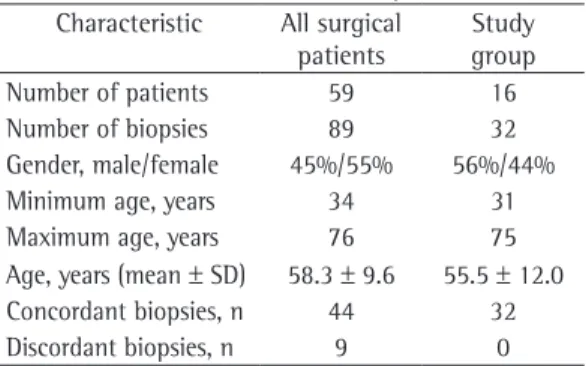
The sensitivity and specificity of surgical biopsy are, on average, over 90%.(14) The
objec-Table 1 - Characteristics of the sample. Characteristic All surgical
patients
Discussion
In the literature, there is disagreement regarding the site of choice for surgical lung biopsy. Studies conducted in the 1960s and 1970s(9,10,12,20,21) described pathological sequelae,
such as fibrosis and vascular alterations, in the lung interstitium in biopsies of the middle lobe and lingular segment. These findings could affect diagnosis and disease staging even in patients ance between this technique and the classical
technique of quantitative morphometry. Other research centers in Brazil have published their experience using the same score, confirming previously published findings.(18,19)
For the use of this score, three main criteria for quantification were established: a) exuda-tive/inflammatory changes; b) fibrotic/reparative changes; and c) airway changes. These altera-tions were quantified using a scale ranging from 0 to 5 for criteria a and b, together with a scale ranging from 0 to 2 for criterion c.
The level of statistical significance was set at 5% (α = 0.05). The program Statistical Package for the Social Sciences, version 13.0 (SPSS Inc., Chicago, IL, USA) was used for this analysis.
Results
Of the 59 patients selected, 28 were excluded for having had only one site biopsied. Another 9 patients had discordance between the lobes: 3 due to nonspecific interstitial pneumonia; 3 due to an unclassifiable pattern; and 3 due to the fact that they had only emphysema in one of the lobes. Of the remaining 22 patients, 4 had had two specimens collected from the same lobe and 2 were excluded for having been submitted to biopsies that were performed on the same side (the right) but did not include the middle lobe.
Therefore, 16 patients (32 biopsies) fully met the inclusion criteria established. The statistical analysis was performed during this patient selec-tion, and patients were divided into four groups according to the biopsy site.
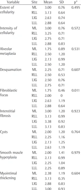
There were no significant differences between the sides (right or left lung) in terms of age, gender or other characteristics of the patients (Table 1). Regarding the other variables, the lobes studied proved to be statistically different only in terms of the presence of fibroblastic foci (p = 0.011, Table 2). We found statistically significant differences for the variable “fibrob-lastic focus”, this finding being significantly more common in the left lower lobe samples than in those obtained from the other sites.
No significant differences were found in terms of the other factors evaluated, regardless of the biopsy site. The differences found did not alter the overall disease staging.
Table 2 - Analysis of the statistical significance of the differences among all of the lung lobes biopsied.
Variable Site Mean SD p* Extent of
cellularity
ML 3.00 0.76 0.495 RLL 3.13 0.64
LIG 2.63 0.74 LLL 2.88 0.64 Intensity of
cellularity
ML 3.00 0.76 0.572 RLL 3.25 0.71
LIG 2.75 0.71 LLL 2.88 0.83 Alveolar
metaplasia
ML 1.75 0.89 0.531 RLL 2.50 1.20
LIG 2.13 0.99 LLL 2.50 1.20
Desquamation ML 2.25 0.71 0.607 RLL 2.50 0.53
LIG 2.50 0.76 LLL 2.75 0.71 Fibroblastic
focus
ML 1.75 0.46 0.011 RLL 2.00 0
LIG 2.63 1.19 LLL 2.88 0.64 Interstitial
fibrosis
ML 3.00 1.20 0.923 RLL 3.13 0.99
LIG 3.38 0.92 LLL 3.13 0.83
Cysts ML 2.00 1.20 0.764
RLL 2.25 1.16 LIG 2.13 1.25 LLL 2.63 1.19 Smooth muscle
hyperplasia
ML 2.00 1.41 0.979 RLL 2.13 0.99
LIG 2.25 1.04 LLL 2.25 0.89 Myointimal
thickening
ML 2.38 1.19 0.604 RLL 3.13 0.35
LIG 2.88 0.83 LLL 3.00 0.93
in subsequent studies,(13,20,22) it was stated that
there was no unequivocal scientific evidence for this restriction, the prejudice against biopsies of such sites remained.
The semiquantitative technique used in the present study, a technique previously described by Cherniak et al.(15,16) and Hyde et al.,(17) is
widely accepted among pulmonary pathologists, being the only one developed for this purpose that has been validated in multiple centers. The choice of this technique had a negative impact on the study: it limited the study to only the UIP pattern, requiring that the pattern be neces-sarily that of UIP for the comparison of the two sites studied. This led us to exclude many patients with discordant findings in view of the technical impossibility of assigning this score to other findings, such as nonspecific interstitial pneumonia.
The histological diagnosis of UIP is based on a series of characteristics of distribution of the disease, defining a histopathological pattern.(23) The size of the lung tissue specimen
is related to the depth of the sample in the lung parenchyma. A lung biopsy should present the peripheral, subpleural region as well as the central or medullary region of the lung. Another recommendation is to obtain a sample of the area of transition between the clearly diseased region seen on HRCT scans and the apparently normal region.(24) An excellent evaluation of the
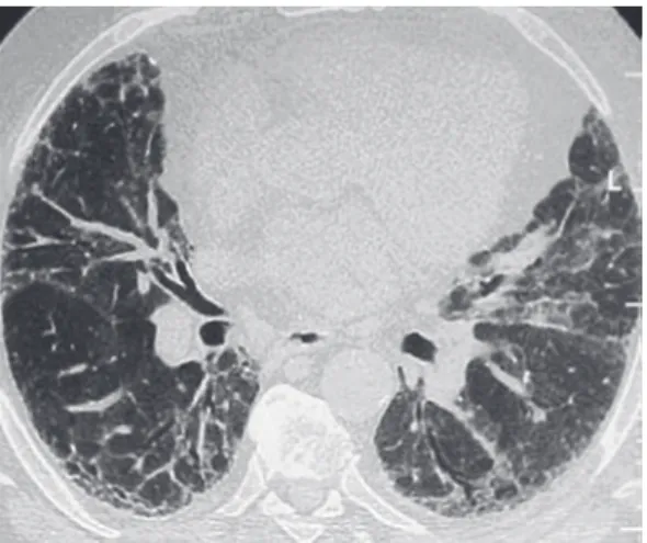
entire lung parenchyma is possible using HRCT, which is routine practice in the initial evaluation of interstitial lung diseases.(25) Currently, HRCT is
used as a routine test in the preoperative period of any surgical lung biopsy (Figure 1). With reasonable accuracy, HRCT allows the selection of ideal sites at distinct stages of the disease in the lung parenchyma to be biopsied.
For most thoracic surgeons, the middle lobe and the lingular segments are anatomically easy to resect, either by mini-thoracotomy or by video-assisted thoracoscopy. In the other lung segments, the collection of samples involving the central and peripheral regions is made difficult by the need to cut an appropriately sized wedge in the parenchyma. In order to obtain a standard sample of this type, the wedge should reach a depth of more than 2 cm, the mean thickness of the peripheral lung region (Figure 2).
The collection of lung tissue from the middle lobe and lingular segment permits samples with without active disease.(10) A comparison of the
distinct biopsied lung sites was not performed, nor was a histopathological analysis to evaluate the effect of these findings on IPF staging. Nevertheless, certain sites were disregarded as options for biopsy and began to be excluded by specialists in interstitial diseases. Although,
Figure 1 - HRCT scan of a patient with usual interstitial pneumonia.
health care facilities, the clinical, pathological and radiological diagnosis has been sufficient as the inclusion criterion in clinical studies of IPF. In our opinion, clinical studies should involve patients whose diagnosis was confirmed by surgical biopsy of various lung lobes.
Video-assisted thoracoscopy allows the collection of various satisfactory samples and has lower postoperative morbidity, since it allows earlier hospital discharge at some health care facilities and dispenses with the use of chest tubes in the postoperative period. In the near future, there will be a progressive reduction in the incisions used to access the pleural space in diagnostic procedures and an even greater reduction in hospital stays, since biopsies will be performed on an outpatient basis, as well as a lower rate of chest tube use, contributing to the reduction in morbidity and mortality.(30)
In some situations, biopsies of the middle lobe and lingular segment have advantages over biopsies of other sites due to their anatomical characteristics. The volume and depth of a lung biopsy is known to affect the diagnostic yield and the quality of the biopsy in interstitial lung diseases. In this respect, the middle lobe and the lingula provide adequate volume and depth for histological evaluation.
In our sample of patients submitted to surgical biopsy of the middle lobe and lingular segment, there were no alterations that could be specifically attributed to these sites or that could affect disease staging, based on the semiquanti-tative score used. Therefore, we recommend that the clinical and surgical teams involved in diag-nosing the patient evaluate HRCT scans in order to select the biopsy site during the preopera-tive period. Collecting multiple biopsy samples increases the diagnostic yield and makes it possible to stage the interstitial disease more accurately.
References
1. Demedts M, Costabel U. ATS/ERS international multidisciplinary consensus classification of the idiopathic interstitial pneumonias. Eur Respir J. 2002;19(5):794-6.
2. Tiitto L, Bloigu R, Heiskanen U, Pääkkö P, Kinnula VL, Kaarteenaho-Wiik R. Relationship between histopathological features and the course of idiopathic pulmonary fibrosis/usual interstitial pneumonia. Thorax. 2006;61(12):1091-5.
3. Pereira CA, Malheiros T, Coletta EM, Ferreira RG, Rubin AS, Otta JS, et al. Survival in idiopathic pulmonary
satisfactory volumes to be obtained, and closure typically requires the use of only an endoscopic stapler or a single suture line. The high cost of endoscopic staplers has been a limiting factor for the use of video-assisted thoracoscopy in lung biopsies in our country. An additional advantage described in the literature is the lower inci-dence of postoperative bronchopleural fistulae after biopsies of the middle lobe or lingular segment.(26)
The UIP pattern is quite characteristic, with more severe disease in the subpleural region of the lung, frequently already at more advanced stages of the disease, with areas of honeycombing. Shallow biopsy depth allows only a peripheral representation of the lung parenchyma. In these situations, there is a predominance of chronic alterations that lead the pathologist to define the profile as terminal lung, without histopatho-logical criteria for UIP or any other characteristic pattern that might have led to this situation.
Fibroblastic foci have been reported as an isolated factor for a worse prognosis in IPF.(2,27,28) In this study, this finding was
signif-icantly more common in the left lower lobes. However, one group of authors, having found no such association, questioned the prognostic value of fibroblastic foci in isolation.(7)
In the present study, there was a predomi-nance of males in the group submitted to biopsies on the left side, in which the mean age was also higher, although neither difference was significant. Factors such as age and gender have also been associated with a worse prognosis in IPF,(18,23) and these factors could be the cause of
this finding.
Currently, the objective is to find a way to quantify the findings in UIP and to stage the interstitial lung disease, based on the lung tissue collected in the biopsy. No matter how repre-sentative the parenchyma is in the biopsy, there will always be questions regarding the true stage of the disease.
17. Hyde DM, King TE Jr, McDermott T, Waldron JA Jr, Colby TV, Thurlbeck WM, et al. Idiopathic pulmonary fibrosis. Quantitative assessment of lung pathology. Comparison of a semiquantitative and a morphometric histopathologic scoring system. Am Rev Respir Dis. 1992;146(4):1042-7.
18. Canzian M, de Matos Soeiro A, de Lima Taga MF, Farhat C, Barbas CS, Capelozzi VL. Semiquantitative assessment of surgical lung biopsy: predictive value and impact on survival of patients with diffuse pulmonary infiltrate. Clinics (Sao Paulo). 2007;62(1):23-30.
19. Ferreira RG, Colleta EM, Giannotti Filho O. Avaliação de parâmetros histológicos na pneumonia intersticial usual (fibrose pulmonar idiopática) J Pneumol. 2000;26(6):279-85.
20. Miller JI Jr. The present role and future considerations of video-assisted thoracoscopy in general thoracic surgery. Ann Thorac Surg. 1993;56(3):804-6.
21. Wetstein L. Sensitivity and specificity of lingular segmental biopsies of the lung. Chest. 1986;90(3):383-6. 22. Chechani V, Landreneau RJ, Shaikh SS. Open lung
biopsy for diffuse infiltrative lung disease. Ann Thorac Surg. 1992;54(2):296-300.
23. Dempsey OJ. Clinical review: idiopathic pulmonary fibrosis--past, present and future. Respir Med. 2006;100(11):1871-85.
24. Flaherty KR, Travis WD, Colby TV, Toews GB, Kazerooni EA, Gross BH, et al. Histopathologic variability in usual and nonspecific interstitial pneumonias. Am J Respir Crit Care Med. 2001;164(9):1722-7.
25. Hunninghake GW, Lynch DA, Galvin JR, Gross BH, Müller N, Schwartz DA, et al. Radiologic findings are strongly associated with a pathologic diagnosis of usual interstitial pneumonia. Chest. 2003;124(4):1215-23. 26. Miller RR, Nelems B, Müller NL, Evans KG, Ostrow
DN. Lingular and right middle lobe biopsy in the assessment of diffuse lung disease. Ann Thorac Surg. 1987;44(3):269-73.
27. Enomoto N, Suda T, Kato M, Kaida Y, Nakamura Y, Imokawa S, et al. Quantitative analysis of fibroblastic foci in usual interstitial pneumonia. Chest. 2006;130(1):22-9.
28. Coletta EN, Pereira CA, Ferreira RG, Rubin AS, Villela LS, Malheiros T, et al. Achados histológicos e sobrevida na fibrose pulmonar idiopática. J Pneumol. 2003;29(6):371-8.
29. Churg A, Schwarz M. Transbronchial biopsy and usual interstitial pneumonia: a new paradigm? Chest. 2006;129(5):1117-8.
30. Luckraz H, Rammohan KS, Phillips M, Abel R, Karthikeyan S, Kulatilake NE, et al. Is an intercostal chest drain necessary after video-assisted thoracoscopic (VATS) lung biopsy? Ann Thorac Surg. 2007;84(1):237-9. fibrosis-cytotoxic agents compared to corticosteroids.
Respir Med. 2006;100(2):340-7.
4. Olson AL, Swigris JJ, Lezotte DC, Norris JM, Wilson CG, Brown KK. Mortality from pulmonary fibrosis increased in the United States from 1992 to 2003. Am J Respir Crit Care Med. 2007;176(3):277-84.
5. Fortuna FP, Perin C, Cunha L, Rubin AS. Mortalidade por fibrose pulmonar idiopática no Rio Grande do Sul. J Pneumol. 2003;29(3):1-4.
6. Monaghan H, Wells AU, Colby TV, du Bois RM, Hansell DM, Nicholson AG. Prognostic implications of histologic patterns in multiple surgical lung biopsies from patients with idiopathic interstitial pneumonias. Chest. 2004;125(2):522-6.
7. Hanak V, Ryu JH, de Carvalho E, Limper AH, Hartman TE, Decker PA, et al. Profusion of fibroblast foci in patients with idiopathic pulmonary fibrosis does not predict outcome. Respir Med. 2008;102(6):852-6. 8. Flint A, Martinez FJ, Young ML, Whyte RI, Toews GB,
Lynch JP 3rd. Influence of sample number and biopsy site on the histologic diagnosis of diffuse lung disease. Ann Thorac Surg. 1995;60(6):1605-7; discussion 1607-8.
9. Heath D, Whitaker W. Hypertensive pulmonary vascular disease. Circulation. 1956;14(3):323-43.
10. Newman SL, Michel RP, Wang NS. Lingular lung biopsy: is it representative? Am Rev Respir Dis. 1985;132(5):1084-6.
11. Qureshi RA, Stamenkovic SA, Carnochan FM, Walker WS. Video-assisted thoracoscopic lung biopsy in patients with interstitial lung disease. Ann Thorac Surg. 2007;84(6):2136-7.
12. Ray JF 3rd, Lawton BR, Myers WO, Toyama WM, Reyes CN, Emanuel DA, et al. Open pulmonary biopsy. Nineteen-year experience with 416 consecutive operations. Chest. 1976;69(1):43-7.
13. Temes RT, Joste NE, Allen NL, Crowell RE, Dox HA, Wernly JA. The lingula is an appropriate site for lung biopsy. Ann Thorac Surg. 2000;69(4):1016-8; discussion 1018-9.
14. Flaherty KR, King TE Jr, Raghu G, Lynch JP 3rd, Colby TV, Travis WD, et al. Idiopathic interstitial pneumonia: what is the effect of a multidisciplinary approach to diagnosis? Am J Respir Crit Care Med. 2004;170(8):904-10. 15. Cherniack RM, Colby TV, Flint A, Thurlbeck WM,
Waldron J, Ackerson L, et al. Quantitative assessment of lung pathology in idiopathic pulmonary fibrosis. The BAL Cooperative Group Steering Committee. Am Rev Respir Dis. 1991;144(4):892-900.
About the authors
José Júlio Saraiva Gonçalves
Professor. Universidade para o Desenvolvimento do Estado e da Região do Pantanal – UNIDERP, University for the Development of the State and Region of the Pantanal – School of Medicine, Campo Grande, Brazil.
Luiz Eduardo Villaça Leão
Full Professor. Department of Thoracic Surgery, Universidade Federal de São Paulo/Escola Paulista de Medicina – UNIFESP/EPM, Federal University of São Paulo/Paulista School of Medicine – São Paulo, Brazil.
Rimarcs Gomes Ferreira
Adjunct Professor. Department of Pathology, Universidade Federal de São Paulo/Escola Paulista de Medicina – UNIFESP/EPM, Federal University of São Paulo/Paulista School of Medicine – São Paulo, Brazil.
Renato Oliveira
Attending Thoracic Surgeon. Department of Thoracic Surgery, Universidade Federal de São Paulo/Escola Paulista de Medicina – UNIFESP/EPM, Federal University of São Paulo/Paulista School of Medicine – São Paulo, Brazil.
Luiz Hirotoshi Ota
Adjunct Professor. Department of Thoracic Surgery, Universidade Federal de São Paulo/Escola Paulista de Medicina – UNIFESP/EPM, Federal University of São Paulo/Paulista School of Medicine – São Paulo, Brazil.
Ricardo Sales dos Santos