Indirect bone resorption in orthodontic
movement: when does periodontal
reorganization begin and how does it occur?
Alberto Consolaro*, Lysete Berriel Cardoso**, Angela Mitie Otta Kinoshita***, Leda Aparecida Francischone***, Milton Santamaria Jr****, Ana Carolina Cuzuol Fracalossi*****, Vanessa Bernardini Maldonado******
Tooth movement induced by orthodontic ap-pliances is one of the most frequent therapeutic procedures in clinical dental practice. The search for esthetics and functionality, both oral and den-tal, demands orthodontic treatments, which are often associated with root resorptions that may, in extreme cases, lead to tooth loss, periodontal damage, or both.
The knowledge of induced tooth movement biology, based on tissue, cell and molecular phenomena that take place on each day during movement progression, enable us to act safely and consciously when using drugs, procedures and interventions to optimize orthodontic treat-ment and patient comfort, to reduce or avoid root resorptions and to treat systemically com-promised patients.
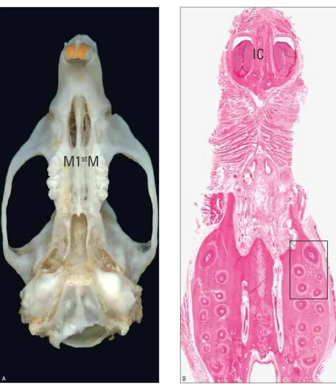
The experimental model of induced tooth movement described by Heller and Nanda5 has been widely adopted3,10 because results can be ex-trapolated to orthodontic clinical practice (Fig 1). Standardization and detailed descriptions of this experimental model ensure greater applicability and easier result extrapolations. The improvement of this model may provide further knowledge about the biology of induced tooth movement.3,10
In general, experimental times were 5 to 7 days in the first studies.7,8,9,13 However, it remains un-clear what tissue phenomena take place in murine maxillary first molar roots that received intense forces and produce indirect bone resorption. Sev-eral questions raised in previous studies4,6,10,11 using this model have not been answered to this date:
» Is the root resorption associated with ex-perimental induced tooth movement more closely related with frontal or undermining bone resorption?
» How long does it take to eliminate the hya-line areas, and when does the periodontal ligament begin its reorganization?
» When and how is the reabsorbed cortical bone replaced to reinsert the periodontal ligament?
» Do the hyalinized areas of connective tissue undergo phagocytosis, resorption or circum-scription?
» Where does root resorption occur, immedi-ately next to or away from hyaline areas? » When indirect bone resorption is suspected,
do microscopic data suggest the adoption of a greater interval for the reactivation of the orthodontic appliance?
* Head Professor, School of Dentistry of Bauru (FOB) and Graduate Program of School of Dentistry of Ribeirão Preto (FORP), University of São Paulo (USP), Brazil. ** Professor, Histology, Anhanguera School, Bauru, Brazil.
*** Professor, Oral Biology Program, Sagrado Coração University, Bauru, Brazil. **** Professor, Orthodontics Program, Araras University, Araras, Brazil.
***** MSc in Oral Pathology from FOB. PhD from Federal University of São Paulo, São Paulo, Brazil. ****** MSc in Pediatric Dentistry from FORP-USP.
IC
A B
FIGURE 1 - Murine skull where molars and incisors (IC) are seen, particularly maxillary first molar (M1stM) after movement by appliance designed by Heller and Nanda.5 Microscopic cross-section (B) shows tooth roots, particularly M1stM, in cervical plane.
A
B
C
anchorage in maxillary premolars promotes hyalinization of the periodontal ligament on the buccal face. Forces dissipate and the process ends when the midpalatal suture is separated. Does indirect bone resorption begin long before that? When does it actu-ally begin, at 3, 5, 7 or 9 days?
Few studies investigated the chronology and sequential events of indirect bone resorption and the consequent periodontal reorganization resulting from it. Microscopic analyses of the events induced by intense forces on teeth that undergo experimentally induced movement in murine models contributed to answer some of the questions raised, such as in the study con-ducted by Cardoso,2 together with Consolaro, Kinoshita, Francischone, Santamaria Jr., Fracalos-si and Maldonado. Their most interesting find-ings were the late results, when the periodontal ligament is reorganized and root resorptions are more active and intense (Figs 6, 7 and 8).
In patients, delayed events and periodontal reorganization occur at each activation time, between 15 and 21 days. At the end of six to twelve years, the resulting sum of the several orthodontic appliance activation times may be demonstrated by radiographic and CT images of periodontal tissues and tooth roots. Knowing each activation time and its beginning, middle and end substantially increases our chances of acting to reduce unwanted consequences.
Some of the interventions that orthodontic specialists may choose, based on results of ex-perimental studies, are:
1) Defining plans to prevent root resorption and bone loss.
2) Distributing the application of forces on tooth structure to reduce patient pain and dis-comfort.
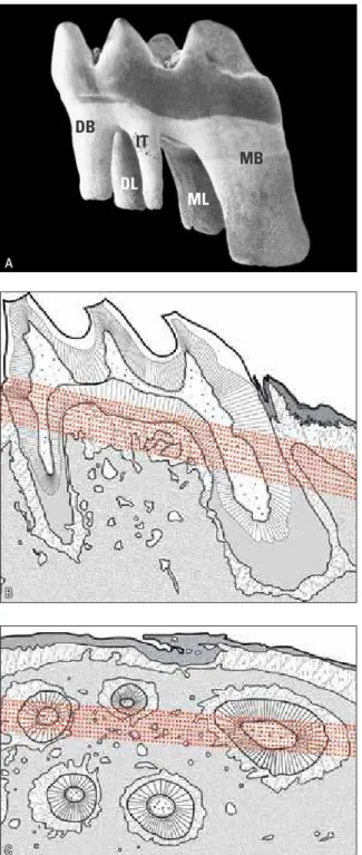
Ligament hyalinization reduces or blocks tooth movement and may also be associated with root resorption. Knowledge about tissue, FIGURE 2 - In A, murine first molar and its five roots. In the
mesio-buccal (MB) root, forces dissipate along its larger and longer struc-ture. In other roots (distobuccal, intermediate, distolingual and me-siolingual), delicate structures clearly show effects of forces on periodontal tissues. In B, red lines show cross-sections at cervical
level in schematic drawing of a longitudinal section of murine first molar. In C, red lines correspond to longitudinal views in
cross-section of murine maxillary first molar (A: modified from Alatli-Kut
et al.1; B and C: of Fracalossi4).
DB IT
MB
FIGURE 3 - Normal periodontal structures on the mesial face of murine M1stM distobuccal root, which received intense forces in the experimen-tal model designed by Heller and Nanda.5 B = alveolar bone; PL = peri-odontal ligament; C = cement; D = dentine; P = tooth pulp; V = vessels; Cb = cementoblasts; Ob = osteoblasts; ECM = extracellular matrix. (HE;10X).
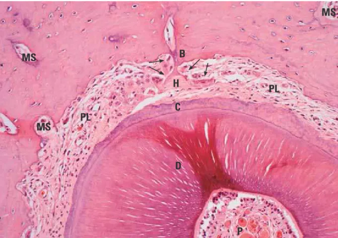
FIGURE 5 - Indirect bone resorption on mesial face of murine M1stM distobuccal root after application of intense forces for 5 days. Hyalinized periodontal ligament (H) and clastic activity (circle) surround it. B = alveolar bone; PL = periodontal ligament; C = cement; D = dentine; P = tooth pulp; MS = marrow space. (HE; 10X).
FIGURE 4 - Incipient indirect bone resorption on mesial face of murine M1stM distobuccal root after application of intense forces for 3 days. Hyalinized periodontal ligament (H) and initial clastic activity (circle) surround it. B = alveolar bone; PL = periodontal ligament; C = cement; D = dentine; P = tooth pulp. (HE; 10X).
B
v v
PL
ECM
D
P C
Cb Ob
v B
H
C
D
H
P PL
H
MS
MS
B
H
H
H
D
C
PL
MS
P
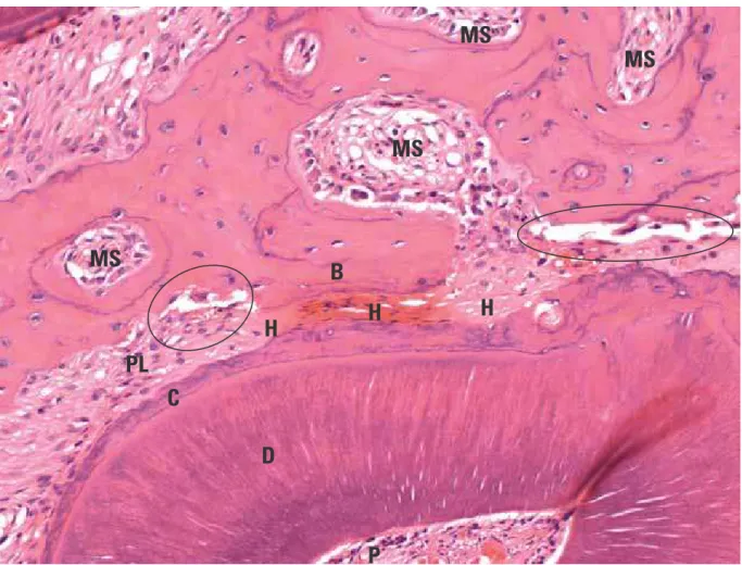
FIGURE 6 - Indirect bone resorption (arrows) on mesial face of murine M1stM distobuccal root after application of intense forces for 7 days. Hyalinized periodontal ligament (H) and clastic interaction with hyalinized areas surround it. Root surface exposure due to root resorption induced by death of cementoblasts; several associated bone re-modeling units (circles). B = alveolar bone; PL = periodontal ligament; C = cement; D = dentine; P = tooth pulp; MS = marrow space. (HE; 10X).
FIGURE 7 - Indirect bone resorption (arrows) on mesial face of murine M1stM distobuccal root after application of in-tense forces for 9 days. Ligament is reorganizing and frontal bone resorption is already visible on periodontal surface of cortical plate (circle). Hyaline areas remaining from previously hyalinized periodontal segment (H) are associated with clastic activity. Root resorption (RR) is seen in cement and dentine, together with active bone remodeling units. B = alveolar bone; PL = periodontal ligament; C = cement; D = dentine; MS = marrow space. (HE;10X).
MS
B
C
D
P
H
H
PL
PL
MS
MS
MS
MS
MS
B
H
H
H
C
D
RR
RR
cell and molecular phenomena involved in in-duced tooth movement may provide a basis for clinical procedures.
Murine molars have 5 roots,3,5,12 and the ex-perimental orthodontic appliance (Fig 1) de-signed by Heller and Nanda5 applies intense forces on four roots: distobuccal, intermediate, distolingual and mesiolingual (Fig 2). In the me-sial or mesiobuccal root, the forces applied by the appliance dissipate along larger and longer root structures, which affect periodontal tissues
similarly to the application of slight or moder-ate forces. Because of these characteristics, in the experimental model the effects of two types of forces may be analyzed at the same time accord-ing to their intensity: mild/moderate or intense.
The distolingual root, according to the study
by Cardoso,2 may show morphological changes
associated with indirect buccolingual bone re-sorption in cross-sections of the cervical region of the root and the alveolar bone process, as il-lustrated in Figures 3, 4, 5, 6, 7 and 8.
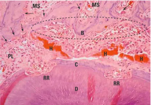
FIGURE 8 - Indirect or undermining bone resorption (arrows) on mesial face of murine M1stM distobuccal root after application of intense forces for 9 days, and more advanced reorganization than in Figure 7. Periodontal ligament is reorganizing together with remnants of cortical bone. Hyaline areas remaining from previously hyalinized periodontal segment (H) are associated with clastic activity. B = alveolar bone; PL = periodontal ligament; C = cement; D = dentine; P = tooth pulp; MS = marrow space. (HE;10X).
H
B
PL
MS
C
D
P
PL
The conclusions of the study discussed here showed that:
1. At 3 days of tooth movement induced by in-tense forces, indirect bone resorption had not begun in most of the specimens analyzed, but some showed discrete points of bone remod-eled units (Fig 4).
2. Only at 5 days were osteoclasts from bone remodeled units seen on adjacent bone sur-faces and around hyaline areas. At this time, root resorption was still incipient and limited to cement (Fig 5).
3. At 7 days, there was clear indirect bone resorp-tion on trabecular bone and cortical surfaces, but is far from the cortical bone associated with the segment of hyalinized periodontal ligament (Fig 6). Bone resorption in
orth-moderate forces, occurs on the surface of cor-tical bone in front of the area of periodontal ligament compression and is, therefore, called frontal bone resorption. In indirect bone re-sorption, the connections of cortical bone with adjacent and underlying bone are under-mined by numerous bone remodeling units. Root resorptions are active, occur in a larger extension and affect dentine more deeply. 4. At 9 days of tooth movement induced by
in-tense forces, the hyaline areas are under par-tial resorption (Figs 7 and 8). The periodon-tal ligament is under reorganization. Only isolated signs of the previous undermined cortical plate remain because it is undergo-ing complete remodelundergo-ing. Root resorptions are still actively occurring.
1. Alatli-Kut I, Hultenby K, Hammarstrom L. Disturbances of cementum formation induced by single injection of 1-hydroxyethylidene-1,1-bisphosphonate (HEBP) in rats: light and scanning electron microscopic studies. Scand J Dent Res. 1994;102(5):260-8.
2. Cardoso LB. Análise morfológica da evolução da reabsorção óssea à distância na movimentação dentária induzida em molares murinos [dissertação]. Bauru: Universidade Sagrado Coração; 2011.
3. Consolaro A. Reabsorções dentárias nas especialidades clínicas. 2ª ed. Maringá: Dental Press; 2005.
4. Fracalossi ACC. Análise da movimentação dentária induzida
em ratos: inluência do alendronato nas reabsorções dentárias,
estudo comparativo em cortes transversais e longitudinais e avaliação microscópica em diferentes períodos de observação [dissertação]. Bauru: Universidade de São Paulo; 2007. 5. Heller IJ, Nanda R. Effect of metabolic alteration of periodontal
ibers on orthodontic movement: an experimental study. Am J
Orthod. 1979;75:239-58.
6. Maldonado VB. Efeitos microscópicos do ácido salicílico (aspirina) e do acetaminofeno (tylenol) na movimentação dentária induzida e nas reabsorções radiculares associadas [dissertação]. Ribeirão Preto: Universidade de São Paulo; 2009. 7. Mazziero ET. Bisfosfonato e movimentação dentária induzida:
avaliação microscópica de seus efeitos [tese]. Bauru: Universidade de São Paulo; 1999.
8. Ortiz MFM. Inluência dos bisfosfonatos na movimentação dentária induzida, na frequência e nas dimensões das
reabsorções radiculares associadas [tese]. Bauru: Universidade de São Paulo; 2004.
REFERENCES
9. Pereira ACC. Inluência da gravidez e dos anticoncepcionais
na reabsorção radicular e na remodelação óssea consequente à movimentação dentária induzida: avaliação microscópica [dissertação]. Bauru: Universidade de São Paulo; 1996. 10. Ren Y, Maltha JC, Kuijpers-Jagtman AM. The rat as model for
orthodontic tooth movement: a critical review and proposed solution. Eur J Orthod. 2004;26:483-90.
11. Santamaria Jr M. Biologia da movimentação dentária induzida e
das reabsorções radiculares associadas. Inluência do gênero e
dos bisfosfonatos [tese]. Bauru: Universidade de São Paulo; 2009.
12. Schour I, Massler M. The teeth. In: Farris EJ, Grifith JK. The
rat in laboratory investigation. 2nd ed. New York: Hafner;
1963. p. 104-65.
13. Vasconcelos MHF. Análise morfológica comparativa do periodonto de sustentação submetido a forças biologicamente excessivas em ratas adultas sem e sob o uso de anticoncepcionais e ratas prenhas [dissertação]. Bauru: Universidade de São Paulo; 1996.
Contact address
Alberto Consolaro