Analysis of the correlation between mesiodistal
angulation of canines and labiolingual inclination
of incisors
Amanda Sayuri Cardoso Ohashi*, Karen Costa Guedes do Nascimento*, David Normando**
Objective: To assess the degree of correlation between canine angulation and incisor inclination. Methods: Mesiodistal angulation of canines and labiolingual inclination of incisors were obtained by means of digital graphics software (ImageTool®) from
standard-ized photographs of the casts of 60 patients. Incisor inclination was also assessed by lateral cephalometric radiographs. Results: Random error showed a variation of around 2° in measurements made on the casts (1.8-2.5), while systematic error, measured by the in-traclass correlation test, displayed excellent reproducibility for both methods used in this study (p<0.001, r=0.84-0.96). Linear correlation tests revealed a significant positive cor-relation between canine angulation and incisor inclination in the maxillary arch (r=0.3, p<0.05) and even more significantly in the mandibular arch (r=0.46 to 0.51, p<0.001), when both were measured on the casts. When incisor inclination was examined by cepha-lometrics, correlation level was statistically insignificant for maxillary incisors (r=0.06 to 0.21, p>0.05) and varied widely in the mandibular arch (r=0.14 to 0.50). Conclusions: The introduction of changes in the angulation of canines with the aim of monitoring compensations observed in incisor inclination is warranted, especially in the lower arch. Abstract
Keywords: Malocclusion. Canines, angulation. Incisors, inclination.
* Dental Surgeon - Intern, Discipline of Orthodontics, School of Dentistry, Pará State Federal University.
** MSc in Integrated Clinic (FOUSP). PhD in Orthodontics, Rio de Janeiro State University (UERJ). Professor of Orthodontics, School of Den-tistry (UFPa). Coordinator of the Specialization Program in Orthodontics (ABO-Pa).
intROduCtiOn
Tooth inclination and angulation have long been investigated in orthodontics. In 1928, Angle3
system-ized orthodontic treatment by developing the Edge-wise appliance, whereby tooth inclination and angu-lation were produced through bends placed in the leveling archwire and inserted in the bracket slots.
Andrews1 published a study in 1972 to
per-form an in-depth examination of the character-istics of normal, optimal occlusion and identi-fied six features shared by all the study casts. The author then introduced “The Six Keys to Optimal Occlusion” and suggested that attain-ing these morphological features was the goal of
orthodontic treatment. Andrews noted that the long axes of all teeth were mesially tipped to varying degrees, depending on the group of teeth examined, and termed this feature as the 2nd key
to normal occlusion. The 3rd key outlined by
An-drews was crown inclination (torque), defining a positive value for the upper incisors (buccal crown torque) and negative, or lingual for the remaining teeth. The angulation and inclina-tion values observed in the Andrews study2 were
instrumental for the invention of the Straight-Wire appliance.
A few years later, changes were made to the inclination of incisor brackets to compensate for the skeletal discrepancies that were not addressed in its entirety during orthodontic treatment.2 In
the case of Class III malocclusion, incisors were tipped more buccally in the maxillary arch and more lingually in the mandibular arch, while in Class II the opposite was implemented. It later became evident that, in fact, lower incisor inclina-tion was strongly influenced by the relainclina-tionship between apical bases in the sagittal plane, which played an important role in achieving a normal8
incisal relationship in the same manner that changes in upper incisor inclination significantly affect posterior occlusion.12
In addition to the compensatory inclinations designed for the incisor region,2 whose effects on
the arch length were eventually scientifically prov-en10 a few years later, changes were incorporated in
canine angulation with the purpose of monitor-ing the compensations built into incisor brackets.4
Thus, the mesiodistal angulation of canines would be increased whenever orthodontic treatment aimed to incline incisors labially, and be decreased when the goal was either to incline incisors lingual-ly or maintain an existing lingual compensation.
The impact exerted by changes in incisor an-gulation on arch length had been previously as-sessed by a mathematical model,7 which showed
that these alterations caused only small changes in the dental arch. However, no study seems to have
examined the effects of mesiodistal angulation of canines on incisor inclination (torque).
In an orthodontic treatment geared toward case individualization, such as skeletal malocclusion cases treated with compensations, it is extremely important to recognize the natural features of each patient’s compensations. As well as the role played by incisors, canine angulation needs to be investi-gated since these teeth are positioned in an impor-tant area within the geometric design of the dental arch. The first step seems to consist in examining whether such canine angulation compensations are indeed present in the several skeletal altera-tions in the face — a fact which was confirmed in a previous study9 — and also whether or not such
compensations and changes exhibit a significant correlation, which is the purpose of this study.
MAtERiAL And MEtHOdS
This study comprised a sample of 60 pa-tients in the stage of permanent dentition, from the private practice of one single orthodontist, with the aim of establishing a correlation analy-sis between mesiodistal canine angulation and labiolingual incisor inclination and anteroposte-rior position.
The sample comprised individuals with differ-ent types of malocclusion as determined by molar relationship: Class I (n=20), Class II (n=20) and Class III (n=20) without previous orthodontic treatment. Patients who presented with tooth loss, agenesis, moderate or severe crowding and/ or syndromes were immediately excluded since these factors might interfere with canine and inci-sor inclination.
c
b
d
a were placed on a glass plate (Fig 2a), at a distance
of 20 cm from the camera (Fig 2b). At the bottom of each model a black device was placed with a marking in the center, used as reference to cen-tralize the teeth that would be photographed (Fig 2c), as described in a previous study.9
The camera lens was propped on a utility wax plate to optimize lens direction (Fig 2d). Canines and incisors were clearly centered at the time the photographs were taken. To better visualize the long axes of canines, these teeth were positioned so that their labial surface faced the examiner (Fig 3A). Incisors were positioned in lateral view so as to render visible their inclination (Fig 3B).
A total of 360 photographs were taken and later exported to a computer program (Adobe Photoshop 7.0®) where the occlusal plane was
traced (Fig 4). Those images were subsequently imported into an image editing program (Image Tool® – www.imagetool.com) where canine
angu-lation and incisor inclination were measured.
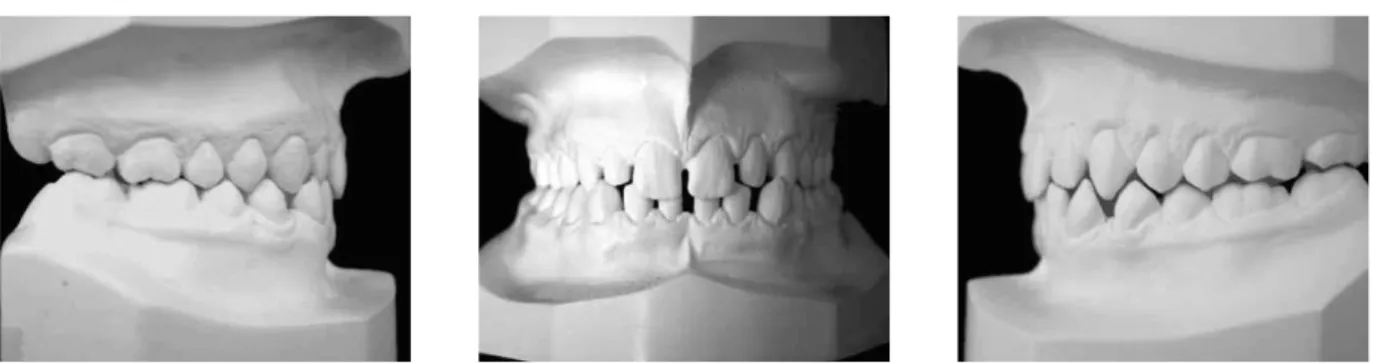
When necessary, photograph brightness and contrast were adjusted in order to enhance visual-ization of structures, thereby providing a sharper outline of the teeth. The occlusal plane was traced from the incisal surface of the central incisors to the mesiobuccal cusp of the first permanent mo-lar to determine both canine and incisor inclina-tion. Canine angulation measurements were then FIGURE 1 - Plaster casts of an individual with Class I malocclusion used in the sample.
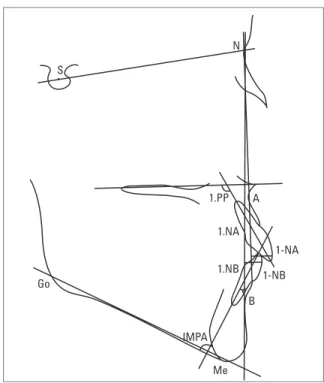
FIGURE 2 - Method used to standardize how photographs of plaster models were taken to determine canine angulation.
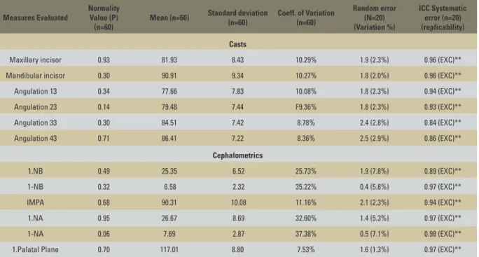
S
N
1.NA
1.NB
1-NA
1-NB
B
IMPA
Me Go
1.PP A
performed using the same graphics software used for tracing the long axes of canine crowns. Based on the intersection of these two lines the value of the angle of the clinical crown of the canine was obtained in the plaster casts. The same program was used to measure incisor inclination by draw-ing a line tangent to the center of the right central incisor crown, which intersected the previously outlined occlusal plane (Fig 3B).
Incisor inclination was measured using lateral cephalometric radiographs of the sample (Fig 4). The angle formed between the long axis of man-dibular incisors and the manman-dibular plane (IMPA) and the angle formed between the long axis of the maxillary incisors and the palatal plane (1.PP) were also examined. Incisor inclination was also assessed using measures 1.NA and 1.NB and dis-tances 1-NA and 1-NB.
Radiographs were traced manually and points were digitized using an 1812 series Genius Tab-let. Tracings were performed by one of the re-searchers and checked by an orthodontist. Mea-surements were obtained by means of SMTC (Sistema de Medição e Traçado Cefalométrico) computer software.
Statistical analysis
After obtaining cephalometric and dental cast measurements, the D’Agostino-Pearson test was employed to analyze normal data distribu-tion. Method error study was performed by re-assessing 20 cases. Random error was reviewed by Dahlberg’s formula while systematic error was analyzed by intraclass correlation test. Cor-relation analysis between the measurements was performed using Pearson’s linear correlation test. The confidence level used throughout the analy-sis was 5% (P<0.05). Calculation for determining sample size was performed assuming the use of Pearson’s linear correlation test with 5% alpha level, 80% power and a minimum correlation co-efficient (r value) of 0.35. The sample size found for such conditions comprised 63 individuals.
RESuLtS
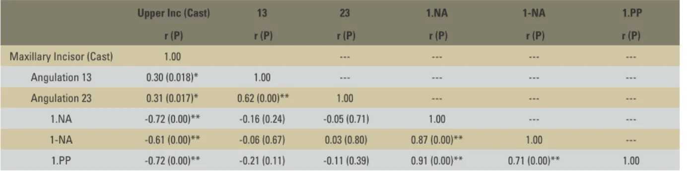
Data analysis (Table 1) indicated that the samples showed normal distribution (P>0.05). It was also noted that the variation coefficient was approximately 10% for measurements made on the dental casts, and cephalometric angles of the incisors relative to the basal bone (1.PP, IMPA). These values, however, were higher than 25% when the cephalometric measurements related the incisors to a reference line joining their re-spective basal bones to the nasion point (1.NA, 1-NA, 1.NB, 1-NB).
Random error analysis of measurements made on the casts ranged from 2% to 2.9% of the mean. Cephalometric measurements that examined the incisors relative to a reference line on the cranial base (1.NA, 1-NA, 1.NB, 1-NB) showed a random error greater than 5% of the mean, while for the angle formed between the long axis of the teeth and their apical base (IMPA and 1.PP) error was about 2%. However, for all measures examined by analysis of systematic error using intraclass
correlation coefficient, the level of replicability was excellent (Table 1).
The results in Table 2 show no significant corre-lation between the position of the incisors, as mea-sured on the radiographs (1.NA, 1.PP and 1-NA), and canine angulation, as examined on the casts. As can be observed, there was a weak but significant positive correlation between the position of the incisors, as measured on the casts, and canine an-gulation (P<0.05). A strong correlation was found between the position of the upper incisors, as mea-sured on the radiographs and on the casts (P<0.01). Table 3 shows a significant correlation between the position of the lower incisors, as measured on radiographs, and canine angulation, as measured on the casts. The only exception was the correla-tion between tooth 33 and 1-NB. When both ca-nine angulation and lower incisor position were measured on the casts, a significant correlation (P<0.01) was found. There was also a significant correlation between lower incisor position as measured on radiographs vs. on models (P<0.01).
The data therefore demonstrate that the me-siodistal angulation of canines tended to follow incisor inclination, when both were measured on the casts, but more significantly in the lower arch than in the upper arch.
diSCuSSiOn
This study lends support to the notion that changes induced in the mesiodistal angulation of canines aimed at monitoring sagittal compensa-tions observed in incisors allow an increase or de-crease in dental arch perimeter. Despite this asser-tion, some important details should be pointed out in the results, especially with regard to differences in the degree of correlation between these mea-surements when the upper and lower dental arches are analyzed separately, as well as the method used to measure incisor inclination (Tables 2 and 3).
The results revealed that, in general, when canines are more mesially tipped, the incisors tend to follow this angulation and become more labially inclined. Likewise, when canines
Measures Evaluated
Normality Value (P)
(n=60)
Mean (n=60) Standard deviation (n=60)
Coeff. of Variation (n=60)
Random error (N=20) (Variation %)
ICC Systematic error (n=20) (replicability)
Casts
Maxillary incisor 0.93 81.93 8.43 10.29% 1.9 (2.3%) 0.96 (EXC)**
Mandibular incisor 0.30 90.91 9.34 10.27% 1.8 (2.0%) 0.96 (EXC)**
Angulation 13 0.34 77.66 7.83 10.08% 1.8 (2.3%) 0.94 (EXC)**
Angulation 23 0.14 79.48 7.44 F9.36% 1.8 (2.3%) 0.93 (EXC)**
Angulation 33 0.30 84.51 7.42 8.78% 2.4 (2.8%) 0.84 (EXC)**
Angulation 43 0.71 86.41 7.22 8.36% 2.5 (2.9%) 0.86 (EXC)**
Cephalometrics
1.NB 0.49 25.35 6.52 25.73% 1.9 (7.8%) 0.89 (EXC)**
1-NB 0.32 6.58 2.32 35.22% 0.4 (5.8%) 0.97 (EXC)**
IMPA 0.68 90.31 10.08 11.16% 2.1 (2.3%) 0.94 (EXC)**
1.NA 0.95 26.67 8.69 32.60% 1.4 (5.3%) 0.97 (EXC)**
1-NA 0.06 7.69 2.87 37.38% 0.5 (7.1%) 0.98 (EXC)**
1.Palatal Plane 0.70 117.01 8.80 7.53% 1.6 (1.3%) 0.97 (EXC)**
TABLE 1 - Analysis of normal distribution (D’Agostino-Pearson’s test), mean, standard deviation, coefficient of variation, random error, systematic error (Intraclass correlation-ICC) and level of replicability.
TABLE 2 - Pearson’s correlation matrix (r) and P value (in parentheses) for measurements made in the upper arch.
TABLE 3 - Pearson’s correlation matrix (r) and P value (in parentheses) for measurements made in the lower arch. * P<0.05; **P<0.01.
* P<0.05; **P<0.01.
Upper Inc (Cast) 13 23 1.NA 1-NA 1.PP
r (P) r (P) r (P) r (P) r (P) r (P)
Maxillary Incisor (Cast) 1.00 --- --- ---
---Angulation 13 0.30 (0.018)* 1.00 --- --- ---
---Angulation 23 0.31 (0.017)* 0.62 (0.00)** 1.00 --- ---
---1.NA -0.72 (0.00)** -0.16 (0.24) -0.05 (0.71) 1.00 ---
---1-NA -0.61 (0.00)** -0.06 (0.67) 0.03 (0.80) 0.87 (0.00)** 1.00
---1.PP -0.72 (0.00)** -0.21 (0.11) -0.11 (0.39) 0.91 (0.00)** 0.71 (0.00)** 1.00
Mandibular Incisor (Cast) 33 43 1.NB 1-NB IMPA
r (P) r (P) r (P) r (P) r (P) r (P)
Mandibular Incisor (Cast) 1 --- --- --- ---
---Angulation 33 0.46 (0.00)** 1 --- --- ---
---Angulation 43 0.52 (0.00)** 0.44 (0.00)** 1 --- ---
---1.NB 0.61 (0.00)** 0.29 (0.02)* 0.26 (0.04)* 1 ---
---1-NB 0.43 (0.00)** 0.14 (0.28) 0.26 (0.05)* 0.76 (0.00)** 1
---IMPA 0.69 (0.00)** 0.50 (0.00)** 0.36 (0.00)** 0.73 (0.00)** 0.47 (0.00)** 1
exhibited a smaller mesial angulation, incisors appeared more lingually inclined. This correla-tion, however, was more evident in the lower arch (Table 3) than in the upper arch (Table 2). The authors could not find a logical explanation for this fact, but it is likely that the manner in which the lower arch was restricted by the up-per arch may be related to these results. Data from a previous study9 demonstrate that only
the lower canines showed a significant change in angulation when Class III subjects were com-pared with Class I individuals, corroborating the results achieved in this study.
The idea that changes in tooth angulation could influence tooth inclination (torque) and arch length was investigated through a mathemat-ical model7 that examined the incisors. Moreover,
the compensations observed in incisor inclination and their relationship with the maintenance of
in-cisal contacts in the presence of sagittal skeletal alterations had already been detailed on models.8
In light of the results of this study, however, it is reasonable to believe that the influence of canine angulation seems to be, as yet, an important factor affecting sagittal incisor compensation in skeletal discrepancy cases.
Several methodologies for assessing tooth an-gulation and inclination have recently been de-scribed in the literature. However, these meth-ods typically involve devices not available in the market and require customized fabrication6,11,13
or high cost technologies.5 In this study, the
specifically for this study, which also showed an excellent level of replicability and a random error of about 2% (Table 1).
The correlation between canine angulation and incisor inclination yielded different results de-pending on whether the incisors were examined cephalometrically or on the casts. The maxillary arch (Table 2) exhibited a weak (r=0.3/0.31) but significant correlation (P<0.05) between incisor inclination, as measured on the casts, and the de-gree of canine angulation. Furthermore, there was no statistically significant correlation when incisor inclination was examined with the aid of cephalo-metric measurements (P>0.05).
The lower arch (Table 3) showed a statistically significant correlation every time that canine an-gulation was correlated with incisor inclination, as measured on the casts (r=0.46/0.52, P<0.01) and cephalometrically. The strongest correla-tions (r=0.50/0.36, P<0.01) were obtained for the measurement that reflects the angle formed between the long axes of the incisors and the mandibular plane (IMPA), while the weakest correlations (r=0.14, P>0.05/r=0.26, P<0.05) were found for the measurement that examines (in millimeters) the position of the lower incisors relative to the NB line (1-NB).
The results revealed a different behavior when incisor inclination was examined on the casts vs. cephalometrically. Tables 2 and 3 show that in-cisor inclination, when measured on radiographs and on the casts, showed a significant correlation, i.e., measurements made on the models closely
followed those made on radiographs. However, the strongest correlation for both the upper and lower arches was noted when incisor inclination, as examined on the casts, was correlated with the cephalometric angles used to assess the inclination of the tooth in direct relation to the basal bone (IMPA and 1.PP), while a weaker correlation was found for both arches when incisor inclination, as measured on the casts, was correlated with incisor protrusion on the radiographs (1-NA and 1-NB).
It is also noteworthy that the cephalometric measures that correlated the incisors with their reference line (1.NA, 1-NA, 1.NB and 1-NB) exhibited the highest coefficient of variation (always greater than 25%). This result has led the authors to regard these measurements with utmost caution, given their extremely wide variation (Table 1).
1. Andrews L. The six keys to normal occlusion. Am J Orthod. 1972;62(3):296-309.
2. Andrews L. The diagnostic system: occlusal analysis. Dent Clin N Am. 1976;20(4):671-90.
3. Angle EH. The latest and best in orthodontic mechanism. Dent Cosmos. 1928;70:1143-58.
4. Capelozza Filho L, Silva Filho OG, Ozawa TO, Cavassan AO. Individualização de bráquetes na técnica de straight wire: revisão de conceitos e sugestões de indicações para uso. Rev Dental Press Ortodon Ortop Facial. 1999;4(4):87-106. 5. Capelozza Filho L, Fattori L, Maltagliati LA. Um novo
método para avaliar as inclinações dentárias utilizando a tomograia computadorizada. Rev Dental Press Ortodon Ortop Facial. 2005;10(5):23-9.
6. GhahferokhI AE, Elias L, Jonssons S, Rolfe B, Richmond S. Critical assessment of a device to measure incisor crown inclination. Am J Orthod Dentofacial Orthop. 2002;121(2):185-91.
7. Hussels H, Nanda RS. Effect of maxillary incisor angulation and inclination on arch length. Am J Orthod Dentofacial Orthop. 1987;91(3):233-9.
8. Ishikawa H, Nakamura S, Kim C, Iwasaki H, Satoh Y, Yoshida S. Individual growth in class III malocclusions and its relationship to the chin cap effects. Am J Orthod Dentofacial Orthop. 1998;114(3):337-46.
REfEREnCES
9. Azevedo LR, Torres TB, Normando ADC. Angulação dos caninos em indivíduos portadores de má oclusão de Classe I e de Classe III: análise comparativa através de um novo método utilizando imagens digitalizadas. Dental Press J Orthod. 2010;15(5):109-17.
10. Ohigiins EA, Kirschen RH, Lee RT. The inluence of maxillary incisor inclination on arch length. Br J Orthod. 1999;26(2):97-102.
11. Richmond S, Klufas ML, Syawany M. Assessing incisor inclination: a non-invasive technique. Eur J Orthod. 1998;20(6):721-6.
12. Sangcharearn Y, Ho C. Maxillary incisor angulation and its effect on molar relationships. Angle Orthod. 2007;77(2):221-5. 13. Zanelato ACT, Maltagliati LA, Scanavini MA, Mandetta S.
Método para mensuração das angulações e inclinações das coroas dentárias utilizando modelos de gesso. Rev Dental Press Ortodon Ortop Facial. 2006;11(2):63-73.
Contact address
David Normando
Rua Boaventura da Silva, 567- apt. 1201 CEP: 66.060-060 - Belém / PA, Brazil E-mail: davidnor@amazon.com.br Submitted: August 2008