Prevalence of dental anomalies of number in different
subphenotypes of isolated cleft palate
João Paulo Schwartz1, Daniele Salazar Somensi1, Priscila Yoshizaki1, Luciana Laís Savero Reis1, Rita de Cássia Moura
Carvalho Lauris2, Omar Gabriel da Silva Filho2, Gisele Dalbén3, Daniela Gamba Garib4
How to cite this article: Schwartz JP, Somensi DS, Yoshizaki P, Reis LLS, Lauris RCMC, Silva Filho OG, Dalbén G, Garib DG. Prevalence of dental anomalies of number in different subphenotypes of isolated cleft palate. Dental Press J Orthod. 2014 Jan-Feb;19(1):55-9. doi: http://dx.doi.org/10.1590/2176-9451.19.1.055-059.oar
Submitted: December 01, 2011 - Revised and accepted: April 08, 2012
Contact address: Daniela Gamba Garib
Rua Rio Branco, 19-18 – Bauru/SP – Brazil — CEP: 17.040-480 E-mail: [email protected]
1 Specialist in Orthodontics, Hospital for Rehabilitation of Craniofacial
Anomalies – São Paulo University (HRAC-USP).
2 MSc in Orthodontics, HRAC-USP. 3 PhD in Pediatric Dentistry, HRAC-USP.
4 Full professor and assistant professor of Orthodontics, School of Dentistry —
University of São Paulo/Bauru.
» The authors report no commercial, proprietary or financial interest in the prod-ucts or companies described in this article.
Objective:This study aimed at carrying out a radiographic analysis on the prevalence of dental anomalies of number (agen-esis and supernumerary teeth) in permanent dentition, in different subphenotypes of isolated cleft palate pre-adolescent patients. Methods: Panoramic radiographs of 300 patients aged between 9 and 12 years, with cleft palate and enrolled in a single treatment center, were retrospectively analyzed. The sample was divided into two groups according to the extension/ severity of the cleft palate: complete and incomplete . The chi-square test was used for intergroup comparison regarding the prevalence of the investigated dental anomalies (P < 0.05). Results: Agenesis was found in 34.14% of patients with complete cleft palate and in 30.27% of patients with incomplete cleft palate. Supernumerary teeth were found in 2.43% of patients with complete cleft palate and in 0.91% of patients with incomplete cleft palate. No statistically significant difference was found between groups with regard to the prevalence of agenesis and supernumerary teeth. There was no difference in cleft prevalence between genders within each study group. Conclusion: The prevalence of dental anomalies of number in pre-adolescents with cleft palate was higher than that reported for the general population. The severity of cleft palate did not seem to be associated with the prevalence of dental anomalies of number.
Keywords:Cleft palate. Tooth abnormalities. Panoramic radiograph.
Objetivo:o propósito deste estudo foi avaliar radiograficamente a prevalência das anomalias dentárias de número (agenesias e supranumerários), na dentição permanente, em diferentes subfenótipos da fissura isolada de palato, em pacientes pré-adolescentes. Métodos: foram investigadas, de forma retrospectiva, 300 radiografias panorâmicas de pacientes com fissura palatina (pós-forame), de 9 a 12 anos de idade, matriculados em um mesmo centro. A amostra foi dividida em dois grupos de acordo com a extensão/gravidade da fissura palatina: completa e incompleta. O teste qui--quadrado foi utilizado para comparação intergrupos das prevalências de anomalias avaliadas (p < 0,05). Resultados: a agenesia dentária foi encontrada em 34,14% dos pacientes com fissura pós-forame completa e em 30,27% com fissura pós-forame incompleta. A prevalência de dentes supranumerários correspondeu a 2,43% nos pacientes com fissura pa-latina completa e a 0,91% no grupo com fissura papa-latina incompleta. Não houve diferença estatística significativa entre os grupos quanto à prevalência de agenesias dentárias e supranumerários. Não se observou diferença sexual quanto à prevalência de fissura dentro de cada grupo de estudo. Conclusão: os pacientes pré-adolescentes com fissura palatina apresentam maior prevalência de anomalias dentárias em relação à população em geral. A gravidade da fissura palatina parece não se associar com a prevalência de anomalias dentárias de número.
INTRODUCTION
The embryonic explanation for isolated clet palate is the lack of fusion of the palatal shelves that form the secondary palate. In this type of clet, the palatine pro-cesses do not fuse neither in the midline nor with the nasal septum, keeping the communication between oral and nasal cavities, while the formation of lips and alveolar ridge is processed normally.1
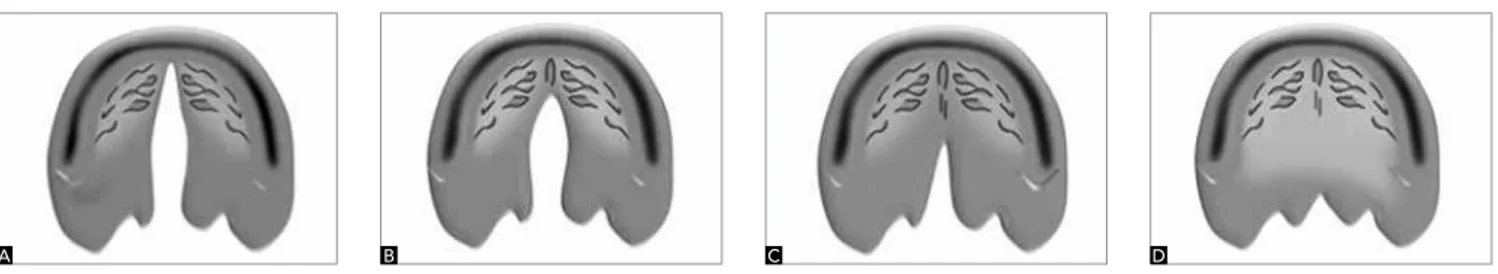
Isolated cleft palate may be complete or incom-plete.1 It is considered complete when it affects the
hard and soft palate, extending to the incisive fora-men (Fig 1A). On the other hand, it is considered incomplete when it partially affects the soft and/ or hard palate, not reaching the incisive foramen (Figs 1B, 1C and 1D).
The prevalence of cleft lip and palate is approxi-mately 1:1000 births.1 In general, individuals of Asian
descent have higher prevalence while those of Afri-can descent have lower prevalence when compared to Caucasian individuals.2 The etiology of cleft lip
and palate is complex, with multifactorial causality, in which case both genetic and environmental factors play a major role in determining the malformation.3
From an embryological standpoint, cleft palate is a disorder that differs from cleft lip and palate. Differ-ences in epidemiology and etiologic factors have also been reported in the literature.3
Similarly to the general population, odontogenic disorders are also found in patients with clefts. It is as-sumed that cleft and dental anomalies present a com-mon or inter-related genetic origin, considering the high prevalence of dental anomalies in cleft patients .4
In other words, patients with clefts present more in-cidence of dental anomalies than individuals without clefts.5 Moreover, the prevalence of dental
anoma-lies seems to be related to the extension/severity of cleft lip and palate.6
The diagnosis of dental anomalies of number is es-sential to deine the treatment plan in the rehabilita-tion process of patients with clet palate, either orth-odontic, with prosthesis or implants. The purpose of this study was to radiographically assess the prevalence of dental anomalies of number in diferent subpheno-types of isolated clet palate.
MATERIAL AND METHODS
This study was conducted in the department of Or-thodontics at the Hospital for Rehabilitation of Craniofa-cial Anomalies, University of São Paulo (HRAC-USP), ater approval by the respective Institutional Review Board. The study analyzed the radiographs of 300 pa-tients from the HRAC-USP iles. The sample com-prised 117 (39%) males and 183 (61%) females who were in late mixed dentition (second transitional period of mixed dentition according to the Van der Linden classii-cation) and early permanent dentition. The patients aged between 9 and 12 years old (chronological age). At this age, the third molars were excluded from the evaluation.
The total sample was divided into two study groups, according to the extension of clet palate indicated in the medical records of patients: Group 1 - complete clet palate; and Group 2 - incomplete clet palate.
The occurrence of permanent teeth agenesis and supernumerary permanent teeth was evaluated in pan-oramic radiographs by a calibrated observer with the aid of a ilm viewer in a room with appropriate lightening. The study included only radiographs with good tech-nical quality that allowed good visualization of teeth, erupted or not, and their surrounding structures.
Ater the prevalence of dental anomalies was calcu-lated in each study group, the chi-square test was used for comparison. It was also used to verify intragroup dif-ferences in the prevalence of anomalies between sexes. The results were considered at a signiicance level of 5%.
Figure 1 - Complete cleft palate invariably extends from the incisive foramen to the uvula (A). Incomplete cleft palate involves the posterior region of the pal-ate without reaching the incisive foramen (B); affects the soft palpal-ate and part of the hard palpal-ate (C); or may affect only the soft palpal-ate (D).
RESULTS
Out of the 300 patients analyzed, 82 (27.33%) had complete clet palate, whereas 218 (72.66%) had incomplete clet palate. Among individuals with complete clet palate, 31 (37.8%) were males and 51 (62.19%) females. As for patients with incomplete clet palate, 86 (39.44%) were males and 132 (60.55%) fe-males. In both groups, the proportion between male and female was approximately 1:2.
The prevalence of dental anomalies in Group 1 (com-plete clet palate) and Group 2 (incom(com-plete clet palate) is expressed in percentage and presented in Table 1. In Group 1, tooth agenesis of permanent teeth excluding the third molars was observed in 28 (34.14%) patients,while su-pernumerary teeth were found in 2 (2.43%). In Group 2, tooth agenesis was observed in 66 (30.27%) patients, while supernumerary teeth was found in 2 (0.91%). No signiicant diference in the prevalence of hypodontia
and supernumerary teeth was found between Groups 1 and 2 (Table 1). Additionally, there was no diference be-tween groups in the prevalence of agenesis for the most commonly afected teeth: second premolars and maxil-lary lateral incisors (Table 1).
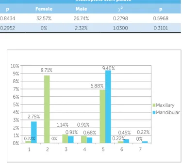
Figures 2 and 3 show the prevalence of agenesis of each permanent tooth in Groups 1 and 2, respectively. With regard to supernumerary teeth, in Group 1 they were found in the region of the maxillary let lateral in-cisor and the maxillary let second premolar, while in Group 2 supernumerary teeth were observed in the re-gion of the maxillary right lateral incisor and between the central incisors (mesiodens).
The distribution of dental anomalies according to sex is shown in Table 2. In both groups, there was no statistically signiicant diference between males and females for the prevalence of tooth agenesis and supernumerary teeth (Table 2).
Figure 2 - Prevalence of agenesis of each permanent tooth (excluding third molars) in the complete cleft palate group (group 1).
Figure 3 - Prevalence of agenesis of each permanent tooth (excluding third molars) in the incomplete cleft palate group (group 2).
G1 + G2 G1 G2 c2 p
Hypodontia 31.33% 34.14% 30.27% 0.0064 0.9359
Supernumerary 1.33% 2.43% 0.91% 0.0000 1.0000
Hypodontia MxLI 8.50% 7.92% 8.71% 1.9340 0.1644
Hypodontia Mx2P 7.50% 9.14% 6.88% 0.0000 1.0000
Hypodontia Md2P 8.66% 6.70% 9.40% 0.2545 0.6139
Table 1 - Prevalence of dental anomalies in groups 1 and 2, and intergroup comparison results (chi-square test).
* MxLI = maxillary lateral incisor, Mx2P = maxillary second premolar, Md2P = mandibular second premolar.
Table 2 - Prevalence of dental anomalies according to sex and intragroup comparison results (chi-square test).
Complete cleft palate Incomplete cleft palate
Female Male c2 p Female Male c2 p
Hypodontia 35.29% 32.25% 0.0390 0.8434 32.57% 26.74% 0.2798 0.5968
Supernumerary 0% 6.45% 1.0960 0.2952 0% 2.32% 1.0300 0.3101
0% 0% 0% 0% 0% 0% 0% 0% 0% 3.65% 3.65%
9.14%
6.70% 7.92%
3.04%
1 2 3 4 5 6 7
1% 2% 3% 4% 5% 6% 7% 8% 9% 10%
Maxillary Mandibular
0% 2.75%
0.22% 0%
0.68% 0.45% 0.22%
0.22% 0% 9.40%
6.88% 8.71%
1.14% 0.91%
0.91%
1 2 3 4 5 6 7
1% 2% 3% 4% 5% 6% 7% 8% 9% 10%
clet subphenotypes. Supernumerary teeth were preva-lent in 2.43% of group 1 and 0.91% of group 2 (Ta-ble 1). In patients with complete palatine clet, super-numerary teeth were found in the region of maxillary let lateral incisors and maxillary let second premolars. In patients with incomplete clet palate, they were found in the region of maxillary right lateral incisors and be-tween the central incisors (mesiodens). The prevalence of supernumerary teeth in adolescents without clets is 1% to 2%.15 A previous investigation found no
super-numerary teeth in patients with isolated clet palate.10
A recent study found that the presence of dental anomalies may represent an additional clinical marker for oral clets, suggesting a common genetic origin for these anomalies.16 The development of tooth germs
and the occurrence of clet palate are closely related during embryological development, both anatomically and chronologically, and many studies have reported the manifestation of dental anomalies associated with various forms of clet lip, clet palate or both.16 It has
been proposed that individuals with clet have higher prevalence of dental anomalies than the general popula-tion, and that the severity of the malformations seems to be directly related to the extension of the clet.16 In
this study, the prevalence of tooth agenesis and the to-tal prevalence of dento-tal anomalies, except for supernu-merary teeth, was slightly higher in female patients, al-though no statistically signiicant diference was found. The same was observed for the occurrence of complete and incomplete isolated clet palate.
CONCLUSION
The prevalence of dental anomalies of number seems not to be related to the subphenotypes of clet palate. Individuals with complete and incomplete clet palate showed a similar prevalence of permanent tooth agen-esis and supernumerary teeth. Further studies are neces-sary to determine the exact inter-relation between clet palate and the prevalence of other dental anomalies. DISCUSSION
Clet palate is more common among female patients.1,7
In this study, both complete and incomplete clet palate prevailed in females. In individuals without clets, ex-cluding the third molars, the prevalence of tooth agenesis in the population varies from 4.3% to 7.8%, primarily afecting the mandibular second premolar, followed by the maxillary lateral incisor and maxillary second premo-lar.8 The present results show that the prevalence of tooth
agenesis is much higher in individuals with clet palate than in the general population.
According to this study, the prevalence of hy-podontia of permanent teeth, excluding the third mo-lars, in patients with clet palate was 31.33% and was similar to that reported in the literature for patients with these malformations. Previous studies reported a prevalence of permanent tooth agenesis in patients with clet lip and palate of 25.5% to 33% in Czech pa-tients,9 30% in Swedish patients,10 25% to 40%11 and
33% in Finnish patients12 and 28.5% in Norwegian
patients.13 Another previous study reported that tooth
agenesis was observed more frequently in patients with complete clet palate than in patients with incomplete clet palate.11 However, our study did not ind any
dif-ference in the occurrence of hypodontia according to the subphenotypes (Table 1).
As for the subphenotypes, in both complete and in-complete clet palate the teeth most afected by hypodon-tia were the maxillary and mandibular second premolars as well as maxillary lateral incisors, particularly the max-illary second premolars in complete clet palate and the mandibular second premolars in incomplete clet palate (Figs 2 and 3). These data are in accordance with oth-er reports in the litoth-erature,9,10,14 and are similar to those
found for the general population.4
1. Silva Filho OG, Freitas JAS. Caracterização morfológica e origem embriológica In: Trindade IEK, Silva Filho OG. Fissuras Labiopalatinas: uma abordagem interdisciplinar. São Paulo: Ed. Santos; 2007. cap. 2, p.17-49. 2. Murray JC. Gene/environment causes of cleft lip and/or palate. Clin Genet.
2002;61(4):248-56.
3. Garib DG. Etiologia das más oclusões: implicações clínicas em ortodontia. In: Lubiana NF, Garib DG, Silva Filho OG. Pro-Odonto Ortodontia: programa de atualização em Ortodontia. Porto Alegre: ABO, Artmed, Panamericana; 2009. cap. 3.1, p. 1-37.
4. Garib DG. Padrão de anomalias dentárias associadas. In: Lubiana
NF, Garib DG, Silva Filho OG. Pro-Odonto Ortodontia: programa de atualização em Ortodontia. Porto Alegre: ABO, Artmed, Panamericana; 2009. cap. 5, p. 59-102.
5. Da Silva AP, Costa B, Carvalho Carrara, CF. Dental anomalies of number in the permanent dentition of patients with bilateral cleft lip: radiographic study. Cleft Palate Craniofac J. 2008;45(5):473-6.
6. Eerens k, Vlietinck R, Heindbünchel K, Van Olmen A, Derom C, Willems G, et al. Hypodontia and tooth formation in groups of children with cleft and, nonrelated controls. Cleft Palate Craniof J. 2001;38(4):374-8.
7. Derijcke A, Eerens A, Carels C. The incidence of oral clefts: a review. Br J Oral Maxillofac Surg. 1996;34(6):488-94.
8. Garib DG, Peck S, Gomes SC. Increased occurrence of dental
anomalies associated with second-premolar agenesis. Angle Orthod. 2009;79(3):436-41.
ReFeRenCeS
9. Jiroutová O. Hypodontia in patients with isolated cleft palate, its relationship to etiopathogenesis. Acta Chir Plast. 1991;33(1):57-63.
10. Larson M, Hellquist R, Jakobsson OP. Dental abnormalities and ectopic eruption in patients with isolated cleft palate. Scand J Plast Reconstr Surg Hand Surg. 1998;32(2):203-12.
11. Ranta R, Stegars T, Rintala A. Correlations of hypodontia in children with isolated cleft palate. Cleft Palated J. 1983;20(2):163-5.
12. Ranta R. A review of tooth formation in children with cleft lip/palate. Am J Orthod Dentofacial Orthop. 1986;90(1):11-8.
13. Andersson EM, Sandvik L, Abyholm F, Semb G. Clefts of the secondary palate referred to the Oslo cleft team: epidemiology and cleft severity in 994 individuals. Cleft Palate Craniofac J. 2010;47(4):335-42.
14. Haataja J, Haavikko K, Ranta R. Hypodontia and supernumerary teeth in Finnish children afected with facial clefts. An orthopantomographic and clinical study. Suom Hammaslaak Toim. 1971;67(6):303-11.
15. Bergström K. An orthopantomographic study of hypodontia,
supernumeraries and other anomalies in schoolchildren between ages of 8-9 years. Swed Dent J. 1977;1(1):45-57.