Post-thoracotomy wound separation (DEHISCENCE):
A disturbing complication
Aydin Nadir, Melih Kaptanoglu, Ekber Sahin, Hakan Sarzep
Cumhuriyet University, School of Medicine Sivas, Department of Thoracic Surgery, Sivas/Turkey.
OBJECTIVES:We described the treatment of dehiscence of thoracotomy incisions in patients who underwent thoracic surgery in the present study.
METHODS:Twenty-four patients with either partial or complete dehiscence of their thoracotomy incisions were included in the study from 2005 to 2010. The patients were evaluated regarding their age, sex, indication for thoracotomy, and surgical approaches. We also described our method of re-closure.
RESULTS: The male/female ratio was 17/7. The youngest and oldest patients were 15 and 75 years old, respectively, and the mean age was 43 years. Among the indications for thoracotomy, empyema was the most common reason (determined in eight [33%] patients), followed by vertebral surgery (determined in six [25%] patients). Bacterial growth was detected in the wound site cultures from 13 (54%) patients. For the patients with dehiscence of their thoracotomy incisions, an en block approximation technique with debridement was performed under general or local anesthesia in 16 (66%) and eight (33%) of the cases, respectively. Three patients exhibited an open thorax with dehiscence of the thoracotomy incision. Thoracoplasty was required in two patients. Using this method, successful closure was obtained in 91.7% (n = 22) of the patients with dehiscence of their thoracotomy incisions.
CONCLUSION: Dehiscence of the thoracotomy incision is an important complication that causes concern in patients and their thoracic surgeons and strongly affects the success of the surgery. An en block approximation technique with significant debridement that enables removal of the necrotic tissues from the wound site can successfully be applied to patients with dehiscence of their thoracotomy incisions.
KEYWORDS: Dehiscence; Failure; Suture; Thoracotomy; Wound.
Nadir A, Kaptanoglu M, Sahin E, Sarzep H. Post-thoracotomy wound separation (DEHISCENCE): A disturbing complication. Clinics. 2013;68(1):1-4.
Received for publication onApril 27, 2012;First review completed onJune 26, 2012;Accepted for publication onJuly 5, 2012 E-mail: [email protected]
Tel.: 00-90-346-2580211
& INTRODUCTION
Wound dehiscence is a surgical complication in which a wound fails to heal or opens along its incision line following surgery. The are many etiologies, including infection, weak tissue or muscle at the wound site, injury to the wound area, and other factors, which are commonly related to poor closure techniques (1).
Articles on thoracotomy wound dehiscence are scarce in the literature (2-4). We could not find the term ‘‘thoracot-omy dehiscence’’ in thoracic surgery textbooks or thoracic surgery articles. The commonly found papers are related to sternal separation after cardiac surgery and dehiscence of laparotomy incisions (5-7). Indeed, we did not find any
manuscript that includes a series of dehiscence of thor-acotomy incisions (DTI).
The incidence of wound dehiscence after cardiac or general surgery is approximately 0.3-5% and 0.25-3%, respectively (1,7). However, the incidence is unclear for thoracic surgery. Our dehiscence rate was 6.6% (24/360 patients) over five years, which alerted us to investigate this relatively high incidence.
Traditional surgical approaches or institution-based tech-niques at surgical clinics are preferred for addressing similar problems (8,9). The publication of these procedures adds to the essential knowledge base. Therefore, we aimed to draw attention to this disturbing complication and publish our experiences. The new term ‘‘thoracotomy dehiscence’’ may potentially be added to the thoracic surgical nomenclature upon recognition of this entity.
& PATIENTS AND METHODS
Twenty-four patients with either partial or complete DTIs were included in the study from 2005 to 2010. This study was approved by the local ethical committee at our university (No: 10/161). The patients were retrospectively
Copyrightß2013CLINICS– This is an Open Access article distributed under the terms of the Creative Commons Attribution Non-Commercial License (http:// creativecommons.org/licenses/by-nc/3.0/) which permits unrestricted non-commercial use, distribution, and reproduction in any medium, provided the original work is properly cited.
No potential conflict of interest was reported.
DOI:10.6061/clinics/2013(01)OA01
CLINICAL SCIENCE
evaluated regarding age, sex, indication for thoracotomy, surgical approach, and re-closure method for the DTI. The separations occurred postoperatively within the first two weeks. The separation included the muscle tissue layers in the complete or partially separated wounds. The length of the separation was between eight and ten centimeters and predominantly located in the anterior section of the incision in the partially separated wounds.
Specimens for cultures and antibiograms were obtained from each wound site. In coordination with the Infection Control Committee, the patients were given either sensitive antimicrobial treatment in the case of growth in their cultures or wide-spectrum antimicrobial treatment other-wise.
The sutures were immediately removed in patients with surgical site drainage and separation. Superficial debride-ments combined with saline irrigation were conducted under local anesthesia. During the early years of the study, this procedure was performed at least once per day until the cultures became negative. However, in later years, the decision was made to close the incisions after sufficient vascularization of the wounds and before waiting for the cultures to become negative.
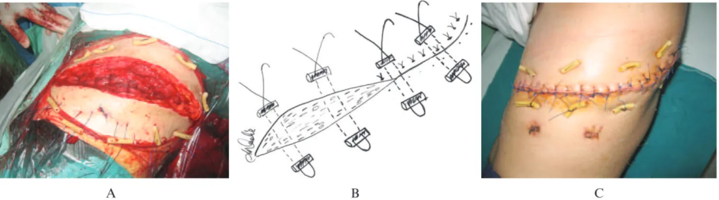
These repairs were performed under general anesthesia. After surgical site disinfection, a large segment of skin, subcutaneous tissue, and muscle was debrided, removing the necrotic tissues (including the wound margins) until fresh bloody tissues were reached. The surgical site was cleansed by scrubbing with povidone-iodine and saline. The skin, subcutaneous tissue and muscular layers were approximated by polypropylene (no:1). To decrease the tension and increase the strength of the sutures, four or five U-shaped retention sutures were inserted based on the size of the wound. To prevent tissue damage, these sutures were supported by Foley catheters that were three centimeters in length (Figure 1A-C). The skin was closed separately by polypropylene sutures (2/0). Dressing of the wounds was performed daily, starting on the second day of the operation. The patients were discharged on the fifth or sixth day after the operation. The sutures were removed on approximately the 15th day.
In patients with open thoraces, the pleural space was cleansed by saline and hydrogen peroxide (3%). After the thickened, necrotic pleural tissues were removed, the thorax was closed.
Vacuum-assisted closure (VAC) was applied in three cases because the wound was too large and suppurative. In eight of the patients who had partial dehiscence, the incision was closed using single polypropylene sutures (2/0) under local anesthesia after sufficient vascularization was observed, and debridement was performed every three to four days, once or twice a given day.
& RESULTS
Seventy percent of the patients in the study were male. The youngest patient was 15 years old, and the oldest was 75 years old, with a mean age of 43 years. Empyema (n = 8) was the most frequent indication for thoracotomy, followed by vertebral fracture (n = 6). Ten (40%) of the patients with either empyema or vertebral fracture had a history of trauma (Table 1). Two of the patients with empyema had concurrent bronchopleural fistulas. Recurrent pneu-mothorax, hepneu-mothorax, lung abscess, solitary pulmonary nodule, mesothelioma, lung carcinoma, and congenital cystic adenomatous malformation were among the other thoracotomy indications.
Bacterial growth was detected in wound cultures from 13 (54%) patients.Staphylococcus epidermidiswas isolated from six patients, but we considered this organism to be contamination from the skin. In total, 25 microorganisms were isolated: one type in 14 patients, two types in four patients, and three types in one patient. Pseudomonas aeruginosa, Staphylococcus aureus, Escherichia coli,
Enterococcus faecalis, and Enterococcus cloacea were other responsible microorganisms that were isolated in four, three, two, two and two patients, respectively.
The first result observed in the DTI patients was an adequate closure of the skin, except at the site of the separation. However, upon removing the sutures one by one and exploring the wound, we noticed that the subcutaneous tissue and muscular layers were all separated and that the exudate had spread throughout the incision (Figure 2).
Almost all the patients exhibited surgical site infections. However, there were no serious purulent massive dis-charges in these wounds requiring dressings two-three times per day, despite the full-thickness involvement of the thoracotomy incisions—that is, the majority of our patients presented non-suppurative surgical site dehiscence and/or infection.
A B C
Figure 1 - A)The photomicrograph displays the retention sutures that were placed after debridement in a patient who had complete dehiscence.B)The drawing depicts the placement of sutures during our re-closure technique.C)Retention and skin sutures are shown in a patient during the early postoperative period.
Dehiscence of the Thoracotomy Incision
Nadir A et al. CLINICS 2013;68(1):1-4
An en block approximation technique combined with debridement was performed under general anesthesia in 16 (66%) of the DTI patients. In two patients, superficial general anesthesia was provided because these individuals did not appear to tolerate intratracheal general anesthesia because of their poor general health status. Debridement and suturation were performed in eight patients with local anesthesia. Pathogenic microorganisms were isolated from the cultures of six of these eight patients. The improvement was complete in the patients whose interventions were conducted under local anesthesia. Successful results were obtained in 14 (87.5%) of the 16 patients whose en block approximation was performed under general anesthesia.
Wound site healing was not achieved in a patient with empyema and sepsis and in another patient who underwent spinal surgery for Pott’s disease; VAC was also applied to the latter patient. In another two patients with large wound defects that ruled out primary closure, VAC was applied, enabling primary closure in one patient and closure using a skin flap in the other patient. There were open thoraces in three patients with wound separation; thoracoplasty was required in two of these patients.
Three patients in this series died. The first patient (75 years old), who exhibited empyema and a bronchopleural fistula, suffered an additional lung collapse. This subject had to receive mechanical ventilation due to respiratory failure that developed after thoracomyoplasty, although the thoracotomy incision was successfully closed. He died from cardiac arrest during follow-up in the intensive care unit. Another patient (53 years old) with poor wound healing was lost because of disseminated pneumonia and sepsis. The third patient (63 years old) had Pott’s disease. He had undergone vertebral surgery in another center and was admitted to our clinic with a diagnosis of open thorax and empyema. Despite repetitive debridements and irrigations, the treatment for empyema failed, and the patient expired due to the emerging sepsis.
& DISCUSSION
Wound dehiscence is a mechanical failure of wound healing, which is a significant problem that can be affected by multiple factors.
Reports reveal a significantly higher incidence of wound dehiscence after emergency surgery than after elective surgery (7). Trauma was the most common etiology (n = 10) in our patients. It is possible that contusion, edema, and extravasation into deeper layers due to the intensity of the trauma were responsible for the delayed wound healing. However, we experienced the same problem in our
Table 1 -Characteristics of the patients.
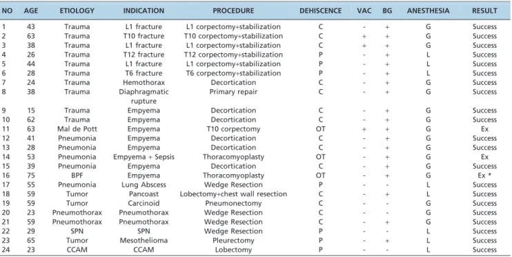
NO AGE ETIOLOGY INDICATION PROCEDURE DEHISCENCE VAC BG ANESTHESIA RESULT
1 43 Trauma L1 fracture L1 corpectomy+stabilization C - + G Success
2 63 Trauma T10 fracture T10 corpectomy+stabilization C + + G Success
3 38 Trauma L1 fracture L1 corpectomy+stabilization C + + G Success
4 26 Trauma T12 fracture T12 corpectomy+stabilization P - + L Success
5 44 Trauma L1 fracture L1 corpectomy+stabilization P - + L Success
6 28 Trauma T6 fracture T6 corpectomy+stabilization P - + L Success
7 24 Trauma Hemothorax Decortication C - + G Success
8 38 Trauma Diaphragmatic rupture
Primary repair C - + G Success
9 15 Trauma Empyema Decortication C - + G Success
10 62 Trauma Empyema Decortication C - + G Success
11 63 Mal de Pott Empyema T10 corpectomy OT + + G Ex
12 41 Pneumonia Empyema Decortication C - + G Success
13 28 Pneumonia Empyema Decortication C - + G Success
14 53 Pneumonia Empyema+Sepsis Thoracomyoplasty OT - + G Ex
15 39 Pneumonia Empyema Decortication C - + G Success
16 75 BPF Empyema Thoracomyoplasty OT - + G Ex *
17 55 Pneumonia Lung Abscess Wedge Resection P - - L Success
18 59 Tumor Pancoast Lobectomy+chest wall resection C - + L Success
19 59 Tumor Carcinoid Pneumonectomy C - - G Success
20 23 Pneumothorax Pneumothorax Wedge Resection C - - G Success
21 59 Pneumothorax Pneumothorax Wedge Resection C - + G Success
22 29 SPN SPN Wedge Resection P - - L Success
23 65 Tumor Mesothelioma Pleurectomy P - + L Success
24 23 CCAM CCAM Lobectomy P - - L Success
BPF: Bronchopleural fistula,SPN: Solitary pulmonary nodule,CCAM: Congenital cystic adenomatous hyperplasia,L: Lumbar,T: Thoracic,C: Complete
P: Partial,OT: Open thorax,BG: Bacterial growth,G: General,L: Local. (*): Re-closure was successful.
Figure 2- A patient’s necrotic and suppurative tissues, including the skin, subcutaneous tissue and muscle layers, were observed before debridement. Only the pericostal sutures were intact.
CLINICS 2013;68(1):1-4 Dehiscence of the Thoracotomy Incision
Nadir A et al.
non- traumatic cases. Notably, DTIs were observed in four consecutive patients during seasonal transition periods, from summer to winter or vice-versa. Thereupon, the ‘‘Infection Control Committee’’ of the hospital traced the etiology. Specimens for culture were obtained from the surgical team and the surgical environment, including the operating room, surgical sets, and suture materials. Unfortunately, this procedure did not reveal a responsible factor.
Separation or dehiscence of thoracotomy incisions is rare, and the treatment is undetermined. Similar phenomena, such as post-thoracotomy empyema or open chest, are managed either with long-lasting dressings on an open chest window or with repeated scheduled mechanical debridements and packing of the pleural cavity with povidone-iodine-soaked dressings and temporarily closing the chest after each session until final closure (8,9). We preferred local dressings, followed by large-scale debride-ment and final closure in a single session.
The quality and nature of the suture material is critical, and failure of the suture material is a risk factor. Published articles have compared various suture materials or suturing techniques; however, the various brands of materials have not been compared based on their tensile strength or structural integrity (10-11). We noticed that the structural material of the polyglactin sutures, which were used for closure of either subcutaneous or muscular layers, deformed prematurely and could not retain the ability to hold the tissue layers together. Although we returned the suture materials to the manufacturer, we have not received a response. We speculate that inexpensive suture materials with toxic or allergic side effects may be responsible for this complication. In fact, we have not encountered this problem since purchasing a different brand of sutures.
The vacuum-assisted closure system was introduced into clinical practice in 1997 and has evolved as the standard of care in the treatment of sternal wound infections during recent years (12). Articles on the use of VAC in thoracotomy wounds first appeared in the year 2006 and increased thereafter (3,4). VAC application in patients with large defects enables the approximation of the wound margins and a decrease in the bacterial load at the infection site. This technique can be used either before approximation or to prepare for reconstruction of failed cases. Although it is a time-consuming (2-4 months) and costly procedure, VAC is a safe and effective alternative, especially in patients with large defects or associated comorbidities.
Surgical site infection is a serious complication that greatly affects the success and cost of the surgery. An immediate en block approximation technique, combined with a wide debridement of the infected and necrotic tissues, may be successfully performed in the majority of cases.
& AUTHOR CONTRIBUTIONS
Nadir A and Kaptanoglu M organized and conducted the study and provided substantial scientific contributions. Nadir A also illustrated the picture of the surgical technique. Sahin E and Sarzep H contributed to the data collection.
& REFERENCES
1. Ridderstolpe L, Gill H, Granfeldt H, Ahlfeltdt H, Rutberg H. Superficial and deep sternal wound complications: incidence, risk factors and mortality. Eu J of Cardiothorac Surg. 2001;20(6):1168-75, http://dx.doi. org/10.1016/S1010-7940(01)00991-5.
2. Welvaart WN, Oosterhuis JWA, Paul MA. Negative pressure dressing for radiation- associated wound dehiscence after posterolateral thor-acotomy. Interact Cardiovasc and Thorac Surg. 2009;8(5):558-60, http:// dx.doi.org/10.1510/icvts.2008.196485.
3. Varker KA, Ng T. Management of Empyema cavity with the vacuum-assisted closure device. Ann Thorac Surg. 2006;81(2):723-5, http://dx. doi.org/10.1016/j.athoracsur.2004.10.040.
4. O’Toole MJ, Kolb JE, Lindblad WJ, Cohen IK, McKneally MF. Pneumothorax and wound dehiscence related to collagenase deregula-tion: treatment with diphenylhydantoin. Ann Thorac Surg. 1996; 61(6):1646-50, http://dx.doi.org/10.1016/0003-4975(96)00212-3. 5. Fawzy H, Alhodaib N, Mazer CD, Harrington A, Latter D, Bonneau D,
et al. Sternal plating for primary and secondary sternal closure; can it improve sternal stability? J Cardiothorac Surg. 2009;4:19, http://dx.doi. org/10.1186/1749-8090-4-19.
6. Wynne R, Botti M, Stedman H, Holsworth L, Harinos M, Flavell O, et al. Effect of three wound dressings on infection, healing comfort, and cost in patients with sternotomy wounds. A randomized trial. Chest 2004;125(1):43-9.
7. Spiliotis J, Tsiveriotis K, Datsis AD, Vaxevanidou A, Zacharis G, Giafis K, et al. Wound dehiscence: is still a problem in the 21th century: a retrospective study. World J Emerg Surg. 2009;4:12.
8. Deschamps C, Allen MS, Miller DL, Nichols III FC, Pairolero PC. Management of postpneumonectomy empyema and bronchopleural fistula. Seminars Thorac Cardiovasc Surg. 2001;13(1):13-9.
9. Schneiter D, Grodzki T, Lardinois D, Kestenholz PB, Wojcik J, Kubisa B, et al. Accelerated treatment of postpneumonectomy empyema: A binational long-term study. J Thorac Cardiovasc Surg. 2008;136(1):179-85, http://dx.doi.org/10.1016/j.jtcvs.2008.01.036.
10. Karabay O, Fermanci E, Silistireli E, Aykut K, Yurekli I, Catalyurek H, et al. Intracutaneous versus transcutaneous suture techniques. Tex Heart Ins J. 2005;32(3):277-82.
11. Durkaya S, Kaptanog˘lu M, Nadir A, Yılmaz S, C¸ ınar Z, Dog˘an K. Do absorbable sutures exacerbate presternal scarring? Tex Heart Inst J. 2005;32(4):544-8.
12. Fleck T, Kickinger B, Moidl R, Waldenberger F, Wolner E, Grabenwoger M, et al. Management of open chest and delayed sternal closure with the vacuum assisted closure system: preliminary experience. Interac Cardiovasc and Thorac Surg. 2008;7(5):801-4.
Dehiscence of the Thoracotomy Incision
Nadir A et al. CLINICS 2013;68(1):1-4