439
BRAZILIAN JOURNAL OF OTORHINOLARYNGOLOGY 72 (4) JULY/AUGUST 2006 http://www.rborl.org.br / e-mail: revista@aborlccf.org.br
Surgical maneuvers
performed on rhinoplasty
procedures carried out at an
otorhinolaryngology residency
program
Summary
Lucas Gomes Patrocínio1, Paulo Márcio CoelhoCarvalho1, Hélio Muniz de Souza1, Hugo Gonçalves
Couto1, José Antônio Patrocínio2
1 MD, Otorhinolaryngology Resident - Federal University of Uberlândia.
2 Full Professor, Head of the Otorhinolaryngology Department - Federal University of Uberlândia.
Department of Otorhinolaryngology - Medical School - Federal University of Uberlândia - MG. Mailling Address: Lucas Gomes Patrocínio - Rua XV de Novembro 327 apto. 1600 Uberlândia MG 38400-072.
Tel/Fax: (0xx34) 3215-1143 - E-mail: lucaspatrocinio@triang.com.br
Paper submitted to the ABORL-CCF SGP (Management Publications System) on April 13th, 2005 and accepted for publication on June 2nd, 2006.
R
hinoplasty is one of the most challenging surgical procedures, due both to the diversity of the techniques and to the difficulty in foreseeing long-term outcomes. Each patient has a different nasal anatomy, dictated by genetic inheritance - race, thus requiring a different technique for each case. The international literature emphasizes the techniques used for the Caucasian nose, which is rarely seen in our region. Aim: Evaluate and discuss surgical maneuvers used on rhinoplasty procedures performed on local patients at our ENT residency services. Materials and Methods: We evaluated the operative notes from all patients submitted to rhinoplasty at the Residency Program on Otorhinolaryngology at the Federal University of Uberlândia, from December 2003 to June 2004. Results: One hundred and sixty-six patients were submitted to rhinoplasty, in which marginal incisions were performed in 118 (71.1%), with the delivery technique performed on the inferior lateral cartilages and some procedures carried out on them (strut, sheen, sutures, etc). Only 45 patients (27.1%) were submitted to basic rhinoplasty and 3 (1.8%) to open rhinoplasty. Conclusion: Most of our patients demanded additional procedures, and the “basic rhinoplasty”, commonly performed on the Caucasian nose was an exception on our patients.Keywords: graft, incisions, ethnic nose, osteotomy, rhinoplasty.
ORIGINAL ARTICLE
440
BRAZILIAN JOURNAL OF OTORHINOLARYNGOLOGY 72 (4) JULY/AUGUST 2006 http://www.rborl.org.br / e-mail: revista@aborlccf.org.br
INTRODUCTION
Rhinoplasty is a surgical procedure of which tech-nique depends on the anatomy of the nose to be operated upon. Since there are not two noses alike, there are not two identical techniques. The technique varies according to the possible anatomic variations, making it the most
challenging of the cosmetic surgeries1-3.
Since it is a surgery that is highly dependent on the anatomic alterations found, the technique to be used will depend on the type of nose that will be operated. This is influenced by hereditary patterns and, consequentely, by race4,5.
International literature emphasizes the Caucasian
nose1-3. In the present study we aim at assessing and
dis-cussing the most frequent maneuvers carried out in the patients of our region, located in the Triângulo Mineiro, a place of important race mixing between blacks, whites and Indians (also common to other regions of the country).
MATERIALS AND METHODS
We have retrospectively assessed the charts of 166 patients who underwent rhinoplasties from December of 2003 to June of 2004, in the Otorhinolaryngology Depart-ment of the Medical School of The Federal University of Uberlândia (FAMED-UFU).
From the operation chart of each patient we filled out a form with the following details: access incisions to the nasal bone-cartilage skeleton, maneuvers performed on the inferior lateral and superior cartilages, implants and/or grafts utilized, procedures made to the nasal base, types of osteotomies and number of concomitant septoplasties. All the patients were operated under local anesthesia with
intravenous sedation6.
The results were plotted on tables and graphs. In percentages, each surgical maneuver was separately ana-lyzed, the need for grafts or incision in relation to the total number of patients operated, thus allowing for a numeri-cal analysis of the approaches carried out in rhinoplasty procedures performed in our department.
RESULTS
Results are depicted on Tables 1 through 9, in rela-tion to the total number of patients operated (166).
The procedures were performed through three approaches: “delivery” (71.1%), closed (27.1%) or open (1.8%) (Table 1).
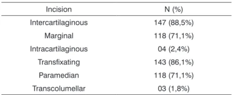
As to the incision (s) used to approach the nasal tip, the intercartilaginous was performed in 147 (88.5%) patients; the marginal in 118 (71.1%) and the intracarti-laginous in 4 (2.4%) (Table 2).
The most commonly performed maneuvers on the inferior lateral cartilages were the ones used to reduce their
Table 1. Surgical approach used in the rhinoplasties carried out in the department of Otorhinolaryngology - FAMED-UFU, from December, 2003 to June, 2004.
Approach N (%) Delivery 118 (71%)
Closed 45 (27,1%) Open 03 (1,8%)
Total 166 (100%)
Table 2. Types of incisions used in the rhinoplasties carried out in the department of Otorhinolaryngology - FAMED-UFU, from December, 2003 to June, 2004.
Incision N (%) Intercartilaginous 147 (88,5%)
Marginal 118 (71,1%) Intracartilaginous 04 (2,4%)
Transfixating 143 (86,1%) Paramedian 118 (71,1%) Transcolumellar 03 (1,8%)
volume and enhance nasal tip contour, with a resection of the cephalic portion (58.4%) and interdomal suturing (45.1%), followed by those used to increase support and enhance tip projection, such as the placement of a
post-graft (24.1%) and use of a Sheen7 shield graft (21.6%)
(Table 3).
There was a high incidence of patients who con-currently underwent nasal septoplasty (30,7%), aiming at
Table 3. Maneuver performed on the inferior lateral cartilage in the rhinoplasties carried out in the department of Otorhinolaryngology - FAMED-UFU, from December, 2003 to June, 2004.
Maneuver N (%) Resection of the cephalic portion 97 (58,4%)
Domes lateralization 11 (6,6%) Interdomal suture 75 (45,1%)
Post 40 (24,1%) Sheen shield cartilaginous graft 36 (21,6%) Seagull wing 08 (4,8%)
preserving respiratory function after rhinoplasty. Other maneuvers commonly performed on the nasal septum were its shortening (38.5%) or enlargement, with a carti-laginous graft (4.2%) (Table 4).
441
BRAZILIAN JOURNAL OF OTORHINOLARYNGOLOGY 72 (4) JULY/AUGUST 2006 http://www.rborl.org.br / e-mail: revista@aborlccf.org.br
Among grafts and implants used, the most frequent was septal cartilage (31.9%), followed by Dacronâ (13.8%)
Table 4. Procedures used on the nasal septum in the rhinoplasties carried out in the department of Otorhinolaryngology - FAMED-UFU, from December, 2003 to June, 2004.
Procedure N (%) Septoplasty 51 (30,7%)
Shortening 64 (38,5%) Augmentation (Graft) 07 (4,2%)
Table 5. Procedures used on the nasal dorsum in the rhinoplasties carried out in the department of Otorhinolaryngology - FAMED-UFU, from December, 2003 to June, 2004.
Procedure N (%) Lowering 126 (75,9%)
Graft 11 (6,6%)
Table 6. Osteotomies performed in the rhinoplasties carried out in the department of Otorhinolaryngology - FAMED-UFU, from December, 2003 to June, 2004.
Osteotomy N (%) Lateral 125 (75,3%) Medial 14 (8,4%) Frontal 01 (0,6%)
and pinna cartilage (4.8%) (Table 7). Most of the patients who underwent procedures on the anterior nasal spine required an augmentation on this region (13.8%) and a lesser number required its removal (6.6%) (Table 8).
The most frequently performed procedure on the wide alar base was its narrowing through resection and suturing (26.5%), followed by an “interalar” suturing (3.6%)
Table 7. Grafts used in the rhinoplasties carried out in the department of Otorhinolaryngology - FAMED-UFU, from December, 2003 to June, 2004.
Graft/Implant N (%) Nasal septum cartilage 53 (31,9%)
Pinna cartilage 08 (4,8%) Dacron® 23 (13,8%)
Table 8. Procedure performed on the nasal spine in the rhinoplasties carried out in the department of Otorhinolaryngology - FAMED-UFU, from December, 2003 to June, 2004.
Procedure N (%) Removal 11 (6,6%)
Graft 23 (13,8%)
(Table 9).
Table 9. Procedures performed on the alar base in the rhinoplasties carried out in the department of Otorhinolaryngology - FAMED-UFU, from December, 2003 to June, 2004.
Procedure N (%) Alae closure 44 (26,5%)
Stitches 06 (3,6%)
DISCUSSION
Every rhinoplasty requires access incisions. Proper exposure is as important in rhinoplasties as they are in other types of surgery; therefore, the incisions should be selected as to specific indications. In “Basic Rhinoplasty” the following incisions are performed: transfixating, intercartilaginous and paramedian on the upper lateral cartilage (used to separate the upper lateral cartilage from the septum).
According to Toriumi e Becker1, incisions are
meth-ods used to give access to bone-cartilaginous structures within the nose, and include the following: transcartilagi-nous, intercartilagitranscartilagi-nous, marginal and transcolumellar. We also add the paramedian incision on the upper lateral
cartilage to these ones8.
Patrocínio et al.8 reported that every rhinoplasty
requires a careful and precise analysis of what must be
corrected. According to Tebbetts2, one should use as many
incisions as are necessary in order to guarantee ideal exposure and control. The accuracy of the incision may substantially influence its closure quality and subsequent scarring. A precise incision requires tissue stabilization, exposure, planning and an accurate technique.
Incisions for surgical approaches change according to the defect to be corrected. Some authors advocate the
transcolumellar external access in 100% of the cases1. We
used this access in only 1.8% of the cases. In the major-ity (71.1%) we used marginal incisions, with a “delivery” approach to the inferior lateral cartilages. The low rate of closed rhinoplasties (27.1%) shows the high incidence of deformities on the inferior lateral cartilages that need
surgical correction4,5.
Trans/intracartilaginous access in primary rhinoplas-ties, in which all we need is to reduce the tip volume, is carried out through a 4 to 6mm incision caudal to the cephalic border of the lateral crura. In this procedure, what matters is what we leave behind, and not what we take from the cartilage. This access allows for tip reduction and
refining, without the risk of altering the nasal valve3.
inter-442
BRAZILIAN JOURNAL OF OTORHINOLARYNGOLOGY 72 (4) JULY/AUGUST 2006 http://www.rborl.org.br / e-mail: revista@aborlccf.org.br
cartilaginous incisions8. Almost all the procedures carried
out by open rhinoplasty can be performed through this access, without leaving a columellae scar. Open rhino-plasty was used in only 3 of our cases. The advantage of this procedure is direct visualization of the alar cartilages, making it easier to place sutures and grafts, besides facili-tating teaching. It is well indicated for fissure noses and tertiary or quaternary rhinoplasties, those with important
pinching and assymetries4,5. Most secondary rhinoplasties
are lesser surgeries than the first ones, and are used to correct subtle defects and, therefore, do not require the open procedure.
As to the actions to be performed on the lower lat-eral cartilages, the cephalic portion was resected in order to better define the tip in 58.4% of the patients. Interdomal suturing, post placement, Sheen shield cartilaginous graft placement, domes lateralization and “seagull wing” type of graft were performed in 45.1%, 24.1%, 21.6%, 6.6% and
4.8% patients, respectively. According to Sheen7, the four
points that define nasal tip are: supratip breakpoint, right “domes”, left “domes” and lobe-columellar junction; and the abovementioned maneuvers aimed at acting on these points. Maybe it is because we are otorhinolaryngolo-gists that we had so many nasal septoplasties (30.7%), allowing not only a functional improvement, but also the
supply of grafting cartilage9. In only 4.2% of the cases a
graft was placed on the causal septum because of a small naso-labial angle.
Lateral osteotomy was used in 75.3% of the cases, and this may be explained by the large number of noses
with broad bone base in our region4,5. For men we used
the 3mm osteotome and for women, the 2mm, with an access that is lateral and superior to the head of the inferior nasal conchae, with a dotted-line type of fracture in an ascending fashion to the naso-maxillary angle.
Nasal septum cartilage grafting was used in 31.9% of the patients, pinna cartilage in 4.8% and Dacronâ in 13.8%. Septal cartilage was used mainly to make the post and the Sheen shield grafts, and the pinna was used for
dorsum grafts10; “seagull wing” and Dacronâ were used
for the anterior nasal spine. The latter, since it is a soft material, it is not used for support, but as filling substance only, and it is easy to obtain in our settings, because since all we need is a small quantity, we use the sterilized remains of what is used by the Vascular Surgery Depart-ment. There are other types of implants described in the literature, such as Gore-texâ, Supramidâ, Proplastâ and
hydroxyapatite11-13.
Access to the anterior nasal spine, for resection in 6.6% of the cases or grafting in 13.8%, was carried out by the columellar transfixating incision and pouch creation. In these cases, the most frequently used material was Dacronâ (soaked in Clindamycin), placed at the end of the procedure and through a very well closed incision.
It is preferable to use cartilage; however, due to a great need for augmentation, we use Dacronâ in order to avoid a new incision for graft removal, in this case, from the
other ear11,12.
As to the nasal base, we approached it in 30.1% of
the patients, either by base resection with suturing4,5,13 in
26.5% and by interalar stitches in 3.6%, which is indicated when there is a subtle increase in the interalar distance.
According to Daniel3, the lateral alar incision is not usually
necessary, and it always leaves a scar behind. We did not use this type of incision on our patients.
CONCLUSION
In the present study, we carried out maneuvers that complement the ones used in basic rhinoplasty. Most of the patients underwent some kind of procedure on their inferior lateral cartilages, and a large number of them underwent lateral osteotomy, nasal alae closure, graft and/or implants placement and other maneuvers. Basic rhinoplasty, usually carried out in Caucasians, was an exception in our settings.
REFERENCES
1. Toriumi DM, Becker DG. Rhinoplasty Dissection Manual. Philadel-phia: Lippincott Williams & Wilkins; 1999. p. 37-57.
2. Tebbetts JB. Primary Rhinoplasty. Saint Louis: Mosby; 1998. p. 61-86.
3. Daniel RK. Aesthetic Plastic Surgery - Rhinoplasty. Boston: Little, Brown and Company; 1993. p. 283-318.
4. Patrocínio JA, Mocellin M, Patrocínio LG, Mocellin M. Rinoplastia a céu aberto para correção do nariz tipo negróide brasileiro. In: Maniglia AJ, Maniglia JJ, Maniglia JV (editores). Rinoplastia: estética, funcional e reconstrutora. Rio de Janeiro: Revinter; 2002. p. 204-12. 5. Patrocínio JA, Mocellin M, Patrocínio LG. Rinoplastia no Nariz Ne-gróide. In: Campos CAH, Costa HOO (editores). Tratado de Otor-rinolaringologia. Volume 5 - Técnicas Cirúrgicas. São Paulo: Roca; 2002. p. 717-26.
6. Patrocínio JA, Patrocínio LG, Ramin SL, Souza DD, Maniglia JV, Maniglia AJ. Anestesia. In: Maniglia AJ, Maniglia JJ, Maniglia JV (edi-tores). Rinoplastia: estética funcional e reconstrutora. Rio de Janeiro: Revinter; 2002. p. 62-8.
7. Sheen JH. Aesthetic Rhinoplasty. Saint Louis: Mosby; 1978. 8. Patrocínio JA, Sousa AD, Coelho SR. Incisões para inserção de
im-plantes no nariz. Acta AWHO 1986;5(2):45-52.
9. Mocellin M, Maniglia JJ, Patrocínio JA, Pasinato R. Septoplastia Técnica de Metzembaum. Rev Bras Otorrinolaringol 1990;56:105-10. 10. Patrocínio JA, Patrocínio LG. Nariz em sela. In: Campo CAH, Costa
HOO (editores). Tratado de Otorrinolaringologia. Volume 5 - Técnicas Cirúrgicas. São Paulo: Roca; 2002. p. 727-38.
11. Patrocínio LG, Patrocínio JA. Uso de enxertos na rinoplastia. Arq Otorrinolaringol 2001;5(1)21-5.
12. Patrocínio LG, Patrocínio JA. Atualização em enxertos na Rinoplastia. Rev Bras Otorrinolaringol 2001;67(3):394-402.

