84
1. PhD in Thoracic Surgery – Federal University of the State of Rio de Janeiro – UNIRIO (Thoracic Surgeon of the Andaraí Hospital – RJ, Ministry of Health, Head of the Thoracic Surgery Division of the Thoracic Diseases Institute – Federal University of Rio de Janeiro).
2. Thoracic Surgeon of the Andaraí General Hospital – RJ (Thoracic Surgeon of the Andaraí General Hospital – RJ).
3. Head of the Service of Thoracic Surgery of the Andaraí General Hospital – RJ.
4. Thoracic Surgeon of the Andaraí General Hospital – RJ.
Giovanni Antonio MARSICO1, André Luiz de ALMEIDA2, Dirceo Edson de AZEVEDO3, Ivam MATHIAS FILHO4 Rev Bras Cir Cardiovasc 2009; 24(1): 84-87 CASE REPORT
RBCCV 44205-1057
Projétil intrapericárdico móvel
Mobile intrapericardial bullet
Abstract
Patients with bullets in the pericardial sac without myocardial injuries are rare, and most commonly are associated with significant trauma. The diagnosis of an intrapericardial foreign body can be difficult. Its removal is always indicated because it prevents pericarditis, either sterile or infectious, with potential for other significant complications. The authors present two cases of a meandering bullet in the pericardial sac and propose approach and perform review of the literature.
Descriptors: Pericarditis. Pericardial effusion. Pericardium/injuries. Wounds, gunshot.
Resumo
São raros os pacientes com projéteis localizados no saco pericárdico sem que ocorra lesão miocárdica associada, comumente estão relacionados a trauma importante. O diagnóstico de corpo estranho intrapericárdico pode ser difícil. A remoção está sempre indicada, pois previne o surgimento de pericardite estéril ou infecciosa e outras complicações significativas. Os autores apresentam dois casos de projétil de arma de fogo livres no saco pericárdico, sugerem a conduta e fazem a revisão da literatura.
Descritores: Pericardite. Derrame pericárdico. Pericárdio/ lesões. Ferimentos por arma de fogo.
This study was carried out at Andaraí General Hospital - Ministry of Health – Rio de Janeiro – RJ.
Correspondence address: Giovanni Antonio Marsico
Hospital Geral do Andaraí - Cirurgia Torácica Rio de Janeiro Rua Leopoldo 280 – 7º andar – Andaraí. CEP 21541-170.
E-mail: marsicog@gbl.com.br
Article received on July 15th, 2008
Article accepted on December 22nd, 2008
INTRODUCTION
With no associated injury in the myocardium the finding of meandering bullet alone in the pericardial cavity is rare. In this condition the evolution is unpredictable, because while some of the patients remain asymptomatic, pericarditis occurs in the others, with or without pericardial effusion, tamponade, complaints of chest pain and major psychological problems. In asymptomatic patients, the removal of the bullet from the pericardial sac also raises questions [1-3]. We report the removal of a meandering bullet from the pericardial sac in two patients and performed a literature review.
CASE REPORTS
Case 1
85 MARSICO, GA ET AL - Mobile intrapericardial bullet Rev Bras Cir Cardiovasc 2009; 24(1): 84-87
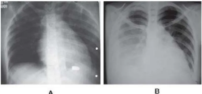
In a retrospective analysis of chest radiographies, performed during the first consultation in the local hospital, was observed (in the PA and lateral views), meandering bullet on the heart silhouette at left, located in the anterior costodiaphragmatic recess (Figure 2A). A simple abdomen radiograph showed meandering bullet located in the pelvis. On new radiographs performed after a week of hospital stay, there was pleural effusion at right and change in the position of the bullet was observable, now located at right cardiophrenic region (Figure 2B). Then the patient was referred to our hospital due to right pleural complication.
With the suspicion of free meandering bullet in the pericardial cavity, the patient was studied using computed tomography. The examination showed meandering bullet near the right atrium and pleural effusion and blood clots in the right pleural cavity. In order to solve the pleural complication and remove the bullet located in the pericardial cavity, the patient underwent then longitudinal right axillary thoracotomy. After total release of the lung with the removal of fibrin and clots, the pericardium was opened anteriorly and longitudinally to the phrenic nerve. There was leaving of about 20 ml of clear yellow liquid, and then the bullet was easily removed. The pericardium was partially closed and the pleural cavity was drained. The patient was discharged from hospital on the eighth day of hospital stay. In the third postoperative month the patient was well. in the bullet position occurred (Figure 1. A and B). The
electrocardiogram was normal. The chest radioscopy confirmed the mobility of the bullet in the pericardial sac.
On the third day of hospital stay, under general anesthesia, left submammary thoracotomy was performed entering the pleural cavity in the fifth intercostal space. The pericardium was opened longitudinally and anterior to the phrenic nerve. Cloudy yellow liquid leaving of around 110 ml. The bullet was free in the pericardial sac and was easily removed. There was no damage to the myocardium. The pericardium was left partially opened and the pleural cavity was drained. In the study of pericardial fluid there was 50% of neutrophils and did not occur growth of bacteria in the cultures performed. The patient was discharged from hospital on the fifth postoperative day and after eight months of follow-up presented asymptomatic.
Fig. 1 – A) PA Chest radiography, performed with the patient
standing, showing the meandering bullet located in the topography of the heart, B) – AP chest radiography with the patient in dorsal decubitus position, showing the change of the free bullet position in the pericardial cavity
Fig. 2 – Chest radiographies show the change of bullet position
and right pleural effusion
Case 2
20-year-old female patient, referred to our hospital without reporting of surgical procedures that had been undergone. The patient reported aggresion by firearm 16 days before. On physical examination several wounds in final stage of healing were found in the left hemithorax and left flank; some wounds were infected and the largest measured about 2 cm in length, located between the third and sixth intercostal space, limited to the middle and posterior axillary line. In examining the abdomen, we found median longitudinal xypho-pubic incision and colostomy at left. The patient reported closed tubular drainage in the left hemithorax.
DISCUSSION
86
In 1939, Decker [2] studied 100 patients with penetrating cardiac wounds, of which 11 presented free bullets in the pericardial sac. Among these patients, nine were meandering bullet and two were fragments of grenada. Seven of the bullets were removed and four were not and all evolved satisfactorily. The author concluded that intrapericardial foreign bodies can be removed safely, although not necessarily when considering only their presence in such location. However, the authors considered that the early removal of large foreign bodies was essential. Watts and Toone, [4] in the year 1945, after five months of follow-up removed grenade fragment from the pericardial cavity of two soldiers. During this period, there were intermittent episodes of pain in the left hemithorax that spred to the shoulder on the same side. In electrocardiogram changes were observed indicating progressive damage in the myocardium. The symptoms disappeared after removal of the bullet that was found attached between the right atrium and pericardium.
In 1955, Valle [3] examined 42 soldiers with metallic foreign bodies retained in the heart, mediastinum and pericardial cavity. In the first six weeks, all soldiers developed significant symptoms as severe chest pain, fever, tachycardia and difficulty breathing. Among those who developed a pericardial effusion, some were treated with pericardiocentesis and antibiotics and, in others, the foreign body was removed. In 50% of patients were identified pyogenic germs in the pericardial fluid. The author highlights a subgroup of 12 patients with intrapericardial metallic foreign bodies that developed pericardial effusion between four and 26 months after the trauma. Foreign bodies were not removed initially because they were smaller than 0.5 cm. The author concluded that the free foreign bodies in the pericardial cavity should be removed, regardless of their size.
Symbas et al. [5] evaluated 49 patients with retained bullets in the pericardial cavity. In 42 (87.5%) of these patients the bullets were removed and no deaths occurred. Of the seven patients who were followed-up, three remained asymptomatic, one developed chest pain, two pericarditis and one died at 11st postoperative day after failed attempt to remove the
foreign body. Four complications occurred in patients with fragments of grenade in the pericardial cavity.
Most free foreign bodies in the pericardial cavity is metallic and penetrate through the chest wall. However, entering of the foreign body can be caused by erosion in the esophagus or tracheobronchial tree after the aspiration or ingestion of foreign bodies such as teeth, dentures, needles, pins and others. [1,3,6]
McLaughlin et al. [6] treated a patient who had suffered aggression by firearms three days before and they removed from him a free 44 caliber bullet that was in the pericardial cavity. In this period of follow-up occurred progressive
pericardial effusion and cardiac tamponade. After opening the pericardium during a left thoracotomy, the bullet was found floating in the pericardial fluid. In the cultures of the liquid did not occur growth of microorganisms.
The patient of case 1 underwent surgery after four days of the bullet remain in the pericardial sac and presented 110 ml of intrapericardial liquid. Probably, the trend would increase the volume. In researches perfomed the growth of germs did not occur. In case 2, with twenty days of evolution, about 20 ml of pericardial fluid with normal appearance was found and no bacteriological study was performed.
The simple chest radiographies, computed tomography and fluoroscopy, performed with the patient in various positions, often define the diagnosis of free metallic foreign body in the pericardial cavity. The suspicion is confirmed when, by the action of gravity and cardiac movements, the bullets change their position under mediastinal topography. Computed tomography is able to detect non-radiopaque foreign bodies. The differentiation between free foreign body in the cardiac cavities, pericardial cavity and heart wall is important to define the approach. Other exams capable of providing additional information are: the echocardiogram, echotransesophageal and esophagography. Moreover, such exams may exclude or diagnose potential associated injuries, especially in the heart [3-7].
Burkhart et al., [1] in 1998, operated a patient who developed pericardial effusion caused by the presence of a free 45 caliber bullet in the pericardial cavity. The bullet removal was performed by subxiphoid approach. In reviewing the literature, the authors compiled 31 cases of foreign bodies retained in the pericardial sac. They found that in the ten patients who had undergone early removal, all evolved well. Of the 21 who initially were followed-up, the removal was necessary in 15. These patients presented various symptoms attributed to the presence of foreign bodies. The authors concluded that the “large” and free foreing bodies in the pericardial cavity, almost always cause symptoms and should be removed. The authors recommend the early removal of contaminated intrapericardial foreign bodies. They emphasize that the conservative approach must be adopted with extreme caution, only in selected cases, such as smooth metallic foreign bodies smaller than 1 cm, which are minimally contaminated and cause no symptoms.
In most reports [1-7], the removal of the free foreign body in the pericardial cavity was performed by thoracotomy. However, the procedure was performed by subxiphoid approach, during surgery guided by fluoroscopy to locate the bullet. If necessary, the subxiphoid incision can be extended to longitudinal sternotomy. Currently, the videothoracoscopy should be used for the removal of free foreign bodies in the pericardial cavity.
87
REFERENCES
1. Burkhart HM, Gomez GA, Jacobson LE, Broadie TA, Tarver RD. Meandering bullet in the pericardial sac: to remove or not remove. Am Surg. 1998;64(4):341-3.
2. Decker HR. Foreign bodies in the heart and pericardium: should they removed? J Thorac Surg. 1939;9(1):62-79.
3. Valle AR. War injuries of heart and mediastinum. AMA Arch Surg. 1955;70(3):398-404.
4. Watts T, Toone EC. Successful removal of foreign bodies within the pericardium: a report of two cases. Surgery. 1945;17:685-95.
5. Symbas PN, Picone AL, Hatcher CR, Vlasis-Hale SE. Cardiac missiles: a review of the literature and personal experience. Ann Surg. 1990;211(5):639-48.
6. McLaughlin JS, Herman R, Scherlis L, Yeager G. Sterile pericarditis from foreign body: acute tamponade one month following gunshot wound. Ann Thorac Surg. 1967;3(1):52-6.
7. Davis RE, Bruno AD 2nd, Larsen WB, Sugimoto JT, Gaines RD. Mobile intrapericardial bullet: case report and review of the literature. J Trauma. 2005;58(2):378-80.
could have been performed using videothoracoscopy or subxiphoid pericardial window. In the patient of case 2, the approach chosen for the removal of the intrapericardial bullet was by right longitudinal axillary thoracotomy in order to solve at the same time the pleural complication of this side.
CONCLUSIONS
We found some successful reports of conservative treatment in cases of free bullets in the pericardial sac, however, the percentage of patients who develop clinical manifestations and pericarditis is high. Generally, the removal of free foreign body in the pericardial sac is indicated. The possible damage caused by removal of the bullet, compared to the potential risk of its stay in the pericardial sac should be considered and evaluated individually. It is important to analyze the size, composition, location of the foreign body and the symptoms caused. Potentially, it can develop cardiac tamponade, pericarditis with sterile (or not) effusion, and other significant complications. Indeed, the outcome of conservative treatment for intrapericardial foreign bodies is unpredictable, often associated with complications [1-7].