THE COCKROACH, PERIPLANETA AMERICANA, AS A VECTOR OF PATHOGENIG ORGANISMS. I. THE
ACID-FAST ORGANISMS A PRELIMINARY REPORTO
By ALBERT LEIBOVITZ~
Captain, Medical Service Corps, United States Army
Of about 800 species of cockroaches known to man, only a few are domestic (1). Of the domestic species, the three most common are Bluttu
yermanica (Linn.), Periplaneta orientalis, Linn., and Periplaneta ameri- cana (Linn.). During the past three centuries these roaches have become world-wide in their distribution, but are most heavily concentrated in the tropical and sub-tropical regions of the world. One of the most com- mon roaches found in the San Antonio area is P. americana, and the present discussion will revolve primarily around this species.
The cockroach, as we al1 know, is a very efficient scavenger and will eat anything from our staple food supplies; such as, flour, bread, cheese, bacon, and sugar, to such body wastes and exudates as feces, sputum, and pus. Considering that this night marauder travels from place to place in the constant search for food, usually from the sewer to the kit- chen, it is not surprising that the cockroach has long been suspected as a possible carrier of disease-causing organisms. What is surprising is the relatively meager work that has actually been done to prove that this insect can be a vector of agents pathogenic to man. Until rather recently al1 experimental work along this line consisted of feeding various patho- gens to the roach and noting whether the agent could either pass un- harmed through the intestinal tract of the roach or actually infect the roach. Cao (24), an Italian, in 1898 was probably the first to publish in detail a series of experiments in which various agents were fed to the roach. Steinhaus (5), in having Cao% work translated, found that he had successfully passed the following disease-causing organisms: M. tuber- culosis, B. anthrax, Clostridium chauvei (Feseri), Clostridium sporogenes,
Clostridium tetani, Klebsiella pneumoniae, Sarcina albus, Sarcina lutea,
and Pasteurella avicidi. Cao’swork was coníirmed byseveral otherworkers (1, 6,7), and they added M. Zeprae (7), V. cholera (8), various staphylo- cocci present in pus (l), C. diphtheriae (6), and D. pneumoniae (6). In 1930, the next step to prove the roach as a vector of pathogenic or- ganisms was taken when Antonelli (9), another Italian, in trying to de- termine the cause of two small outbreaks of typhoid fever, found that the 1 Presented at United States-Mexico Border Public Health Association Meet- ing at Chihuahua City, Chihuahua, Mexico on 12 April 1950.
2 Fourth Army Area Medical Laboratory, Brooke Army Medical Center, Fort Sam Houston, Texas.
January 19611 PERIPLANETA AMERICANA 31 disease was probably being spread by the cockroach, P. orientaEs, from the open latrines of the affected families. The typhoid bacillus was found on the feet and bodies of the roaches. Bacteriological examination of the water supplies of the affected families was negative and flies were ab- sent. Finding no other means of communication between the two out- breaks, Antonelli offered strong evidente that the cockroach was the means of the spread of the disease from one family to the other. In 1933, Beck and Coffee (10) of the Texas State Board of Health Department found that an epidemic of Salntonella typhimurium infections in their guinea pig colony was probably being disseminated by the cockroach,
P. americana. Roaches infesting the animal cages were found to be har- boring the organisms both on their appendages and in their intestines. It is of interest that species of B. germa&a, which were also found in the infected animals’ cages, were not found to be harboring the Salmonella. Mackerras and Mackerras (ll), in Australia, captured a number of roaches on a ward devoted to treatment of gastro-intestinal diseases and found these insects to be harboring a species of Salmonella (8. botiis- mo&@cans). The authors point out that a large epidemic due to t.his organism was then in progress and the roaches were probably picking up the organisms from the sinks in which the soiled napkins were washed before they were sent to the laundry. Mackerras and Pope (12), using the excellent controlled feeding technique advocated by Wedberg and his co-workers (13-14) at the University of Connecticut, showed that one strain of Xalmonella was harbored as long as 42 days in the intestines of the roach. In this series of experiments outlined above, roaches had been caught at the scene of an outbreak of human disease and it was shown that the insects can harbor the organisms both on their appendages and within their intestines.
In 1948 Bitter and Williams (15) took the next step: to show that ‘(wild roaches,” not necessarily directly associated with an outbreak of active disease, can be harboring pathogenic organisms and thus be a potential source of the start of an outbreak. The authors captured their roaches from the sewers in San Antonio and found that four of 41 roaches collected contained one or more species of Xalmonella.
Believing that the surface has been but barely scratched in demon- strating that the wild cockroach can disseminate pathogenic microor- ganisms to man, we have spent the past winter at the Fourth Army Area Medical Laboratory studying the techniques of capturing the roach, of dissection, and of controlled feeding. Although our work with the acid- fast group of organisms is but one phase of our project, it will serve to bring out our experiences in setting up this project.
MATEFUALANDMETHODS
32 BULLETIN OF THE PAN AMERICAN SANITARY BUREAU
(15). The roach was covered with a petri dish and a slip of paper was
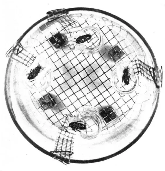
slid underneath to trap the roach. This method is of great value when collecting in a site where roaches are readily accessible, quite numerous, and required in only relatively small numbers. Desiring to collect rather large numbers of roaches, we devised a method, based on the fly-trap, in whioh a suitable funnel was placed in a screw-top Mason or fruit jar. The jar is baited with bread soaked in beer and left overnight in the area from which roaches are desired. Figures 1 and 2 illustrate this type of trap. Traps were placed at weekly intervals for three weeks in four different sewers in the west end of San Antonio in a region known as “Death’s Triangle” due to the large morbidity rate of infant diarrhea and tuberculosis in this area. The traps were allowed to remain in the sewers for 24 hours before they were retrieved. A total of 751 roaches
was caught in the 12 pools; al1 the roaches belonged to the species P.
americana. The yield included 178 adult roaches, 280 nymphs in either their third or fourth molting stage, and 293 nymphs in either the first or second molting stage. The adults and older nymphs were retained for this study and the younger nymphs were discarded.
(2) Preparation of roaches for acid-fast bacteria1 studies.-Theroaches were killed by placing the entire trap in the deep freeze for three to four hours. When necessary, the trap was left in the deep freeze overnight. The adults and older nymphs were sorted out in each pool, respectively, disinfected in an alcohol-bichloride of mercury bath, and their entire intestinal tract removed after the method of Gier (16). The intestines were placed in a 250 CC sterile wide-mouth sturdy glass bottle containing glass beads and capped with sterile rubber stoppers. After macerating the intestines by shaking the bottles in an International Bottle Shaker for one hour, sufficient normal isotonic saline, pH 8.0, was added to yield a 20 per cent (20%) suspension by weight. To an aliquot of this suspen- sion, an equal volume of three per cent sodium hydroxide was added, digestion was allowed to proceed at 37” C for 45 minutes, and then the material was centrifuged at 2,500 rpm for 15 minutes. The supernatant was discarded and the sediment neutralized with three per cent hydro- chloric acid. From this neutralized sediment of each pool, smears were prepared for acid-fast stains, five tubes of Petragnani’s medium were inoculated, and sufficient isotonic saline was added to allow the with- drawal of two ml for guinea pig inoculations.
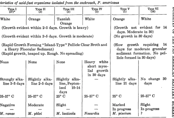
(3) Acid-fast stains.-By the method of Ziehl-Neelsen, acid-fast organisms were noted in the smears prepared from each of the concen- trates, usually in large numbers. The organisms differed in morphology from short, thick, solid staining organisms to long, thin, beaded rods demonstrating palisade formations. The finding from each pool is sum- marized in Table 1.
January 19611 PERIPLANETA AMERICANA 33 centrate in isotonic salme were inoculated into the right groin muscle of the guinea pig. As half of the guinea pigs so inoculated died in three days due to the presente of Clostridium perfr&gens in the suspension, the remaining four pools were inoculated in one ml amounts into the right thigh. This method proved to be more satisfactory in that none of these guinea pigs died of intercurrent infections. Of the eight guinea pigs that survived for the six-week observation period, none were demon- strated at autopsy to have tuberculosis by macroscopic and microscopic studies. See Table 1.
TABLE 1
Studies on acid-fast orqanisms in the “wild” cockroach (P. americana)
Po01 1 2 3 4 5 6 7 8 9 10 ll 12 - _- - SeW.3 A B c D A l3 c D A B C D
TP
maches 23 14 17 32 53 64 26 27 43 75 39 45 Ziehl-Neelsen acid-Iast stain+++z 4-f-f ++ +:+ +++ +i- +++ +++ ++ +++ +++
6-P3 Negative for tuberculosis S-P Died in 3 day@ 10 Negative for tuberculosis
5-P Died in 3 day+
12 Negative for tuberculosis 7-P Died in 3 days4 6-P Died in 3 day@ 1-P Died in 3 days4 10 Negative for tuberculosis
4 Negative for tuberculosis 8-P Negative for tuberculosis 6 Negative for tuberculosis
Guinea pig inoculations* Survivors autopsied in six weeks
1 Guinea Pigs, Pools l-8, received 2 ml concentrate into right femoral muscle. Guinea Pigs, Pools 9-12, received 1 mi of concentrate into right thigh. z + present in small numbers; ++ present in moderate numbers; +++ present in large numbers.
* P signifies medium was proteolysed. Usually completely proteolysed in 3 weeks.
4 Deaths due to Clostridium perfringens.
Characteristic
TABLE II
Characteristics of acid-fast organisms isolated from the cockroach, P. americana
Pigment ... Glycerol Agar. ... Nutrient agar ... TB Broth Base** without Tween
80. ... TB Agar without** Tween 80
3,e.I
White
(Growth evident within 2-5 days. Growth is heavy) (Growth evident within 2-5 days. Growth is moderate)
(Rapid Growth Forming “Island-Type” Pellicle Clear Broth and a Heavy Floccular Sediment)
(Rapid growth, heaped up. Rough. No spreading) Mycelium Formation ... None
Litmus milk.. . . . . . .
Optimum Temperature.. . . . Guinea Pig intradermal skin
inoculation . . . _ . . . Rabbit intravenous inoculation. Probable Specie . . . _ . . .
- -_
-
T eI 73) Orange TY#II Tannish Orange FkN White
Strongly alka- line 2-3 days
25-37” C Negative
- M. ranae
None
Slightly alka- line 2-5 days
25-37” C Moderate - M. phlei
None
Slightly alka- line, Pepton- ized lo-14 days 25’ C Slight - M. lacticola
Heavy white short myce- lia1 growth in 30 days
-
25-37” C - - Nocardia
%
T eV 7%
l
Orange
/ White
(Growth not evident for 14 E days. Moderate in 30) % (No growth in 30 days)
(Slow growth requiring 14 ii days for moderate granular sediment formation. No pel- licle f ormed ir
Slightly alka- line 21 days
25’ C Marked In progresa M. piscium
L 30 days)
ti B 0 5 No change 30 u1
days !s
3 B+
25-37” C $
Slight
In progress 3
? $
9 * Number of species isolated in each category.
January 19511
PERIPLANETA
AMERICANA35
FIGCRE l.-Roach trap, assembled.
36
BULLETIS OF THE PAS AMERICAN SANITARY BUREAUthe cultural characteristics of the acid-fast organisms outlined in Bergey’s
Manual (17), the 83 strains isolated from al1 12 pools were divided into
six types. The majority
of the rapid growing organisms are classified
under Type 1 and probably are strains of M~ycobacterium ranae; the ma-
jority of the slower growing organisms are placed in Type VI and at the
present time the characteristics of this organism do not fit any of the
descriptions outlined in Bergey’s *Manual (17). At,tempts to classify the
organisms were based, tentatively, upon their pigment production, rapid-
ity of growth, ability to grow on nutrient agar, reaction in litmus milk,
January 1951]
PERIPLANETA
ilMERICANA37
may be &W. phlei, Type III may be M.
Zacticoìa,Type IV is a member of
the Nocardia group and its virulence is being tested in guinea pigs by
intraperitoneal
inoculations. Type V may be M.
is entirely an unknown to date. See Table II.
piscium
and Tvpe VI
s
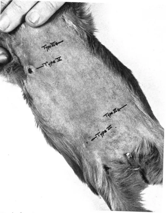
FIGURE 4.-Intradermal skin test in guinea pigs to determine virulence of acid- fast organisms.
Attempts
to classify the saprophytic
acid-fast organisms bv their
biochemical (21) and antigenic (22) characteristics as well as Optima1
temperature for growth (23) leave much to be desired. The phage tech-
nique of typing (24) may be the answer to this problem.
The differentiation
of the virulent from the non-virulent
strains of
38
BULLETIN OF THE PAN AMERICAN SANITARY BUREAUmedium to enable this classification by artificial means. Figure 3 demon-
strates the growth of saprophytic strains on this medium. Miss Lester
in advocating an intradermal skin test in guinea pigs, has demonstrated
a way in which many animals may be saved where large numbers of
organisms are to be tested. Figure 4 demonstrates this technique with the
organisms isolated from the roach. It is of interest that the marked re-
actions noted in guinea pigs were obtained with organisms which pro-
duce orange pigment. This observation duplicates that of Miss Lester.
Control guinea pigs inoculated with virulent strains of human tubercle
bacilli demonstrate the same picture as our Type V, but upon ulceration
of the nodule the lesion continues to drain.
FIGURE 5.-Method of feeding cockroaches under controlled conditions. DISCUSSIOX ASD SUMMilRY
January 19611 PERIPLANETA AMXRICANA 39
1. The feeding of pathogenic organisms to the cockroach and noting whether the organisms are capable of enduring the passage through the roach and for what period of time.
2. The isolation of specific pathogenic organisms from the appendages or the intestines of roaches captured in the vicinity of an outbreak of disease caused by these pathogens.
3. The isolation of organisms capable of causing disease in man from roaches captured in areas in which there is no known outbreak of the disease at
that time.
We believe that a combination of the above criteria offers a logical approach to incrimination of the cockroach as a vector of many patho- genic organisms, including protozooan, bacterial, and vira1 agents. Recently Hurlbut (25) found that cockroaches injected into the hemocele with the Lansing strain of poliomyelitis retained the virus in its body in a viable state for at least 15 days. In this preliminary report, we have attempted to outline our experiences in methods of collection of roaches and the preparation of material for the study of the acid-fast group of micro-organisms.
The cockroach, P. americana, has been shown to be a vector of many types of acid-fast. organisms. Although most of these organisms, if not all of them, are saprophytic by our present criteria, it is of importance to know that the cockroach is capable of transmitting them. Our future work wil1 entail further capturing of roaches from areas not connected with any specific outbreak of disease and from areas where definite out- breaks of disease are occurring. Complete bacteriological and vira1 stud- ies will be made in attempts to isolate agents pathogenic to man. In addi- tion, feeding experiments using a slight modification of the feeding jar of Wedberg (See Figure V) will be conducted.
Eighty-three strains of acid-fast organisms were isolated from the in- testinal tracts of 548 roaches. These were classified into six types based on their cultural characteristics. Tentative identifkation of these types, based on Bergey’s Manual, Sixth Edition, is as follows: Type 1, M. ranae, Type II, M. phlei; Type III, M. ZactzYcoZa, Type IV, Nocardia, Type V, M. pixium, Type VI, unknown.
ACKNOWLEDGMENTS
The author gratefully acknowledges the many suggestions and gnidance of- fered by Colonel Dwight M. Knhns, MC, Commanding, Fourth Army Area
Medical Laboratory and Drs. Oscar B. Williams and Charles E. Lankford of the
University of Texas; to Sgt. Juan Torres, Cpl. George Lestander, and Cpl. George Ritchie of the 212th Malaria Snrvey Detachment for their excellent
assistance and suggestions in the collection and dissection of the roaches; to
40 BULLETIN OF THE PAN AMEiRICAN SANITARY BUREAU BIBLIOGRAPHY
(1) Morreii, C. C. 1911. The bacteriology of the cockroach. British Med. Jour., 2: 1531-1532.
(2) Cao, G. 1898. Sui passagio dei microorganiismi a traverso l’intestino di alcuni insetti. Ufficiale Sanit. Riv. Igiene Med., Patrica, ll: 337- 348,385-397.
(3) Cao, G. 1906. Nouve osservazioni su1 passagio dei microorganismi a tra- verso l’intestino di alcuni insetti. Ann. Igiene Sper., 16: 339-368. (4) Cao, G. 1906. Sui passagio dei germi a traverso le larvi di alcuni insetti.
Am. Igiene Sper., 16: 645-664.
(5) Steinhaus, E. A. 1946. Insect microbiology. Comstock Publishing Company, Ithaca, New York.
(6) Longfellow, R. C. 1913. The common house roach as a carrier of diiease. Am, J. Pub. Health, S: 58-61.
(7) Macfie, J. W. S. 1922. Observations on the role of cockroaches in disease. Am. Trop. Med., 4: 441-448.
(8) Barber, M. A. 1914. Cockroaches and ants as carriers of the vibrios of asiatic cholera. Phiippine J. Sci., 9: 14.
(9) Antonehi, G. 1930. La blatta neiia igiene domestica. Gior. d. r. Sec. ital. d’ig., 62: 132-142.
(10) Beck, O., and Coffee, W. B. 1943. Observationson SaZmcneZZa typhimur&m. J. Bact., 46: 200 (Abstract).
(ll) Mackerras, M. J., and Mackerras, 1. M. 1948. Salmoneiia infections in Australian cockroaches. Austraiian J. Sci., 10: 115.
(12) Mackerras, 1. M., and Pope, P. 1948. Experimental Saimonelia infections in Austraiian cockroaches. Austraiian J. Exper. Biol. & M. Sci.,
16’: 465-470.
(13) Wedberg, S. E., and Clarke, N. A. 1947. A simple method for controiied experimentation on the passage of micro-organisms through the digestive tract of insects. J. Bact., 64: 447-450.
(14) Wedberg, S. E., Brandt, C. D., and Heimboldt, C. F. 1949. The passage of micro-organisms through the digestive tract of Blaberus cranifer mounted under controlied conditions. J. Bact., 68: 573-578. (15) Bitter, R. S., and Wiiiiams, 0. B. 1949. Enteric organismsfrom theAmerican
cockroach. Jour. Infec. Dis., 86: 87-90.
(16) Gier, H. T. 1947. Intraceiiular bacteroids in the cockroach (Periplaneta ameticuna, Li). J. Bact., 65: 173-189.
(17) Breed, R. S., Murray, E. G. D., and Hitchens, A. P. 1948. Bergey’s Manual of determinative bacteriology. Sixth Edition. Wiliiams and Wiins Company, Baltimore, Md.
(18) Middiebrook, G., Dubos, R. J., and Pierce, C. 1947. Viruience and morpho- logical characteristics of mammaiian tubercle baciiii. Jour. Exper. Med., 86: 175-184.
(19) Dubos, R. J., and Middiebrook, G. 1947. Media for tubercle baciiii. Am. Rev. Tuberc., 66: 334-345.
(20) Lester, Vera. 1939. Saprophytic acid-fast baciiii as a source of error in diagnostic work. Acta. Tuber. Stand., 251-285.
Enero 1961] PERIPLANETA AMERICANA 41 (22) Mudd, Stuart: 1925. A study by new methods of the surfaces of norma1
and sensitized acid-fast bacteria. Proc. Soc. Exper. Biol. & Med. 23’5: 569-572.
(23) Gordon, R. E., and Hagan, W. A. 1938. The classification of acid-fa& bacteria. II. J. Bact., 36: 3946.
(24) Dr. Karl Meyer (Personal communication).
(25) Hurlbut, H. S. 1950. The recovery of poliomyelitis virus after parenteral introduction into cockroaches and houseflies. Jour. Inf. Dis., 86: 103-104.
LA CUCARACHA, PERIPLANETA AMERICANA, COMO VECTOR DE MICROORGANISMOS PATOGENOS. 1. MICROORGANISMOS
ACIDORRESISTENTES, INFORME PRELIMINAR (Sumario) Durante los dltimos 50 anos, a intervalos irregulares, se ha tratado en dife- rentes ocasiones de señalar a la cucaracha como vector de microorganismos patógenos. Los experimentos anotados en la literatura pueden ser divididos en tres categorfas: (1) Alimentación de microorganismos patógenos a la cucaracha, observando si estos sobreviven al pase y durante qué tiempo. (2) Aislamiento de microorganismos patógenos especfficos hallados en los intestinos o apéndices de cucarachas capturadas en zonas inmediatas a brotes de enfermedades causadas por ellos. (3) El aislamiento de microorganismos patógenos para el hombre, hallados en cucarachas capturadas en zonas libres de epidemias durante ese tiempo.
El A. considera que la combinación de estos factores pueden 16gicamente señalar a la cucaracha como vector de muchos microorganismos patógenos, incluyendo agentes protozoarios, bacterianos y virales. Hurlbut descubrió re- cientemente que al inocular la cepa Lansing de poliomielitis en el hemoceloma de las cucarachas, el virus se conservaba vivo por 15 dfas. En este informe preliminar, el A. ha tratado de esbozar los metodos utilizados por ellos para capturar cucarachas, y la preparación del material usado para el estudio de los microorganismos del tipo acidorresistente.
Se ha demostrado que la Periplaneta ameticana es vector de muchos micro- organismos ácidorresistentes. Aunque la mayorfa de ellos, si no todos, son saprofíticos según el criterio actual, tiene importancia el saber que la cucaracha es capaz de transmitirlos. Las investigaciones futuras del A. comprender& la captura de cucarachas en zonas donde no han ocurrido brotes, y donde haya brotes en evoluci6n. Realizarán estudios bacteriológicos y virales completos con el prop6sito de aislar agentes patógenos al hombre. Asimismo, realizaran pruebas de alimentación utilizando el jarro de Wedberg, con una pequeña modificación.