2017
UNIVERSIDADE DE LISBOA
FACULDADE DE CIÊNCIAS
DEPARTAMENTO DE FÍSICA
Novel Fluorescent Cell-Based Sensors for Detection of Viral
Pathogens
Daniela Filipa da Cruz Freitas
Mestrado Integrado em Engenharia Biomédica e Biofísica
Perfil em Biofísica Médica e Fisiologia de Sistemas
Dissertação orientada por:
Doutor Nuno Miguel Matela
ii
UNIVERSIDADE DE LISBOA
FACULDADE DE CIÊNCIAS
DEPARTAMENTO DE FÍSICA
Novel Fluorescent Cell-Based Sensors for Detection of Viral
Pathogens
Daniela Filipa da Cruz Freitas
Mestrado Integrado em Engenharia Biomédica e Biofísica
Perfil em Biofísica Médica e Fisiologia de Sistemas
Dissertação orientada por:
Doutor Nuno Miguel Matela
iii
ACKNOWLEDGEMENTS
First, I would like to express my gratitude to all people directly or indirectly involved in this thesis.
To Dr. Paula Alves, for giving me the opportunity to do my master thesis at Animal Cell Technology Unit at IBET/ITQB NOVA, for the good working conditions offered and for being a strong example of leadership.
To Dr. Ana Sofia Coroadinha, my external advisor, for giving me the opportunity to join the Cell Line Development and Molecular Biotechnology Laboratory, the supervision and availability.
To Dr. Nuno Matela, my internal advisor, for the guidance and help always demonstrated during my course.
A special acknowledgment to Miguel Guerreiro, for his guidance, constant support and encouragement, patience throughout the time and help. It was such a great pleasure to share this last year with such a hard-working scientist. I learnt so much with you, I don’t have words to thank you for that.
To all my Animal Cell Technology Unit colleagues, for good work environment and help. A special thanks to all my colleagues of Cell Line Development and Molecular Biotechnology Lab for all the encouragement in the good and bad moments, fruitful discussions and help. To Daniel Mestre and Tiago Vaz for all the support, laughs and always having the right words to say.
Um grande e especial agradecimento a toda à minha família, por todo o apoio e paciência, por acreditarem sempre em mim e, acima de tudo, por não permitirem que eu deixasse de acreditar em mim. Ao Pedro, por todo o carinho e apoio incondicional, por estares sempre ao meu lado nos bons e maus momentos. Um simples obrigado não é o suficiente para agradecer tudo o que fizeram por mim todos estes anos.
Aos meus amigos, Inês Palma, João Mendes, Joana Dias e Raquel Maio, pelo companheirismo e amizade, por todo o apoio e ajuda durante estes anos. You’re the best!
Às minhas amigas de sempre, que a distância nunca irá afastar. Tenho-vos a agradecer todo apoio e encorajamento, compreensão, amizade e por todos os momentos engraçados. Sempre juntas!
iv
ABSTRACT
Viruses are major pathogenic agents that can cause a variety of serious diseases. Indeed, the establishment of cell culture techniques and recombinant virus manipulation contributed for the development of viral-based biotherapies, like gene therapy or vaccines, which require accurate and fast quantification of virus. Despite the numerous titration methods existing nowadays, the majority of them are not able to provide a robust and fast quantification, essential for their clinical application. Moreover, most of them provide indirect measurements of infectious particles, over-estimating virus infectivity, and some rely on virus modification, e.g. by making use of reporter genes (labelled-viruses), which are not allowed when using those virus for clinical applications. As so, the development of a new system capable to cope these drawbacks is of paramount importance for research, diagnostics and industry.
In this work, genetically encoded switch-on fluorescent mammalian cell-based assays for detection and quantification of label-free Adenoviruses, using the adenoviral protease (Adenain) as a trigger of the sensor, were developed. Three different main strategies were designed based on structural distortion of a fluorescent protein (GFP – Green Fluorescent Protein), preventing fluorescence emission: GFP VISENSOR (cGFP), Embedded Split-GFP VISENSOR (eS11) and Circular Split-GFP VISENSOR (cS11). Upon Adenain proteolytic processing, structural distortion is relieved and fluorescence emission is reconstituted.
VISENSORS performance was assessed by optimizing the best combination of backbone structure and cleavage site, initially by a transient screening and later confirmed on a more biological context, where cells stably expressing the sensor were infected by human Adenovirus serotype 5. Despite eS11 and cGFP displaying similar signal to noise ratio (SNR) performances, cS11 strategy seems the most promising, reaching a signal to noise ratio of 3.12 at 72 hours post-infection. Virus detection was accomplished as soon as 24 hours post-infection in all strategies. Moreover, this work validated the use of VISENSORS as an Adenain dependent sensor and specific for Adenovirus. An attempt to reach maximum distortion and improving SNR performances, a parallel strategy was implemented by structurally distorting both split-GFP fragments. However, the results were not promising. A detailed characterization of the best strategy will be performed as future work, using cell clones stably expressing the sensor to assess VISENSORS’ applicability to Adenovirus quantification.
VISENSORS show great potential to deliver a fast, reliable and accurate method for virus and viral vector detection and quantification, much needed not only in the development of viral based-biotherapies, but also for diagnostic and clinical applications.
Keywords: Virus detection and quantification, Adenovirus, Label-free virus, Cell based-sensors,
v
RESUMO
Os vírus constituem um dos principais agentes patogénicos responsáveis por uma grande variedade de doenças graves. O estabelecimento de técnicas de cultura de células e a manipulação génica de vírus contribuíram decisivamente para o desenvolvimento de bioterapias baseadas em partículas virais, como a terapia génica ou a vacinação, que requerem uma quantificação precisa e rápida da carga viral. Apesar dos inúmeros métodos de titulação existentes hoje em dia, a sua maioria não é capaz de fornecer uma quantificação robusta e rápida, essencial para sua aplicação clínica. Além disso apresentam uma série de desvantagens, tais como o facto de na sua maioria fornecerem medidas indiretas do número de partículas infeciosas, sobrestimando a infecciosidade do vírus; são morosos e com reduzido potencial de processamento rápido; outros dependem de modificação génica, envolvendo por exemplo o uso de genes repórter (vírus com marcação), o que não é permitido em aplicações clínicas. Como tal, os biossensores virais representam uma excelente alternativa aos métodos de titulação tradicionais, sendo amplamente utilizados em biomedicina no diagnóstico de doenças infeciosas e desenvolvimento de medicamentos. De todos os biossensores existentes, os sensores baseados em células constituem como uma das estratégias mais promissoras, devido à capacidade das células em responder às mudanças ambientais externas de forma rápida e precisa. Tomando partido das células como elementos de deteção, torna-se possível o desenvolvimento de sensores de elevada especificidade e sensibilidade a um grande número de agentes externos, como os vírus, fornecendo assim uma medida direta da sua infecciosidade. Na construção de um biossensor, para além do detetor é também necessário um transdutor do sinal. Para tal, a fluorescência é amplamente usada como transdutor devido à sua alta sensibilidade e seletividade, suficiente resolução espaço-tempo e baixo custo. Assim, e tendo em conta a necessidade de desenvolver um novo sistema capaz de lidar com as desvantagens apresentadas pelos métodos de titulação atuais, uma estratégia que tire partido de proteínas fluorescentes associadas a células para deteção e quantificação de vírus sem marcação revela-se extremamente promissora.
Este trabalho teve como objetivo o desenvolvimento de um biossensor de fluorescência para deteção e quantificação de vírus sem marcação (de acrónimo VISENSORS), tomando partido da protease viral, componente responsável pela maturação e processamento das proteínas virais, como ativador do sensor. Como modelo de estudo, e para prova de conceito, foram usados Adenovírus devido à sua importância no desenvolvimento de vacinas e uso como vetor viral em aplicações de terapia génica. Três estratégias de biossensores foram implementadas tendo por base a distorção estrutural de uma proteína fluorescente, a GFP, incapacitando-a de emitir fluorescência até que seja aliviada a distorção pela protease adenoviral. A primeira estratégia (denominada Circular VISENSOR, cGFP) consiste na circularização da superfolder-GFP, tomando partido da singular característica das inteínas (porções de proteína) de se libertarem, fundindo os segmentos a qual se encontravam ligadas. Na tentativa de alcançar máxima distorção para um melhor desempenho do sensor, duas outras estratégias foram desenvolvidas tendo por base a transcomplementação da split-GFP, criando distorção estrutural apenas ao nível do fragmento S11 da GFP. Desta forma, a segunda estratégia (denominada Embedded split-GFP, eS11) consistiu na inserção do fragmento S11 no loop de uma pequena proteína. Por sua vez, a terceira estratégia (denominada Circular split-GFP, cS11) consistiu na circularização do fragmento S11, mediada por inteínas, de forma semelhante à cGFP. A fluorescência é reconstituída quando a protease reconhece uma sequência especifica de corte e, por consequência, alivia a distorção estrutural permitindo a transcomplementação dos fragmentos de GFP e a emissão de fluorescência.
vi A aplicação de diferentes distorções estruturais no biossensor pode alterar o seu desempenho quer ao nível da emissão de fluorescência quando não ativado ou após a sua ativação. Assim, para avaliar o desempenho do sensor começou por se otimizar a melhor combinação de estrutura de sensor e local de clivagem, primeiramente de forma transiente e posteriormente confirmando num contexto mais biológico, infetando com adenovírus células que expressam de forma estável os sensores. Os resultados obtidos confirmaram esta hipótese. Através da adição de glicinas (aminoácido de pequenas dimensões e elevada flexibilidade) verificou-se um aumento da emissão de fluorescência em todas as estratégias, possivelmente provocado por relaxamento da distorção estrutural. Por outro lado, a utilização de sequências de clivagem constituídas na sua maioria por aminoácidos mais hidrofóbicos demonstraram uma diminuição de fluorescência, contrariamente às sequências que possuem na sua constituição aminoácidos maioritariamente hidrofílicos. Sugere-se assim que as diferentes hidrofilicidades das sequências de corte podem ter um impacto ao nível da eficiência de clivagem por parte da protease adenoviral e/ou da correta maturação do cromóforo da proteína fluorescente. Surpreendentemente, em todas as estratégias, foi observado uma diminuição da emissão de fluorescência por parte das células não infetadas ao longo do tempo, o que pode dever-se ao fato das células não infetadas atingirem a fase estacionária (ao contrário de células infetadas cujo ciclo celular é interrompido) em que a expressão de proteína (tal como o sensor) diminui e, consequentemente, a emissão de fluorescência também diminui. Assim, para a performance Sinal/Ruído por parte de cada sensor é de se considerar não só uma contribuição do aumento de emissão de fluorescência devido à ativação do sensor pela protease adenoviral (Sinal), mas também uma contribuição da diminuição da fluorescência por parte das células não infetadas que são usadas como controlo negativo (Ruído). Comparando as três estratégias desenvolvidas, eS11 e cGFP demonstraram possuir desempenhos semelhantes ao nível da razão Sinal/Ruído, enquanto que a estratégia cS11 parece ser a mais promissora, atingindo uma razão Sinal/Ruído de 3.12 às 72 horas após a infeção. Adicionalmente, demonstrou-se que o sensor é especifico, sendo apenas ativado na presença da protease de Adenovírus e não de outras proteases virais.
Para além das três estratégias principais, foi desenvolvida em paralelo uma nova abordagem baseada na distorção estrutural de ambos os fragmentos da split-GFP. Esta foi alcançada através da circularização do fragmento S10 da GFP (cGFPS10) em combinação com as distorções realizadas nas estratégias eS11 e cS11. Os resultados obtidos, no entanto, não se mostraram promissores em parte devido à incompatibilidade das estratégias usadas. Por exemplo, a circularização dos fragmentos S11 e S10 da GFP (cS11 e cGFPS10) apresentou uma razão Sinal/Ruído menor do que a distorção apenas do fragmento S11 (cS11). Este fenómeno poderá ser explicado pelo uso em ambos os processos de circularização do mesmo tipo de inteínas, não evitando assim a trans-circularização de S11 e S10 e a formação de uma GFP funcional, sem ser necessária a ativação por parte da protease viral.
Este trabalho foi pioneiro na implementação em células de mamíferos de estratégias baseadas em distorção estrutural de proteínas fluorescentes como biossensores para a deteção e quantificação de Adenovírus sem marcação. Durante esta tese de mestrado foram implementadas, com sucesso, três diferentes estratégias para deteção de Adenovírus. A melhor das estratégias, a cS11, será agora alvo de uma caracterização detalhada, usando clones celulares a expressarem de forma estável o sensor por forma a validar as otimizações realizadas e a avaliar a sua aplicabilidade como método de quantificação de Adenovírus.
vii Os VISENSORS demonstram assim grande potencial tendo em vista o desenvolvimento de um método rápido, fiável e específico para a deteção e quantificação de vírus e vetores virais sem marcação, bem como uma enorme aplicabilidade não só no estudo e desenvolvimento de bioterapias baseadas em vírus, mas também com aplicações clínicas e em diagnóstico.
Palavras-chave: Deteção e quantificação de vírus, Adenovírus, Vírus sem marcação,
Biossensores
viii
PREFACE
This master thesis is within the scope of the project PTDC/EBB-BIO/118615/2010, PTDC/EBB-BIO/118621/2010 and SFRH/BD/90685/2012 (PhD grant); entitled “Development of fluorescent-cell based systems for the detection and quantification of label-free virus for research and diagnostics” funded by the Portuguese Fundação para a Ciência e Tecnologia (FCT).
ix
LIST OF CONTENTS
1. Introduction ... 1
1.1. An Historical Perspective ... 1
1.2. Methods for Virus Detection and Quantification ... 2
Detection and Quantification of Infectious Virus Units ... 3
Quantification of Viral Nucleic Acid ... 4
Quantification of Virus Proteins... 4
Quantification of Virus Particles ... 5
1.3. Biosensors for Virus Detection and Quantification ... 5
Fluorescent Cell-Based Sensors ... 6
1.4. First Steps Towards Label-free Virus Detection ... 8
1.5. Biology of Adenoviruses ... 9
2. Aim and Strategy ... 11
3. Materials and Methods ... 12
3.1. Plasmids ... 12
Circular GFP VISENSORS ... 12
Embedded SPLIT-GFP VISENSORS ... 12
Circular Split-GFP VISENSORS ... 12
Viral Proteases... 13
3.2. Cloning Procedures ... 13
3.3. Bacterial Strains and Culture Media ... 14
3.4. Plasmid Purification and Quality control ... 14
3.5. Cell Lines and Culture Conditions ... 14
3.6. Cell Concentration and Viability ... 14
3.7. Transient Transfections for VISENSORS’ Characterization ... 14
3.8. Protein Extraction, Quantification and Western Blot Analysis ... 15
3.9. Transient Lentiviral Vector Production ... 15
3.10. Lentiviral Vector Titration ... 16
3.11. Establishment of Stable Cell Lines Expressing the VISENSORs ... 16
3.12. Adenovirus Stock Preparation and Titration ... 16
3.13. Adenovirus Infection for VISENSORS’ Characterization ... 17
3.14. Statistical Analysis ... 17
4. Results ... 18
4.1. Circular GFP VISENSOR ... 18
Optimization of Backbone and Cleavable Sequence ... 18
Validation of cGFP System ... 19
HEK 293 Cell Line Stably Expressing cGFP System ... 21
4.2. Embedded Split-GFP VISENSOR ... 22
Optimization of Backbone and Cleavable Sequence ... 22
HEK 293 Cell Line Stably Expressing eS11 System ... 23
x
Optimization of Backbone and Cleavable Sequence ... 25
HEK 293 Cell Line Stably Expressing cS11 System ... 25
4.4. Combined Distortion of GFPS10 and GFPS11 Fragments ... 27
5. Discussion and Conclusions ... 28
xi
FIGURE INDEX
Figure 1.1 - Evolution of viral vectors used in gene therapy clinical trials. ... 2
Figure 1.2 - Fluorescence cell-based systems already developed for virus detection ... 8
Figure 1.3 - Schematic representation of the Single Step Cloning Screening Method. ... 9
Figure 1.4 - Schematic representation of adenovirus structure ... 9
Figure 2.1 - Different strategies considered for VISENSORS development.. ... 11
Figure 4.1 - Circularization process of cGFP biosensor ... 18
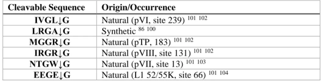
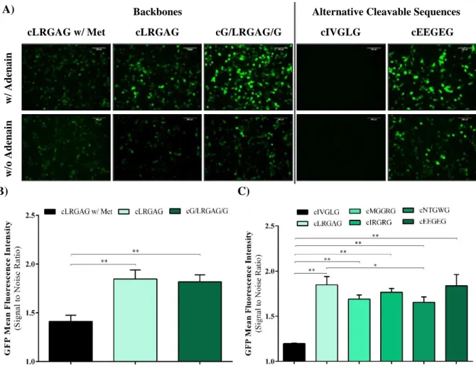
Figure 4.2 – Backbones and alternative cleavable sequences optimization for cGFP system .... 20
Figure 4.3 - Proteolytic processing analysis of the cGFP sensor. ... 20
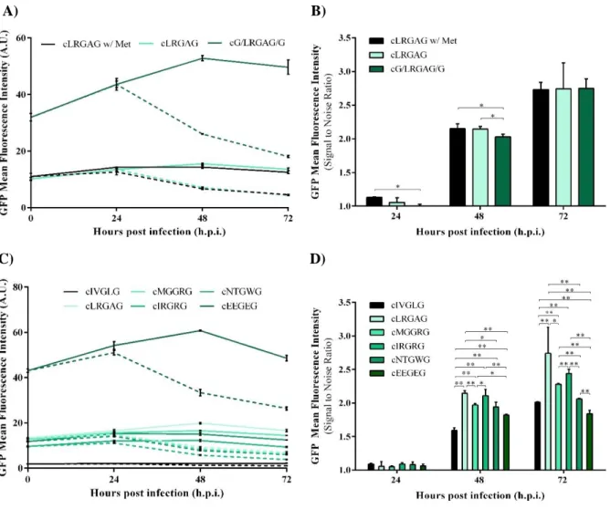
Figure 4.4 - Characterization of HEK 293 cells stably expressing cGFP sensor. ... 21
Figure 4.5 - Implementation of the eS11 system... 23
Figure 4.6 – Backbones and alternative sequences optimization of eS11 system. ... 23
Figure 4.7 - Characterization of HEK 293 cells stably expressing eS11 sensor. ... 24
Figure 4.8 - Circularization process of cS11 system. ... 24
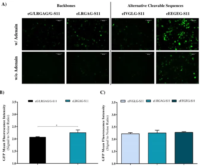
Figure 4.9 – Backbones and alternative sequences optimization and characterization of HEK 293 cells stably expressing cS11 sensor. ... 26
xii
TABLE INDEX
Table 4.1 Cleavable sequences recognized by Adenoviral protease. ... 19 Table S1 List of PCR primers, templates and restriction sites of plasmids constructed for cS11 strategy. ... 36 Table S2 List of PCR primers, templates and restriction sites of plasmids constructed for viral proteases. ... 36
xiii
LIST OF ABBREVIATIONS
Abs Absorbance
AdV Adenovirus
AdV5 Human Adenovirus serotype 5
ANOVA Analysis of variance
BSA Bovine serum albumin
cGFP Circularized superfolder-GFP strategy
CMV Cytomegalovirus
CPE Cytopathic effects
cS11 Circularized GFPS11 fragment strategy
DMEM Dulbecco’s modified eagle’s medium DMSO Dimethyl sulfoxide
DNA Deoxyribonucleic acid
E. coli Escherichia coli
eGFP Enhanced GFP
ELISA Enzyme-linked immunosorbent assay
EMCV.IRES Encephalomyocaerditis virus-internal ribosome entry site
eS11 Embedded GFPS11 fragment strategy
FBS Fetal bovine serum
FDA Food and Drug Administration
gag Group-specific antigen gene
GaLV Gibbon ape leukemia virus
GFP Green fluorescent protein
GFPS10 S1-S10 fragment of Split-GFP
GFPS11 S11 fragment of Split-GFP
HCV Hepatitis C virus
HEK Human embryonic kidney
HIV-1 Human immunodeficiency virus type 1
h.p.i. Hours post-infection
HSD Honest significant difference
IF Immunofluorescence
IFA Immunofluorescence Foci Assay
I.P. Infectious particles
IPS-1 Mitochondrially-tethered IFN-β promoter stimulator protein 1
ITR Inverted terminal repeat
LB Luria broth
LV Lentivirus
MLV Murine leukemia virus
MOI Multiplicity of infection
M-PER Mammalian protein extraction reagent
NAAT Nucleic acid amplification tests
Npu DnaE Nostoc punctiforme DnaE NTA Nanoparticle tracking analysis
PBS Phosphate buffer saline
PCR Polymerase chain reaction
xiv
PFU Plaque-forming units
PI Propidium iodide
pol Polymerase gene
pro Protease gene
pVIc Cleaved 11-residue peptide from C terminus of precursor protein VI
qPCR Quantitative polymerase chain reaction
Rev Regulator of expression of viral proteins
RNA Ribonucleic acid
RRE Rev responsive element
RSV Rous sarcoma virus
RT-PCR Reverse transcription-PCR
SEAP Secreted alkaline phosphatase
sfGFP Superfolder-GFP
SIN Self-inactivated
SNR Signal to noise ratio
SPR Surface plasmon resonance
TAE Tris-Acetate-EDTA
TBST Tris buffered saline with Tween 20
TCID50 Tissue Culture Infectious Dose – 50
VBBs Viral-based biopharmaceuticals
VLPs Virus-like particles
VSV-G Vesicular stomatitis virus G glycoprotein
1
1. INTRODUCTION
1.1. AN HISTORICAL PERSPECTIVE
According to World Health Organization (WHO), one of the leading causes of death worldwide, especially in low income countries, is infectious diseases. These are caused by pathogenic microorganisms and can be spread directly or indirectly from one person to another. In 2010 over 15 million people died worldwide due to infectious diseases and it is expected that in 2050 the number of deaths continues to be a challenge with 13 million deaths predicted 1. The
prevalence of infectious diseases led to an increase in vaccine market since vaccination has been a key element in reducing mortality attributed to infectious pathogens. In 2014 the global vaccine market revenue hit the 32.2 billion USD mark and a growth of 27 billion USD is estimated until 2020 2.
The establishment of cell culture techniques and recombinant virus manipulation contributed decisively to the development of viral-based biopharmaceuticals (VBBs). VBBs comprise virus-derived components or virus-based particles able to be used for therapeutic purposes, which includes viral vectors for gene therapy, oncolytic virotherapy and viral vaccines
3. A vaccine consists of a pathogen-based preparation able to provide acquired immune protection
against specific diseases and can be categorized into: live attenuated, inactived and subunit. Live attenuated vaccines make use of live mutated viruses, inept to cause any disease but still able to infect target cells. Inactivated vaccines use killed viruses treated by chemical (formalin or formaldehyde) or physical means (heat or radiation); therefore, the virus is no longer infectious but its immunogenicity persists. Subunit vaccines consist of parts of the virus comprising specific antigens known to induce high immune responses 4. Recently, a novel type of subunit vaccines
has been approved and commercialized, named like particles (VLPs). VLPs combine virus-derived structural antigens with the lack of genetic material, which makes them non-infectious particles 45.
VBBs had also a huge impact on the development of gene therapy field. As stated by Jayanant Iemsam-Arng et al.6, gene therapy can be described as a procedure aimed at replacing,
manipulating or supplementing non-functional genes with healthy ones. For gene therapy success, an effective delivery system should be developed without causing major side effects. One extremely effective method for gene delivery takes advantage of viral vectors, since viruses have the ability to express the genes of interest in target cells, using cell’s transcription equipment. Through genetic manipulation, viral vectors comprise only the transgene and the minimal number of genes responsible for viral production and replication, reducing virus infectivity 78. The first
steps in gene therapy started in the 70s and since then gene therapy has faced an exponential progress. Twenty years later, in the 90s, the first human gene therapy clinical trial for therapeutic purposes using viral vectors was approved by Food and Drug Administration (FDA) 9 10.
According to The Journal of Gene Medicine, in 2016 around 2400 approved clinical trials using viral vectors have been performed worldwide. Adenovirus (AdV), adeno-associated virus, lentivirus (LV) and retrovirus are the most used viral vectors in gene therapy clinical trials (Figure
1.1). Specifically, AdV has been the most studied viral vector in gene therapy thanks to their
efficient transduction and transgene expression 11 12. However, these viruses can induce high
immune responses in humans, which might have contributed in the last years to an increased interest in LV vectors (Figure 1.1). In 2004 the State and Food Drug Administration of China approved the commercialization of the Gendicine®, a gene therapy product based on AdV
2 serotype 5 vector with the therapeutic gene p53, for the treatment of head and neck squamous cell carcinoma 1314.
Figure 1.1 - Viral vectors used in gene therapy clinical trials.Data gathered from: The Journal of Gene Medicine, 2016 John Wiley and Sons, Ltd, www.wiley.co.uk/genmed/clinical [Accessed: 08-Aug-2017].
However, and despite the current success of gene therapy, its application must be carefully planned. Indeed, in 1999 gene therapy has experienced a terrible incident with the death of an 18 years old patient. The boy was erroneously administrated with a high titer of AdV vectors, which generated a high immune response leading to his death four days later by multi organ failure 10. This incident showed that dosage is of paramount importance for a safe and successful
application of gene therapy protocols; as such, sensitive and trustworthy titration methods are needed to assure it.
1.2. METHODS FOR VIRUS DETECTION AND QUANTIFICATION
Clinical and research applications based on viruses and viral vectors require reliable, accurate and fast methods for virus detection and quantification. For example, the study of new vaccines designs makes use of virus titration to estimate vaccine efficiency. In gene therapy, it is mandatory having a number of viral genome copies on target cells among a defined and reproducible range. If not, it could lead into an inadequate therapeutic effect or reinforce serious side effects.
Current titration methods for viral vectors can be categorized into: functional and non-functional 15. Functional methods provide information concerning virus functionality, resulting in
a direct estimation of virus infectivity. Virus quantification can be based, for example, on number of viral genome copies on target cells (e.g. Quantitative Polymerase Chain Reaction - qPCR), expression of reporter genes (e.g. Green Fluorescent Protein, GFP) or cytopathic effects (CPE) (e.g. Tissue Culture Infectious Dose 50%, TCID50). Non-functional titration methods provide an indirect estimation of the infectious viruses, which often lead to an over-estimation of virus infectivity. This estimation depends, for example, on the virus component to be measured in the viral preparations. The number of viral genome copies in viral preparations (e.g. qPCR) is an example of such indirect titration methods. Below are described some of the assays widely-used for virus detection and quantification.
0 5 10 15 20 25 30 2005 2006 2007 2008 2009 2010 2011 2012 2013 2014 2015 2016 Nu m b er o f C lin ical T rials
3 DETECTION AND QUANTIFICATION OF INFECTIOUS VIRUS UNITS
Cell culture virus isolation was one of the first methods for virus detection and for many years it was considered an attractive method for the detection of viral human pathogens in vitro
16. Some viruses can cause morphologic changes in host cells, which are named CPE. The virus
identification is accomplished by observing differences on host cells during virus culture, such as rounding cells or texture changes 17. The plaque assay is a widely-used virus titration method
providing a direct and quantitative measurement of virus titer based on the number of plaque-forming units (PFU), specific for lytic viruses 1819. This assay consists on infecting a monolayer
of cells with serial viral dilutions. Infected cells will generate a hole (or plaque) due to the cell lysis promoted by the virus infection. Cells are then fixed and stained and virus titer is determined by counting the number of plaques 1720. The main advantages of the use of this method stands on
the ease to implement, it’s inexpensive and doesn’t require advanced materials. Nevertheless, this approach is laborious, time-consuming (could take several days for plaques to be formed) and is operator error-prone. Moreover, only viruses capable of generating plaques on host cells are suitable to be used. TCID50 it’s another example of an end point assay performed to determine the dilution able to produce CPE in 50% of the seeded cells. Cells are plated in well-plates and infected with serial dilutions of a viral preparation. Virus titer is calculated by determining the last CPE positive dilution, based on the number of positive wells 172021. It’s also time-consuming
(~10 days), and high error due to operator visual inspection. Again, only viruses able to cause CPE can be quantified with this titration method.
Cell culture based techniques can be combined with immunofluorescence (IF) staining for a faster and trustworthy detection. IF consists of a biological assay which makes use of antibodies labelled with a fluorescent dye (also named fluorophore) for detection of specific target antigens under a fluorescent microscope 22 23. So, IF is a fast (requiring only 20 to 30 minutes),
versatile, sensitive and specific method for viral identification. Still, shows some limitations such as variability, because of non-specific binding or cross-reactivity of antibodies, and it is very expensive due to the cost of the antibodies 17. Another extensively used virus infectivity based
method is named immunofluorescence foci assay (IFA). IFA stands as a suitable alternative for the titration of virions in cell lines which do not form plaques or any CPE on host cells, for example the human immunodeficiency virus type 1 (HIV-1) 24. This technique consists on a
modification of the plaque assay, by making use of antibody based staining similar to IF 1720. IFA
shows improved sensitivity and speed (2 days) regarding other TCID50 and plaque assays; however, introduces the same disadvantages as IF assay.
The use of reporter genes for the detection and quantification of virus pathogens is extensively used and easy to perform. Several studies report the use of reporter genes for detection and quantification of multiple virus, such as influenza virus 25, retrovirus 26 or herpes simplex
virus 27. In research laboratories, such as in the Cell Line Development and Molecular
Biotechnology Laboratory (part of ACT Unit IBET/ITQB NOVA), recombinant LV vectors harbouring a fluorescence protein reporter as transgene are regularly titrated by means of flow cytometry. This method allows determining the number of infectious particles depending on the percentage of GFP-positive infected cells. The use of reporter genes provides a fast and sensitive method for detection and quantification of viruses. However, one of the major limitations of this method is the use of reporter genes, which are not tolerated in viral vectors for clinical use, due to safety issues. Additionally, it requires previous virus or viral vector modification, which, besides being time-consuming can alter virus biology and life cycle.
4 QUANTIFICATION OF VIRAL NUCLEIC ACID
Advances in technology have provided new and better tools for the detection of virus, like the introduction of molecular techniques able to detect viral nucleic acid. Polymerase chain reaction (PCR) consists on an enzyme-mediated in vitro assay, where the deoxyribonucleic acid (DNA) polymerase exponentially amplifies a target sequence of DNA for various cycles, using specific primers 28. PCR led to important improvements on the field of molecular diagnosis,
especially on the development of nucleic acid amplification tests (NAAT). NAAT comprises several amplification techniques widely used for virus detection, such as nucleic acid sequence-based amplification, transcription-mediated amplification, qPCR and reverse transcription-PCR (RT-PCR). The latter assays are usually used for assessing viral titers 29. The major advantage of
NAAT is that they require low amounts of viral nucleic acid or even viable viruses to detect viral pathogens. In addition, NAAT have shown higher analytical and clinical sensitivity and specificity than cell culture or IF methods 16. qPCR method is also usually used to determine the
number of integrated proviral DNA copies in target cells, by labelling the amplified DNA portion with fluorescence 1530. This technique is widely used for assessing virus load because it’s a fast,
sensitive, high-throughput and reproducible method. However, imperfect sample purification can lead to PCR inhibition. Besides the quantification of viral DNA, NAAT can also be used for the quantification of viral ribonucleic acid (RNA) by adding a reverse transcriptase enzyme to convert RNA to DNA. The combination of these two approaches, as in RT-PCR, can improve the detection sensitivity 31.
QUANTIFICATION OF VIRUS PROTEINS
The specific relation between antibody and antigen has become a key element to design a diversity of antiviral antibodies detection and quantification assays, for high throughput analysis of several samples, such as immunoblotting and enzyme-linked immunosorbent assay (ELISA). Immunoblotting techniques (also known as Western-Blotting) make use of electrophoresis for the separation of proteins in polyacrylamide gels. The proteins are then transferred onto chemically resilient membranes and incubated with a primary antibody specific for the protein target. Later, the membrane is incubated with the secondary antibody combined with a marker compound for ease detection 32. Thus, this method allows the detection of single
viral proteins from infected cell lysates. This technique is one of the most used confirmatory test for HIV-1 33 and hepatitis C virus (HCV) 34 infection diagnosis, for example.
ELISA is a widely-used method for virus detection and quantification, for example for the diagnose of HIV-1 infection 33. There are two main variations of this technique, regarding
antigen immobilization and detection: direct or indirect. The antigen is immobilized either directly or by a specific antibody (capture antibody). If the primary antibody is conjugated with an enzyme, it’s called direct ELISA. On the other hand, if the primary antibody needs to be combined with an enzyme-conjugated second antibody, it’s named indirect ELISA. The detection of the virus is allowed thanks to the interaction of the enzyme and a substrate, which leads to color formation. The viral quantification is made by the measurement of the optical density, which is proportional to the quantity of antigen 35. Overall, this assay is simple, versatile, sensitive and
specific. However, in some cases is expensive, time-consuming and presents additional variability, due to cross-reactivity and non-specific biding.
5 QUANTIFICATION OF VIRUS PARTICLES
Relating particle size and Brownian motion, nanoparticle tracking analysis (NTA) method is able to determine particle size distribution on viral preparations by laser light scattering microscopy coupled with a camera 36. Kramberger et al. 37 showed fast estimation of virus
concentration in different samples of AdV and influenza virus, however AdV titer was underestimated. So, this method is fast and allows sample visualization during the quantification process; still, provides an indirect measure of infectious particles because it quantifies the number of total virus particles in the sample.
1.3. BIOSENSORS FOR VIRUS DETECTION AND QUANTIFICATION
Conventional systems like the ones presented above have several disadvantages: the majority of them provide indirect measurements of infectious particles, which results in an over-estimation of the virus infectivity; and may rely on vector modification, by making use of reporter genes (labelled-viruses), which is not allowed when using those vectors for clinical trials. Therefore, there’s an evident need for the development of simple, fast and reliable quantification methods for label-free viruses. As so, virus biosensors already represent a promising alternative for traditional titration methods extensively used in biomedicine to diagnose infectious diseases and drug development. A biosensor consists of a device able to convert a biological response into an electrical signal, by a transducer 38. Usually, it involves three basic components: a biological
recognition component (enzyme, antibody, DNA, etc.), a sensor element for signal acquisition (electrical, optical and thermal) and an element for amplification/signal processing 39. The
difference between a biosensor and a regular sensor is the presence of bio-recognition elements (also referred as affinity reagents).
Virus biosensors can be classified according to their affinity reagents and viral targets into: immuno- (antibody-), DNA-, antigen- and cell-based biosensor 40. Immunosensing assays
lean on the specific interactions between antigens and antibodies to get a quantifiable response. Antibodies are produced by the host as an immune response to the presence of viral pathogens, representing one of the most widely used affinity reagent in viral biosensors 41. DNA-biosensors
operation is based on nucleic acid hybridization 42. Recent studies have demonstrated that the use
of peptide nucleic acids exhibit high stability and can accomplished a fast, specific and strong hybridization on viral DNA detection 43 44. Antigen-based biosensors rely on the detection of
whole virus particles and surface antigens, such as the capsid and envelope 40. Like other
serological methods, their performance depends on the number of antibodies produced during the infection. Of all existing virus biosensors, cell-based sensors are the most promising approach. Living cells are capable to preserve vital functions by responding to external environmental changes quickly and precisely. Taking advantage of living cells as sensing elements offers the chance to develop sensors with high specificity and sensitivity to a vast number of external agents, like viruses, responsible for affecting cells’ activity 45 46. Additionally, cell-based sensors can
provide a direct measure of virus infectivity.
There are several transducers utilized on biosensors, which can be divided into three major groups: electrochemical, piezoelectric and optical. Electrochemical biosensors are often used as virus detection methods because they offer a fast, high sensitive, practical, selective and economic detection 47. The bio-recognition process of these biosensors can provide different
responses and according to these, biosensors based on electrochemical transducers are categorized into: conductometry, potentiometry, voltammetry, and impedance 40. The latter ones are the most
6 used for virus detection. Voltammetric biosensors measure the current on varying the potential 40.
Several of voltammetric techniques have been successfully used for virus detection, for example differential pulse voltammetry 48, square wave voltammetry 49 and cyclic voltammetry 50 51.
Impedance-based biosensors have also been extensively used for viral pathogens detection 525354 55. They measure the electrical opposition to current flow at the interface 56. Therefore, impedance
biosensors are a very flexible method, thanks to their ability to detect a diversity of targets just by changing the probe used 57. Additionally, this approach, also named electrochemical impedance
spectroscopy, provides sensitive and non-invasive measurements.
Optical biosensors turned out to be a promising approach for a fast, sensitive and real-time monitoring method for virus detection. Surface plasmon resonance (SPR) is one of the most developed label-free and real time detection methods and relies on the variation of the refractive index of the sample 58. This biosensor owns a series of applications in the detection of several
virus, such as hepatitis B virus 59, HIV-1 60 and dengue virus 61. Despite the advantages, some
limitations should be considered. For example, small molecules generate a weak signal, hampering the differentiation of true signal from noise. In addition, the interpretation of the binding kinetics is not always direct, because in some cases the published biosensor data does not match to the simple bimolecular interaction binding model, which questions the biosensor validity
62. Fluorescence-based biosensors comprise another optical detection biosensor system. Thanks
to its high sensitivity and selectivity, sufficient spatiotemporal resolution and low cost, fluorescence is extensively used in biological imaging 63. Mainly, this method measure the
fluorescent signals from fluorophores conjugated to bio-recognition elements or viral targets 40.
Some of the existing fluorescence biosensors are based on fluorescence resonant energy, which results on great sensitivity for virus detection and quantification 64 65. This phenomenon takes
place only when two fluorophores are in suitable proximity between them and the excitation spectrum of the acceptor is overlapped by the emission spectrum of the donor, resulting on a non-contact, radiationless and distance-dependent energy transfer method 4063.
FLUORESCENT CELL-BASED SENSORS
Recent efforts have been made for the development of fluorescent cell-based sensors, by combining fluorescence elements with molecular biology techniques and protein chemistry. Fluorescent proteins can absorb light and emit it in a longer wavelength. One of the most widely used reporter protein is the GFP from jellyfish Aequorea victoria. This well-known fluorescent protein comprises 238 amino acid residues and two peaks of maximum absorption at 395 and 475 nm. On top of that, GFP exhibits the unique property of generating internally the chromophore by an autocatalytic reaction involving three residues (Ser65-Tyr66-Gly67) 66. This peculiarity allows
GFP to be easily genetically modified and cloned into different biological systems, which seems an attractive approach for biosensors 67. Different versions of fluorescent proteins were developed
since GFP was discovered by Osamu Shimomura et al. 68. The modification of the chromophore
structure or the adjacent environment to absorb and emit light at different wavelengths allows the creation of new variants of fluorescent protein colours 67. Examples of produced colours of
fluorescent proteins based on GFP are blue 69 or yellow fluorescence 70. Moreover, efforts have
been made to improve some GFP properties, like folding, solubility or chromophore maturation. Mutations on the chromophore residue Ser65 to a threonine and Phe64 to a leucine resulted in a
GFP variant with improved brightness, the enhanced GFP (EGFP) 71. Thanks to its enhanced
brightness it is widely used as a fluorescent label for quantitative fluorescence microscopy applications. However, studies have shown that it is not suitable for single-molecule tracking
7 detection for long periods of time 71. Based on EGFP and folding reporter GFP 72, Pédelacq et al.
developed a strongly folded mutant, that folds even when fused to polypeptides, the superfolder-GFP (sfsuperfolder-GFP). This superfolder-GFP variant comprises the cycle-3 mutations from folding reporter superfolder-GFP, the two mutations of EGFP and six new mutations 73. sfGFP displays increased resistance to chemical
denaturation, faster refolding kinetics and high tolerance to circular permutation. Recently, Kamiyama et al. developed a system for protein tagging named split-GFP 74. This system relies
on GFP cleavage into two fragments, GFP S1-10 (GFPS10) and GFPS11. The fragments alone are non-fluorescent since the conserved residue E222 present in GFPS11 is required for chromophore maturation. However, by transcomplementation GFP fluorescence is reconstituted.
Additionally, the use of fluorescence proteins has the advantage of switching from off-to-on mode when structural distortions are performed 4667. Therefore, a fluorescence cell-based
switch off-to-on sensor activated by viral enzymes, like the viral proteases, represents an exciting and promising label-free system for virus titration, without requiring vector modification. There are many viruses in which polyprotein processing and maturation is regulated by viral proteases, which provides several advantages for virus lifecycle. This method of genome organization allows genome condensation by erasing genetic features (e.g. promoters) necessary for individual protein expression as well as regulation of protein activity depending on polyprotein cleavage site 75. The
main function of proteases is hydrolyzing the peptide bond between amino acid residues in a polypeptide chain, which makes them suitable as sensor activator. According to their catalyzation mechanism, proteases can be categorized into: aspartic, metalloproteases, cysteine, serine and threonine. One of the main differences between these proteases is the nucleophile of the active site. In the latter three the nucleophile is an amino acid residue (Cys, Ser or Thr, respectively) from which derives the name, while the others use an activated water molecule 7677. A few switch
off-to-on fluorescent biosensors activated by viral enzymes were already developed and are briefly presented.
In 2009 Schekhawat et al. 78 developed a method based on coiled-coil auto inhibition
embedded in split proteins of firefly luciferase. The biosensor consists of a split reporter protein, an antiparallel heterodimeric coiled-coil and a protease cleavable sequence linker. When the linker is cleaved by the protease, the coiled-coil portions can interact, allowing the transcomplementation of firefly luciferase and, consequently, emission of light (Figure 1.2A).
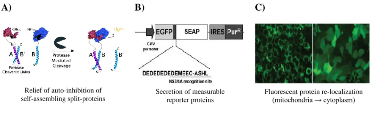
In the same year, Iro et al. 79 developed a cell-based biosensor for HCV, consisting on a
fused EGFP and secreted alkaline phosphatase (SEAP) with a cleavable linker site for HCV protease. After HCV infection, the linker is cleaved and SEAP is released from the fusion protein and secreted into the extracellular supernatant where it can be detected (Figure 1.2B). This system is fast and sensitive however it only allows detection and not virus quantification.
Also for HCV detection, Jones et al. 80 developed a sensitive method able to distinguish
infected cells from non-infected cells, in live or fixed samples, based on relocalization of a genetically encoded fluorescence protein. For this purpose, the system makes use EGFP or red fluorescent protein fused with the carboxy-terminal region of mitochondrially-tethered IFN-β promoter stimulator protein 1 (IPS-1) (a cellular substrate of HCV), comprising the HCV protease cleavage site and a mitochondrial targeting sequence. Upon HCV infection, the reporter protein is cleaved and relocalized from the mitochondria membrane (punctuated pattern) to the cytoplasm (diffuse fluorescence), as depicted in Figure 1.2C. One of the major limitations of this system is not allowing high throughput, because it’s based on single-cell analysis by fluorescence microscopy.
Callahan et al. 81 implemented a method for the detection of HIV-1, based on
transcomplementation of GFP. The GFPS11 was embedded as a surface loop of a small protein, Eglin c, together with a cleavable sequence linker, which leads to its structural distortion and
8 prevents transcomplementation. The fluorescence is reconstituted once the structural distortion is relieved by protease cleavage and transcomplementation of GFPS11 and GFPS10 is allowed.
A) B) C)
Figure 1.2 - Fluorescence cell-based systems already developed for virus detection. A) “Auto-inhibited Coiled-Coil Design Strategy for Split-Protein Protease Sensor”. The C-terminal fragment of firefly luciferase (CFluc) is
attached to one of the coiled-coil portions, A, associated with B’, an amino acid sequence similar to B, and the protease cleavable sequence linker. The B portion of the coiled-coil is coupled with the N-terminal firefly luciferase fragment (NFluc). After cleavage of the linker by the protease, firefly luciferase is able to transcomplement and emit fluorescence. Adapted from S. S. Shekhawat et al. 78. B) Cell-based SEAP reporter assay. EGFP is linked to SEAP by a protease HCV cleavage site and a spacer (DEDEDEDE). Adapted from Iro et al. 79. C) HCV-dependent
fluorescence relocalization. Upon the cleavage of HCV protease, the reporter protein is relocalized from the
mitochondria membrane (punctuated pattern) to the cytoplasm (diffuse fluorescence). Adapted from Jones et al. 80.
The strategies presented above 78 79 81 80, as well as other strategies already developed
present a series of disadvantages that should be considered, such as low sensitivity, low signal to noise ratio (SNR) performance and most are biochemical assays. Additionally, most of them do not allow virus quantification, only detection. Therefore, a robust strategy for detection and quantification of label-free virus and viral vectors is still a current need.
1.4. FIRST STEPS TOWARDS LABEL-FREE VIRUS DETECTION
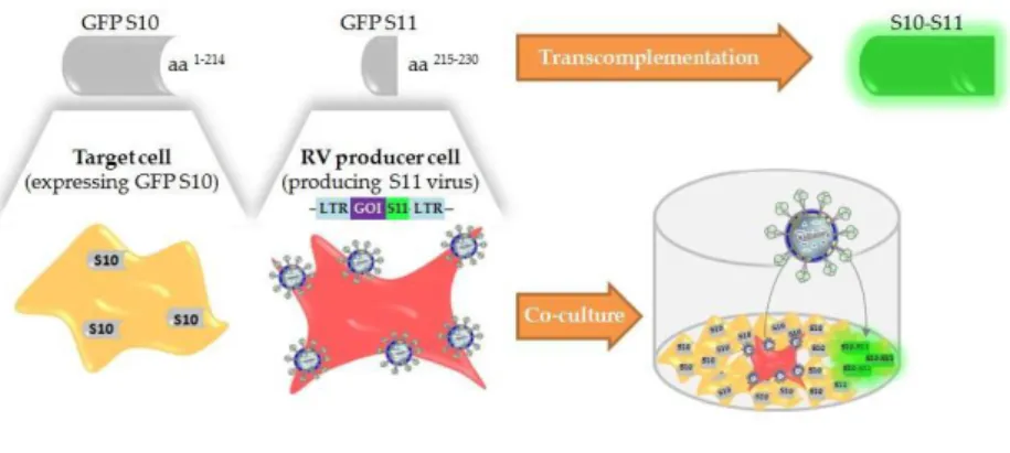
Recently, our group at the Cell Line Development and Molecular Biotechnology Laboratory started to tackle these limitations by developing a new promising titration method for retroviral vectors, named Single Step Cloning Screening Method 26. This method is based on GFP
transcomplementation, and relies on co-culture of cells stably expressing the GFPS10 fragment with cells producing retrovirus coding the GFPS11 fragment in the viral transgene. When GFPS11-coding viruses infect target cells, GFPS10 and GFPS11 transcomplement and fluorescence emission is reconstituted (Figure 1.3). This system is fast, provides high throughput and a direct measure of infectious particles. Despite all the advantages, it’s still not a label-free sensor, since the virus genome encodes the GFPS11 fragment. Therefore, a step further was needed to develop a new method for detection and quantification of label-free viruses, by taking advantage of viral enzymes.
Relief of auto-inhibition of self-assembling split-proteins
Secretion of measurable reporter proteins
Fluorescent protein re-localization (mitochondria → cytoplasm)
9
Figure 1.3 - Schematic representation of the Single Step Cloning Screening Method. Cells stably expressing
GFPS10 fragment (amino acids 1-214) are infected with retrovirus (RV) encoding GFPS11 fragment (amino acids 215-230) fused to a gene of interest. Fluorescence emission is recovered by transcomplementation. GOI, gene of interest; LTR, long terminal repeat. Adapted from A. F. Rodrigues et al. 26.
As previously referred, AdV is still the “gold standard” vector in gene therapy (Figure
1.1). So, it would be of great interest to develop a fluorescent cell-based sensor for detection and
quantification of label-free AdV, by taking advantage of the adenoviral protease as a sensor activator.
1.5. BIOLOGY OF ADENOVIRUSES
Adenoviridae family is composed by more than 55 human serotypes and are known for
causing respiratory, ocular and intestinal infections worldwide. These viruses comprise non-enveloped virions with an icosahedral protein coat surrounding their DNA-containing core (Figure 1.4). The capsid comprises 12 identical fibers each coming from penton base vertices. The genome of the AdV is a linear, double-stranded DNA, coding for two major transcription regions: early and late region. Early region genes encode for non-structural viral proteins responsible for several regulation functions, which will allow late genes’ expression. On the other hand, the late region genes encode structural viral proteins essential to virus particles formation. The linear DNA has a length ranging from 26 to 40 kb and consists of compact nucleosome-like structure which at the extremities contains inverted terminal repeat (ITR) sequences comprising the origin of viral DNA replication 118283 .
10 The AdV replication cycle comprises two phases, the early and late phase. Once the virus interacts with the host cell the early phase begins. The virus enters the cell and carries the viral genome to the nucleus, where the transcription and translation of the early genes occurs. The late phase relies on the expression of the late genes, which allows the assembly in the nucleus of the structural proteins and maturation of infectious virus. AdV are classified as lytic viruses since they induce host cells lysis, starting usually 20 to 24 hours post-infection (h.p.i.), allowing virions to be release 84.
The adenoviral protease – commonly known as Adenain – is essential for virus infectivity and maturation, since it catalysis the processing of six structural viral proteins (three capsid proteins and three DNA-associated core proteins) 85. The processing of these proteins is performed
in consensus cleavage sequences: (M/I/L)XGX↓G (type 1) and (M/I/L)XGG↓X (Type 2) 86.
Additionally, this protease also cleaves the cytokeratin 18 late in adenoviral infection (LAGM↓G)
87, contributing to the cell cytoskeleton dissociation and, consequently, cell lysis. The cysteine
protease Adenain has about 200 amino acid residues and comprises two domains with the active site at the domain interface. The nucleophile of the active site is localized in the Cys122 residue
85. Interestingly, the adenoviral protease is expressed in a nearly inactive form, with its full
activation only occurring in the presence of the two co-factors. The cleaved 11-residue peptide from the C-terminus of precursor protein VI (pVIc) results in a 120-fold increase in protease activity. Adenain interaction with viral DNA rises its activity 3-10 fold 8889. The activation of the
peptide co-factor is performed by Adenain itself, by cleaving pVIc from the precursor protein in a specific cleavage site (IVGL↓GVQS) 85. Of notice, the cytoplasmatic protein actin contains a
C-terminal portion homologous to pVIc 90, that when cleaved can also work as a co-factor.
As of today, there are no approved anti-adenoviral therapeutics available, despite a growing unmet medical need for specific anti-adenoviral drugs, in particular for immunosuppressed patients. Due to its importance in viral maturation and infectivity, Adenain is a viable target for the development of antiviral agents 91.
11
2. AIM AND STRATEGY
The aim of this project was to develop a mammalian cell-based fluorescent biosensor for detection and quantification of label-free viruses, the VISENSORS, taking advantage of the viral protease activity as a trigger of the biosensor. Since several viruses make use of proteases for virus maturation and life cycle, this enzyme seems a suitable sensor activator.
As proof-of-concept, human AdV serotype 5 (AdV5) were used as study model, due to their extensive value in development of vectored vaccines and gene therapy (Figure 1.1).
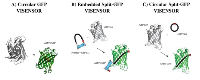
VISENSORS are mammalian cell-based genetically encoded biological sensors that rely on structural distortion of fluorescent proteins, like GFP, to limit its fluorescence emission. Upon recognition of a specific cleavage sequence by the adenoviral protease, when cells are infected with label-free AdV5, fluorescence emission is recovered, thus a switch-on system. Three different strategies were addressed as shown in Figure 2.1.
A) Circular GFP VISENSOR B) Embedded Split-GFP VISENSOR C) Circular Split-GFP VISENSOR
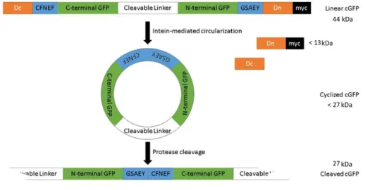
Figure 2.1 - Different strategies considered for VISENSORS development. (A) Circular GFP VISENSOR. Following
Zhang et. al 92 work, this strategy consists on a genetically encoded switch-on fluorescent biosensor whose fluorescence is limited by protein circularization promoted by Npu DnaE intein. Fluorescence is reconstituted once proteolytic cleavage takes place and circular GFP (cGFP) is converted to a linear form, able to complete its final maturation folding. (B) Embedded
Split-GFP VISENSOR. This strategy consists on a mammalian optimized version of Callahan et al. 81, to embed GFPS11 fragment (eS11) as a surface loop of the small Eglin c protein (in blue). This implements a structural distortion on GFPS11 until AdV protease cleaves the cleavable linker (in red). (C) Circular Split GFP VISENSOR. This approach consists on the cyclization of GFPS11 fragment (cS11) in a similar approach followed by Sakamoto et al.93. GFPS11 cyclization is mediated by split-intein protein (Npu DnaE intein) splicing reaction and once the viral protease recognizes a specific cleavable linker (in red), structural distortion is relieved and GFPS11 can transcomplement with GFPS10. Schematic figures adapted from B. P. Callahan et al. 81.
12
3. MATERIALS AND METHODS
3.1. PLASMIDS
All the plasmids used in this work were derived from a lentiviral transgene plasmid – pRRLSin, a self-inactivating (SIN) third-generation lentiviral plasmid kindly provided by Miguel Guerreiro (ACT Unit IBET/ITQB NOVA, Oeiras, Portugal). pRRLSin.CMV.GFPS10.IRES.Zeo.WPRE and
pRRLSin.CMV.GFPS10.IRES.Puro.WPRE take advantage of a cytomegalovirus (CMV) promoter to
drive expression of the gene of interest (GFPS10, for example) and of encephalomyocaerditis virus-internal ribosome entry site (EMCV.IRES) to drive expression of the zeocin or puromycin resistance gene. The original pRRLSin.hPGK.eGFP.WPRE lentiviral transgene was kindly provided by Dr. Didier Trono through Addgene plasmid repository (Addgene, Cambridge, Massachusetts, USA). These lentiviral transgene plasmids were used not only for VISENSORS’ characterization by transient transfection but also for lentiviral production for stable cell lines’ development. Information concerning the PCR primers and plasmids constructed during this work is listed in Tables S1 and S2. The remaining plasmids used during this work were kindly provided by Miguel Guerreiro (ACT Unit IBET/ITQB NOVA, Oeiras, Portugal). Bellow, a brief description of the all the plasmids used is presented.
CIRCULAR GFP VISENSORS
For the construction of cGFP plasmids pRRLSin.CMV.GFPS10.IRES.Zeo.WPRE was used as backbone plasmid, digesting with NheI and BamHI restriction enzymes to replace GFPS10 by the genes coding the different cGFP VISENSORS. As so: pRRLSin_cGFP-LRGAG encodes for the circular version of sfGFP with a LRGA↓G cleavage site; pRRLSin_cGFP-LRGAG_w/_Met and
pRRLSin_cGFP-G/LRGAG/G are similar to pRRLSin_cGFP-LRGAG but with the addition of a
methionine residue at the C-terminal fragment of sfGFP or the addition of one glycine residue at each side the cleavage sequence, respectively; pRRLSin_cGFP-IVGLG, pRRLSin_cGFP-MGGRG,
pRRLSin_cGFP-IRGRG, pRRLSin_cGFP-NTGWG and pRRLSin_cGFP-EEGEG differ from pRRLSin_cGFP-LRGAG only in the Adenain cleavage site (IVGL↓G, MGGR↓G, IRGR↓G, NTGW↓G
and EEGE↓G, respectively).
EMBEDDED SPLIT-GFP VISENSORS
For the construction of eS11 plasmids pRRLSin.CMV.GFPS10.IRES.Puro.WPRE was also used as backbone plasmid, replacing GFPS10 by the genes coding the different eS11 VISENSORS. As so:
pRRLSin_eS11-G/LRGAG/G encodes the GFPS11 fragment embedded in Eglin C’s loop with a
LRGA↓G cleavage site surrounded on each side with one glycine residue; pRRLSin_eS11-LRGAG was constructed by removing the glycine spacers from pRRLSin_eS11-G/LRGAG/G; pRRLSin_eS11-IVGLG and pRRLSin_eS11-EEGEG are based on the construction of pRRLSin_eS11-LRGAG with different cleavable sites (IVGL↓G and EEGE↓G, respectively).
CIRCULAR SPLIT-GFP VISENSORS
For the construction of cS11 plasmids, pRRLSin.CMV.GFPS10.IRES.Puro.WPRE was used as backbone plasmid, replacing GFPS10 by the genes coding the different cS11 VISENSORS. As so:
13
pRRLSin_cS11-LRGAG encodes for the circular version of GFPS11 fragment of Split-GFP with a
LRGA↓G cleavage site; pRRLSin_cS11-G/LRGAG/G and pRRLSin_cS11-GG/LRGAG/GG were derived from pRRLSin_cS11-LRGAG by adding one or two glycine spacers at each side the cleavage sequence, respectively; pRRLSin_cS11-G/IVGLG/G and pRRLSin_cS11-G/EEGEG/G are a modified version of pRRLSin_cS11-G/LRGAG/G, harbouring different cleavage sites (IVGL↓G and EEGE↓G, respectively). All the information concerning the plasmids constructed in this work for this strategy is listed in Table S1.
For the construction of cGFPS10 plasmid, pRRLSin.CMV.GFPS10.IRES.Zeo.WPRE was used as backbone plasmid. pRRLSIN_cGFPS10-LRGAG codes for the circular version of GFPS10 fragment of Split-GFP with a LRGA↓G cleavage site, is an analogous construction to cGFPS11-LRGAG by replacing the GFPS11 with GFPS10 fragment.
VIRAL PROTEASES
pRRLSin.CMV.GFPS10.IRES.Zeo.WPRE was used as backbone plasmid. For the construction
of pRRLSin_Adenain-MVGLG-VIc, encoding the wild-type adenovirus protease fused with a MVGL↓G cleavage site and pVIc (a modified version of Balakirev et al. 94 work), GFPS10 was removed and
replaced with Adenain-MVGLG-VIc coding sequence (often refered as Adenain for simplicity). For the construction of pRRLSin_Adenain-MVGLG-VIc_mCherry, the zeocin resistance gene from
pRRLSin_Adenain-MVGLG-VIc was replaced by the mCherry gene isolated from pPuro_mCherry 95,
kindly provided by Ana Formas-Oliveira (ACT Unit IBET/ITQB NOVA, Oeiras, Portugal).
pRRLSin_Adenain-C104G/C122G_mCherry encodes a non-functional version of the Adenain, due to
C122G (active site nucleophile) 94 and C104G (pVIc binding site) 96 mutations, and lacks pVIc fusion.
All the information concerning the plasmids constructed is listed in Table S2.
3.2. CLONING PROCEDURES
All PCR reactions were performed in a Biometria® T3 Personal Thermocycler (Biometria, Göttingen, Germany) under suitable conditions for amplification, using a proof-reading Phusion® High
Fidelity DNA Polymerase (Finnzymes Oy, Vantaa, Finland). The custom-made oligonucleotides (Table S1 and S2) were synthesised by Sigma-Aldrich (St. Louis, Missouri, USA). All restriction reactions
were incubated at least 1 hour at 37º C using the indicated endonuclease restriction enzymes (New England Biolabs, Ipswich, Massachusetts, USA). DNA fragments were isolated by agarose gel (NZYTech, Lisboa, Portugal) prepared in 1x Tris-Acetate-EDTA (TAE) buffer (Qiagen, Hilden, Germany) and RedSafeTM Nucleic Acid Staining Solution (iNTrON Biotechnology, South Korea). Agarose gel concentration was dependent on the size of the DNA fragments to isolate. Loading buffer (Gel Loading Dye Purple) (New England Biolabs) and a standard NZYDNA Ladder III (NZYTech) were used during gel electrophoresis. Agarose gels were analysed using GelDocTM XR+ system (Bio-Rad, Hercules, California, USA) and purified with Gel Band Purification Kit (GE Healthcare, Little Chalfont, UK). DNA quantification was performed using Nanodrop ND-2000c (Thermo ScientificTM, Waltham,
Massachusetts, USA).
Ligation reactions (Table S1 and S2) were performed using In-Fusion® HD Cloning Kit
(Clontech Laboratories Inc., Mountain View, California, USA) following manufacturer’s instructions, with a molar ratio of insert to vector of 4:1.
14
3.3. BACTERIAL STRAINS AND CULTURE MEDIA
Bacteria transformation was performed using Escherichia coli (E. coli) StellarTM (Clontech
Laboratories Inc.) and One Shot® Stbl3TM (Life Technologies, Carlsbad, California, USA) competent
cells. Agar and liquid cultures were carried in Luria Broth (LB) media (Fast-Media® LB) (Invivogen,
San Diego, California, USA) supplemented with the proper antibiotic and following manufacturer’s instructions.
3.4. PLASMID PURIFICATION AND QUALITY CONTROL
The constructed plasmids were purified at small-scale (yields up to 20 µg of DNA) using
GeneJET Plasmid Miniprep Kit (Thermo ScientificTM) and large-scale (yields up to 500 µg of DNA)
with Genopure Plasmid Maxi Kit (Roche Applied Science, Penzberg, Germany), according to manufacturer’s instructions, and stored at -20º C. Plasmid quantification and purity (evaluated by
Abs260nm/Abs280nm and Abs260nm/Abs230nm ratios) was performed using Nanodrop ND-2000c (Thermo
Scientific). Plasmids restriction pattern was evaluated by enzymatic restriction and agarose gel electrophoresis. Purified plasmids were sequenced using GATC Biotech services (Constance, Germany) for nucleotide sequence validation. Working bacteria banks were established and stored at -80º C in 15% (v/v) glycerol (Sigma-Aldrich).
3.5. CELL LINES AND CULTURE CONDITIONS
HEK 293 derived cell line (ATCC CRL-1573) stably expressing the E1 gene was used for AdV5 production and for the establishment of cell lines stably expressing the VISENSORS. HEK 293T (ATCC CRL-11268) is a HEK 293 derived cell line expressing SV40 large T antigen and was used for transient lentiviral vector production and for transient transfection screenings for VISENSORS’ optimization of backbones and cleavable sequences. HEK 293 FLEX GFPS11 26 is a HEK 293 derived cell line
producing murine leukemia virus (MLV) based vector coding for a GFPS11 gene, pseudotyped with gibbon ape leukemia virus (GaLV) and were used to titrate LV vectors coding for GFPS10. All cell cultures were maintained in Dulbecco’s Modified Eagle’s Medium (DMEM) (GibcoTM, Carlsbad, UK)
supplemented with 10% (v/v) of Fetal Bovine Serum (FBS) (GibcoTM) at 37 ºC inside an incubator with
a humidified atmosphere containing 5% CO2.
3.6. CELL CONCENTRATION AND VIABILITY
Trypan blue exclusion assay was carried to determine cell concentration and viability, using 0.1% (v/v) of Trypan Blue (Sigma-Aldrich) solution prepared in Phosphate Buffer Saline (PBS) (GibcoTM). Cell counting was performed in a Fuchs-Rosenthal haemocytometer (Marienfeld-Superior,
Lauda-Königshofen, Germany) using an inverted microscope (Olympus, Tokyo, Japan).
3.7. TRANSIENT TRANSFECTIONS FOR VISENSORS’ CHARACTERIZATION
Initial characterization of VISENSORS was performed by transient transfection. Briefly, HEK 293T cells were seeded at 8x10⁴ cell/cm2 in 24 well-plates (Nunc, Waltham, Massachusetts, USA). 24
![Figure 1.1 - Viral vectors used in gene therapy clinical trials. Data gathered from: The Journal of Gene Medicine, 2016 John Wiley and Sons, Ltd, www.wiley.co.uk/genmed/clinical [Accessed: 08-Aug-2017]](https://thumb-eu.123doks.com/thumbv2/123dok_br/15185864.1016375/16.892.182.711.181.485/figure-vectors-clinical-gathered-journal-medicine-clinical-accessed.webp)