O R I G I N A L A R T I C L E
Jorge Pala´cios Æ Zı´nia Serafim Æ Maria Jose´ Leal
Mutilations due to medical disorders in children
Accepted: 7 June 2001 / Published online: 28 March 2003
Ó Springer-Verlag 2003
Abstract Soft-tissue and bone necrosis, although rare in childhood, occasionally occur in the course of infectious diseases, either viral or bacterial, and seem to be the result of hypoperfusion on a background of dissemi-nated intravascular coagulation. Treatment consists in correction of septic shock and control of necrosis. Ne-crosis, once started, shows extraordinarily rapid evolu-tion, leading to soft-tissue and bone destruction and resulting in anatomic, functional, psychological, and social handicaps. Ten mutilated children were treated from January 1986 to January 1999 in Hospital de Dona Estefaˆnia, Lisbon, Portugal. One was recovering from hemolytic-uremic syndrome with a severe combined immunodeficiency, another malnourished, anemic child had malaria, and three had chicken pox (in one case complicated by meningococcal septicemia). There were three cases of meningococcal and two of pyocyanic septicemia (one in a burned child and one in a patient with infectious mononucleosis). The lower limbs (knee, leg, foot) were involved in five cases, the face (ear, nose, lip) in four, the perineum in three, the pelvis (inguinal region, iliac crest) in two, the axilla in one, and the upper limb (radius, hand) in two. Primary prevention is based on early recognition of risk factors and timely correc-tion. Secondary prevention consists of immediate etio-logic and thrombolytic treatment to restrict the area of necrosis. Tertiary prevention relies on adequate reha-bilitation with physiotherapy and secondary operations to obtain the best possible functional and esthetic result.
Keywords Gangrene Æ Amputation Æ
Purpura fulminans Æ Septicemia Æ Chicken pox
Introduction
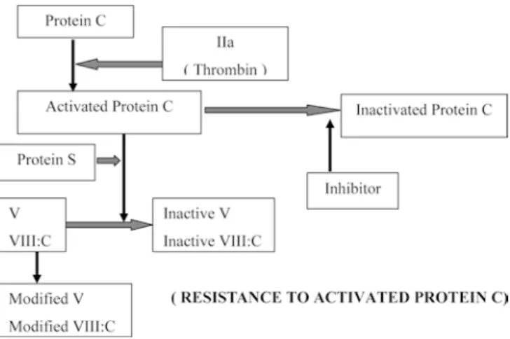
The skin is frequently the site of thrombotic alterations, being an area of diminished vascularization in shock, in which adequate perfusion is restricted to vital zones of the organism (brain, heart and lungs). Proteins C and S, discovered by Mammen in 1960 and DiScipio in 1977, respectively, are powerful inhibitors of coagulation that inactivate factors Va and VIIIa (Fig. 1). These two anticoagulants are synthesized in the liver and are dependent on vitamin K [3].
Hypercoagulability is rare in children and is ex-pressed by recurring venous or arterial thromboses or purpura fulminans (PF), defined as rapidly increasing hemorrhagic cutaneous necrosis associated with seminated intravascular coagulation (DIC). These dis-orders can either be inherited (homo- or heterozygous deficit, dysfunction, or resistance to protein C, protein S, or antithrombin III) or acquired, i.e. in the course of a serious acute infection or without identifiable cause (idiopathic form) [14]. The first type includes hetero-zygotes with autosomal-dominant transmission who develop recurring venous thromboses as young adults and homozygotes with recessive autosomal transmis-sion, who present with neonatal PF.
Acquired deficiency of protein C occurs in cases of shock, DIC, serious infectious purpura, and hepatic disease. The protein C pathway can show a delay in maturation, as in preterm newborns with breathing problems, the newborns of diabetic mothers, and twins. An acquired decrease in protein S has been described in chicken pox, hepatic disease, nephrotic syndrome, pregnancy, and in patients on oral contraceptives.
Serum protein C reaches adult levels in children above the age of 4 years [18]. Therefore, below this age the risk of PF is greatest. The clinical picture and dra-matic evolution of some of these cases are described in publications of a large variety of medical specialities [1, 2, 4, 7, 8, 9, 24]. Bacteria are most frequently involved (Neisseria meningitidis, Haemophilus influenzae,
DOI 10.1007/s00383-002-0779-2
J. Pala´cios (&) Æ Z. Serafim Æ M.J. Leal Hospital de Dona Estefaˆnia,
Rua Jacinta Marto, 1150-192 Lisboa, Portugal E-mail: [email protected]
Streptococcusb-haemolyticus, Pseudomonas aeruginosa), as are viruses (Varicella zoster). The cutaneous lesions are well-defined, star-shaped, purple, and painful or in-durated with a central necrotic area progressing to form a vesicle, ending with a thick scab that, on sloughing sometimes leaves a deep ulcer. The ischemic lesions need not be restricted to the skin, as obstruction of deep vessels can occur in the limbs, leading to gangrene.
Patients and methods
Ten children treated in the Department of Pediatric Surgery of Hospital de Dona Estefaˆnia over a period of 13 years (January 1986 to January 1999), who showed serious mutilations following medical disorders are reviewed. Patients with skin necrosis of limited extent and without major deformities and those with iatrogenic ischemic processes were not included. The following data were analyzed: sex, age, causative medical disease (debilitating factor), period between the beginning of the disease and surgical intervention, type of mutilation, sequelae and secondary opera-tions.
Results
Six patients were females and four were males. Their ages ranged from 6 months to 12 years, and four were
4 years old or less (including two infants of 6 and 9 months). The average age was 3 3/4 years.
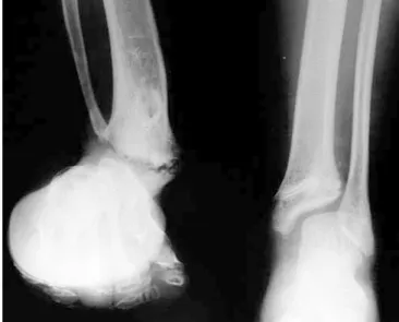
Most patients developed mutilations as a result of septicemia, meningococcal in three cases (plus one a complication of chicken pox) and pyocyanic in two. In the remainder, the causal disease was chicken pox in three cases, malaria in one, and hemolytic-uremic syn-drome in one (Fig. 2 and Fig. 3). Four patients had debilitating factors: serious combined immunodeficien-cy, malnutrition and anemia, second and third-degree burns over 20% of the body area, and infectious mononucleosis in one case each.
The area of necrosis affected the lower limbs in five patients, the face in four, the perineum in three, the abdominal and inguinocrural region in two, the thora-coaxillary region in one, and the upper limb in two (Fig. 3). The interval between the first symptoms of the causal disease and surgical treatment of the affected areas varied between 10 and 30 days, and was not as-certained for one patient. Surgical treatment was un-dertaken once or several times, according to the type and progression of the lesions, consisting of debridement and removal of necrotic tissue with or without amputations or skin grafts. One girl underwent nine surgical inter-ventions. Due to perineal lesions, one patient had a colostomy performed.
Table 1 describes the amputations, deep necroses af-fecting the muscles and/or bones, and resulting unsightly scars. In two patients there was a total of three below-knee amputations. In two others bones were also seri-ously affected; the first required amputation of one finger and seven third phalanges in both hands. The four patients with facial mutilations were 2 years old or less years, and one died before reaching the age of 3 years. The reoperations for correction of sequelae are briefly described in Table 1.
Facial reconstructions had acceptable results in two patients, and mediocre in one, leading to a secondary operation. Both patients who had lower-limb amputa-tions had complicaamputa-tions with the stump, with repeated operations in one patient, who also has serious soft-tis-sue scars that limit movement in both the residual limb and the opposite limb. Both patients have prostheses, but have difficulties in adaptation because of problems with the stumps. In the patients with bone involvement the consequences for growth are visible, especially in the lower limb (Fig. 4). One patient has scars that are be-coming more elastic and may have to be operated upon if they limit movements, one had his colostomy closed after local corrective surgery, one developed a reactive depression and is under psychiatric care.
Discussion
Mutilations due to medical disorders in children result from thrombo-embolic phenomena, leading to more or less extensive necrosis of areas of the body that suffer ischemia during the shock phase (limbs, perineum, nose,
Fig. 1 Protein C pathway
ears). The most frequently implicated disease is septice-mia (meningococcal), exacerbated or not by immuno-depressive factors or diseases.
As clinical symptoms develop rapidly, prompt diag-nosis is essential (history and clinical findings, labora-tory examinations, lumbar puncture (LP), cutaneous microbiologic examination). Knowledge of prognostic factors allows timely hemodynamic stabilization and correct antimicrobial treatment in order to limit the le-sions and eradicate the agent. After the acute phase early rehabilitation should be directed toward the recovery of esthetic, functional, psychological, and social factors for the survivors, whose numbers are increasing due to the efficiency of treatment in intensive care units.
There is still controversy concerning emergency di-agnosis [20, 22], prognosis [6, 16] and treatment [13, 19]
of hypercoagulability syndromes, and more emphasis is being given to the overall rehabilitation of children who survive septicemia [5, 11, 17, 21, 23].
The most frequently involved bacterium in cases of PF is N. meningitidis, with a global mortality of 10%. The clinical signs vary between meningococcemia with-out hemodynamic changes and meningitis or septicemia, the mortality being of no account in the first case and 1%–5% and 30–70%, respectively, in the latter. Two-thirds of the deaths occur during the first 16 h after hospital admission. The need for early diagnosis is therefore evident.
The presence of acute skin lesions should raise the suspicion of a meningococcal infection. Meningococcal meningitis is confirmed by finding gram-negative diplo-cocci in the spinal fluid, a procedure that takes less than
1 h. In meningococcal septicemia, the spinal fluid often does not show any bacteria. As culture is a slow pro-cedure (12–24 h) and other diseases can have similar symptoms, the early treatment of meningococcal septi-cemia is frequently empirical. In cases in which a LP does not give a definite diagnosis, microbiologic exam-ination of the skin is a rapid diagnostic method (<45 min), by either needle aspiration or biopsy. Here the results are not altered by previous antibiotic treat-ment [20], and they distinguish meningitis with or without hemodynamic complications.
The purpose of treatment is the elimination of the etiologic agent with antibiotics, reversal of hypovolemia with fluids and vasopressors, and limitation of necrosis with heparin, fresh frozen plasma, glucocorticoids, and occasionally hyperbaric oxygen in the case of H. influ-enzae or protein C concentrate in the case of S. b-hae-molyticus [14] or N. meningitidis [19]. For diseases like chicken pox, malaria [10], or immunodeficiency syn-dromes, only supportive therapy is available. Age is extremely important [18] (eight children were 4 years old or less), as are pre-existing debilitating factors.
After the almost inevitable appearance of areas of skin necrosis in patients with PF, the need arises to amputate limbs or mutilate other zones of the body
surface, which are usually ischemic, e.g., the limbs, perineum, nose, or ears. Debridement of the affected areas must always be postponed until the limits of the necrotic zone are perfectly defined, which are so often smaller than at first perceived. Secondary operations of the amputation stumps, grafts, and scar correction may be necessary [21]. In cases of asymmetric bone growth resections must be done [5, 7].
There is no doubt that as much viable tissue as pos-sible should be preserved, reducing amputations to a minimum, but sometimes the decision is not easy. This dilemma was evident in one patient who had a viable right foot but exposed bone in the leg and severe tibio-tarsal necrosis. The option of amputation was consid-ered, but ruled out. However, in addition to the nine operations in the initial phase, there was osteomyelitis of the tibia and fibula, serious shortening of the deformed limb, and multiple operations for the bone and skin deformities. We now have the dilemma of a 10-year-old child with a recuperated limb but deplorable esthetic and functional results who is psychologically disturbed after numerous hospital admissions and operations, versus amputation at the age of 2 years and adaptation to a prosthesis. Now, at the age of 10, amputation is refused by both the patient and the parents.
Considering the above complications, it can be stated that amputation is also not the end of the problem. In one case a below-knee amputation caused complications due to poor quality of the surrounding tissue. A free musculocutaneous flap [12] could have protected the stump with preservation of the knee joint. At the time, this was not considered pertinent due to severe scarring of the surrounding tissues.
Special solutions have to be proposed for each par-ticular area, especially on the face and perineum. The impact of deformities of the face, the difficulties of reconstructive surgery, and the social adaptation need to be taken into account. As regards the perineum, in
Table 1 Mutilations and secondary operations for correction of sequelae (A amputation, DN deep necrosis, P plastic procedures, SCstump corrections, VSc vicious scar, D digit, Ph Phalanx) Patient no. Mutilation Secondary operations
1 Nasal wing border, bilateral A Perineum VSc
2 Nasal wing, right A (Guinea-Bissau) Skin flap P
3 Left leg A
Right thigh VSc SC= 7 Left lower limb DN P= 11 4 Axilla DN
Inguinal region DN Perineum VSc
5 Left ear (upper third) A Right leg VSc P= 6 Left foot VSc P= 1 6 Right leg A Left leg A SC= 2 D V (right hand) A P= 4 7 Left forearm DN D V + 3rd Ph D II, III, IV (left hand) A
Making/adjusting prostheses = 4 3rd Ph D II, III,
IV, V (right hand) A
Resection of
radio-ulnar synostosis Right heel VSc
8 P= 2
Right knee, leg and tibiotarsal joint DN
Sequestrectomy Left knee and leg VSc Making/adjusting
prostheses = 6 Osteotomy to correct
right tibia valga
9 Face A Flap
10 P= 1
Perineum DN Anal dilatations Closure of colostomy
addition to reconstructive surgery there are functional repercussions, especially intestinal procedures, as was necessary in one patient. In the long term, these ampu-tations and mutilations disfigure and have anatomic, functional, psychological, and social consequences. Proper and specific treatment is needed, case by case, to achieve rehabilitation [15]. Equally significant is the great number of reconstructive interventions, necessi-tating protracted and repeated hospital admissions.
In conclusion, early evaluation and proper treatment of PF in its acute phase is essential for limiting the area of necrosis and to procure the greatest quantity of viable tissue, keeping in mind that the quality of life in the long term is the main object. Eight of the ten patients de-scribed were aged 4 years or less. Debridement and re-construction of the necrotic areas must preserve the greatest possible amount of viable tissue, each case being unique as regards the anatomic position and extent of damage. For the majority of patients, follow-up implies a large number of secondary operations to correct se-quelae and complications due to growth. A multidisci-plinary approach must always be followed in the early phase as well as in reconstruction and rehabilitation.
Acknowledgements The authors wish to thank the Intensive Care Unit staff and the Department of Physical Medicine and Rehabil-itation of Hospital de Dona Estefaˆnia, Dr. Joa˜o Pascoal, Depart-ment of Orthopedics of Hospital de Dona Estefaˆnia, and Dr. Jorge Roza de Oliveira, Dean of Portuguese Pediatric Surgeons, who translated this paper.
References
1. Adendorff DJ, Lamont A, Davies D (1980) Skin loss in meningococcal septicaemia. Br J Plast Surg 33: 251–255 2. Bass DH, Cywes S (1989) Peripheral gangrene in children.
Pediatr Surg Int 4: 408–411
3. Bick L, Kaplan H (1998) Syndromes of thrombosis and hy-percoagulability: congenital and acquired causes of thrombosis. Med Clin North Am 82: 409–458
4. Campbell WN, Joshi M, Sileo D (1997) Osteonecrosis following meningococcemia and disseminated intravascular coagulation in an adult: case report and review. Clin Inf Dis 24: 452–455 5. Davids JR, Meyer L, Blackhurst DW (1995) Operative
treat-ment of bone overgrowth in children who have an acquired or congenital amputation. J Bone Joint Surg 77A: 1490–1497 6. De La Vega JAB, Calzado AG, Toro MS, Garcia-Mauricio
AA, Cachaza JR, Hachero JG (1993) Enfermidad
menin-goco´cica aguda. Valoracio´n prono´stica. An Esp Pediatr 39: 214–218
7. Farrar MJ, Bennet GC, Wilson NIL, Azmy A (1996) The or-thopedic implications of peripheral limb ischaemia in infants and children. J Bone Joint Surg (Br) 78B: 930–933
8. Fitton AR, Dickson WA, Shortland G, Smithies M (1997) Peripheral gangrene associated with fulminating meningococcal septicaemia. Is early escharotomy indicated? J Hand Surg (Br) 22B: 408–410
9. Gaze NR (1976) Skin loss in meningococcal septicaemia: a report of three cases. Br J Plast Surg 29: 257–261
10. Gear JH (1979) Hemorrhagic fevers, with special reference to recent outbreaks in southern Africa. Rev Infect Dis 1: 571– 591
11. Huang S, Clarck JA (1997) Severe skin loss after meningococcal septicaemia: complications in treatment. Acta Paediatr 86: 1263–1266
12. Huang DB et al (1999) Reconstructive surgery in children after meningococcal purpura fulminans. J Pediatr Surg 3: 595–601 13. Hudson DA, Goddard EA, Millar KN (1993) The management
of skin infarction after meningococcal septicaemia in children. Br J Plast Surg 46: 243–246
14. Karen W (1993) Clotting and thrombotic disorders of the skin in children. Curr Opin Pediatr 5: 452–457
15. Jain S (1996) Rehabilitation in limb deficiency. 2. The pediatric amputee. Arch Phys Med Rehabil 77 [Suppl 3]: S9–13 16. Malley R, Huskins WC, Kupperman N (1996) Multivariable
predictive models for adverse outcome of invasive meningo-coccal disease in children. J Pediatr 129: 702–710
17. Mele JA, Linder S, Capozzi A (1997) Treatment of thrombo-embolic complications of fulminant meningococcal septic shock. Ann Plast Surg 38: 283–290
18. Nardi M, Karpatkin M (1986) Prothrombin and protein C in early childhood: normal adult values are not achieved until the fourth year of life. J Pediatr 109: 843–845
19. Smith OP, White B, Vaughan D, Rafferty M, Claffey L, Lyons B, Casey W (1997) Use of protein-C concentrate, heparin and haemodiafiltration in menincococcus-induced purpura fulmin-ans. Lancet 350: 1590–1593
20. Van Deuren M, Van Dijke BJ, Koopman RJJ, Horrevorts AM, Meis JFGM, Santman FW, Van Der Meer JWM (1993) Rapid diagnosis of acute meningococcal infections by needle aspira-tion or biopsy of skin lesions. B M J 306: 1229–1232
21. Von Wartburg U, Ku¨nzi W, Meuli M (1991) Reconstruction of skin and soft tissue defects in crush injuries of the lower leg in children. Eur J Pediatr Surg 1: 221–226
22. Voss L, Lennon D (1994) Epidemiology, management and prevention of meningococcal infections. Curr Opin Pediatr 6: 23–28
23. Welchon JG, Armstrong DG, Harkless LB (1996) Pedal man-ifestations of meningococcal septicaemia. J Am Pediatr Med Assoc 86: 129–131
24. Woods CR, Johnson CA (1998) Varicella purpura fulminans associated with heterozygosity for factor V Leiden and transient protein S deficiency. Pediatrics 102: 1208–1210