1. Introduction
Veterinary anatomical pathology is a medical specialty which is very similar to human anatomical pathology. This specialty consists in diagnosing diseases based mainly on gross and microscopic examination of organs and tissues obtained by surgical procedure or necropsy. It consists in applying criteria based on knowledge of gross and histological lesions in order to obtain a final pathological diagnosis. The aim of this specific diagnosis is, generally speaking, to find which disease is affecting the animal, if it is alive, or what caused its death. The main difficulties in veterinary pathological diagnosis are related to the various animal species a pathologist has to deal with, the use of exhaustive classifications of different types, costs, subjectivity, including disagreement between the clinical and pathological diagnoses, and the urgency in issuing the report. So, the urgent need to compile all available information to obtain a diagnosis in a short time is the constant challenge professional experts face. Pathology consists in using scientific methods to study structural and functional changes in cells, tissues and organs that underlie disease (Cotran et al., 1999). It is divided into two branches: anatomical pathology, dedicated to examining organs, tissues and cadavers and clinical pathology, dedicated to laboratory analysis of body fluids and/or tissues. The pathologist has the professional expertise devoted to the practice of both, anatomical and clinical pathology (Langone Medical Center, Department of Pathology, 2011).
Past knowledge is an important resource in veterinary pathological anatomy. Professionals often use their previous experience (Dungworth et al., 1999; Goldschmidt et al., 1998; Head et al., 2003; Hendrick et al., 1998; Kennedy et al., 1998; Kiupel et al., 2008; Koestner et al., 1999; Maxie, 2007; Meuten et al., 2004; Misdorp et al., 1999; Scott et al., 2001; Slayter et al., 1994a; Valli et al., 2002; Wilcock et al., 2002)1.
This chapter will be devoted to veterinary anatomical pathology, not in terms of further development of its study methods, extensively detailed in the specialty bibliography, but from a different approach, with the aim of better understanding routine work in the pathology lab. 1The twelve books: (Dungworth et al., 1999; Goldschmidt et al., 1998; Head et al., 2003; Hendrick et al.,
1998; Kennedy et al., 1998; Kiupel et al., 2008; Koestner et al., 1999; Meuten et al., 2004; Misdorp et al., 1999; Slayter et al., 1994a; Valli et al., 2002; Wilcock et al., 2002) were published by World Health Organization (WHO).
How Experience Can Be Useful in
Veterinary Pathological Anatomy
Paulo Tomé
1and Helena Vala
21
Algoritmi Research Center/Polytechnic Institute of Viseu
2Center for Studies in Education, and Health Technologies,
CI&DETS, Polytechnic Institute of Viseu
Portugal
Emphasis will be given to the difficulties faced by the veterinary pathologist every day, in order to justify the development of new solutions to support the issuance of more accurate diagnoses.
The authors describe a framework and a software system that can be useful for diagnosing diseases. The developed framework is based on analysis of the bibliography in this field. Both proposals can be useful to yield better diagnoses. Pathological diagnosis is a supplementary diagnostic test usually ordered by the veterinarian clinician, in order to direct treatment to administer to the sick animal, or living members of a group of animals (e.g. cattle), in which a death occurred. Thus, although the methods used are similar to those used in human medicine, the ends are quite different.
As far as is known, no artificial intelligence system has ever been applied or implemented to areas of diagnosis in veterinary medicine. The system proposed in this chapter is applied to generating solutions for real clinical cases, submitted to complementary pathological diagnosis exams.
In the following sections it will be described pathological diagnosis, as well as the main re-using knowledge mechanisms created in the field of Artificial Intelligence, framework system and the software application. Finally, in section 5 are reported final conclusions and beliefs about the framework and software application.
2. Pathological diagnosis
Veterinary anatomical pathology is quite similar to human anatomical pathology, usually referred as anatomical pathology. It is a medical specialty which diagnoses diseases, based on gross, microscopic, chemical, immunologic and molecular examination of organs and tissues, obtained by surgical procedure and by necropsy (in animal species; autopsy refers to Human species) (Miller et al., 2009).
Anatomical pathology is itself divided in subspecialties, the main ones being:
• surgical pathology, which is the subspecialty pathologists devote more time to. It consists in gross and microscopic examination of surgical specimens, as well as biopsies, submitted mostly by clinicians, dermatologists and surgeons (Grzybicki et al., 2004);
• cytopathology, the branch of anatomical pathology, dedicated to the microscopic examination of cytological specimens, obtained from smears or fine needle aspirates from organs, masses or cysts, used mostly as a complementary diagnosis of the histological exam, since it has more interpretative limitations (Meyer, 2001); • forensic pathology, whose main aim is to determine cause of death of the animal
and is applied mostly in cases of death without previous disease, including criminal causes. This subspecialty is applied to legal purposes. In veterinary medicine, the forensic pathologists are required mainly due to suspicious death by criminal causes (almost always attributed to acts of revenge by neighbors) but in reality, the outcome of most cases culminates in the diagnosis of natural causes, mainly diseases of dietary, infectious or parasitic aetiology, which is very common in domestic animals (Williams et al., 1998);
• necropsy pathology (equivalent to autopsy pathology in human medicine) consists in performing necropsies, i.e. complete and careful examination of the animal cadaver or post-mortem examination, assessing body cavities, liquid presence,
position of organs, gross appearance of organs, sampling injured organs, focus on the transition between normal and injured aspect, in each organ, in order to determine disease factors which caused animal’s death and to contribute to pre-diagnosis of disease.
This subspecialty has a greater importance in veterinary medicine, since it allows other cohabitants of a group (herd or flock) to be saved or an infected animal to be eliminated, acting preventatively to avoid transmission to other animals or humans, contributing favourably to public health. Also, euthanasia is acceptable and legally provided in veterinary medicine, which reinforces the appeal and importance of this branch of anatomical pathology.
In all subspecialties, additional tests from other medical specialities could be required to determine the cause of death or to obtain a definite diagnosis. The most commonly required tests are linked to toxicology, virology, bacteriology and genetics.
Pathological diagnosis is the medical specialty that deals with the examination of gross and microscopic lesions (Miller et al., 2009). It also consists in the further microscopic study of tissues and cells, in order to provide a complementary means for diagnosis. The solid background of expertise is constructed based on medical literature, educational programs, training activities, meetings, technical rules and the cognitive skills of the pathologist which allows him to describe lesions, interpret histological slides and make decisions. In this long and complex process, the pathologist takes into account the animal data, clinical history (including physical exam), results of other complementary exams (including biochemical analysis and x-rays), using a strategy to arrive at a diagnosis which is not qualitatively different from those used by clinicians (Pena & Andrade-Filho, 2009).
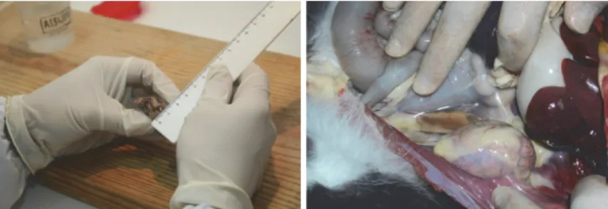
(a) Description and measurement of a surgical specimen
(b) Post-mortem exam of dead animal cadaver
Fig. 1. Gross examination
The most commonly used procedures in the pathological diagnosis processes include gross examination, histopathology or microscopic examination, evaluating histological aspects, including cellular characteristics and organizational patterns, immunohistochemistry and cytopathology, among other more specific tests. Gross examination consists in describing the specimen with the naked eye. It is based on observation, description and measurement of gross lesions, found in a surgical specimen, submitted to the pathology lab (figure 1 a)), or found in animal cadavers, if such is the case, during the meticulous post-mortem exam (figure 1 b)), especially if the animal is injured. It is also during this step, that the pathologist decides which areas and specimens need to be processed for histopathology and microscopic evaluation (University of Utah of Spencer S. Eccles, 2011; Zarbo & Nakhleh, 2009).
(a) Cut of formalin fixed specimens (b) Selection of areas to be processed
(c) Placing the specimen in standard plastic histological cassettes
(d) Paraffin blocks
(e) Execution of histological sections with a rotary microtome
(f) Staining by standard haematoxylin and eosin
(g) Final preparations (h) Microscopic observation
To perform tissue observations under the microscope, a complex and relatively time consuming procedure is performed, in order to obtain very thin sections that can be traversed by the microscope condenser’s light. The microscope is an extension of the pathologist’s vision. It is a fundamental instrument throughout the process of diagnosis and with which the pathologist must feel very comfortable, spending many hours of his life at the microscope. Without a microscope the pathologist is blind and incomplete.
The basis of routine histological technique (figure 2) culminates obtaining histological sections (with 3μm) that will subsequently be stained by standard haematoxylin and eosin or other more specific methods, to obtain a more accurate histopathological diagnosis, namely histochemistry. Histochemistry consists in using chemical reactions to localize chemical compounds of cells and tissues (figure 3 a) and b)) (Pellicciari, 2009) and immunohistochemistry, a technique for identifying cellular or tissue constituents (antigens) by means of antigen-antibody interactions (figure 3 c) and d)). These methods are performed in the pathological diagnostic field, mainly in oncology diagnosis (Miller, 2002).
(a) Histochemistry (b) Histochemistry. Ziehl-Neelsen
Staining. Bar = 25μm
(c) Immunohistochemistry (d) Immunohistochemistry.
Staining with a
peroxidase-labeled-(strept)avidin-biotin
(LAB-SA) method. Bar = 25μm
Fig. 3. Specific histological techniques
Finally, the professional veterinary pathologist produces an understandable and grammatically correct report (Pena & Andrade-Filho, 2009) which includes a complete description of macroscopic and histological observations and a definite diagnosis issued based on the specific knowledge of these observations and statements.
The pathological report must be understood directly by the clinician which operates under the same system of rules (Pena & Andrade-Filho, 2009), without triggering an array of
interpretive questions which will increase the time spent on a particular case. Also, misinterpretation of a pathological diagnosis may lead to an incorrect or surgical therapeutic approach to the lesion concerned (Pena & Andrade-Filho, 2009). So, the development of a system of rules will also help to solve this problem.
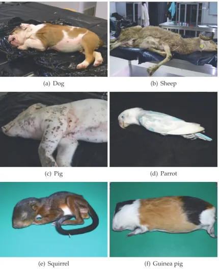
(a) Dog (b) Sheep
(c) Pig (d) Parrot
(e) Squirrel (f) Guinea pig
Fig. 4. Different species of animals to necropsy
One of the major difficulties associated with veterinary medicine is, that unlike human medicine, which is dedicated to a single species, its target is a number of different species of animals to study/diagnose. To overcome this difficulty, the animal species are usually grouped into specialties, based in their affinity (e.g. pets - dogs and cats, ruminants - cows, sheep and goats, swine, equine species, exotic species, etc.) (figure 4). In parallel, veterinary anatomical pathology deals with different animal species, which requires knowledge of the anatomy of each species, species-specific diseases, their lesions and lesion patterns. To achieve a correct and definitive diagnosis per species, the ideal situation would be the constitution of multidisciplinary teams, in which each expert would focus on a single species or group of
species. This may, however, be impossible for smaller institutions wishing to offer a diagnosis service in the field of veterinary pathology.
Also, pathological anatomy uses different types of exhaustive classifications (e.g. infectious diseases, metabolic diseases, endocrine diseases, neoplasm, etc.). It can be highlighted, as reference books to the veterinary pathologist’s diagnostic decisions: Dungworth et al. (1999); Goldschmidt et al. (1998); Head et al. (2003); Hendrick et al. (1998); Kennedy et al. (1998); Kiupel et al. (2008); Koestner et al. (1999); Maxie (2007); Meuten (2002); Meuten et al. (2004); Misdorp et al. (1999); Scott et al. (2001); Slayter et al. (1994a); Valli et al. (2002); Wilcock et al. (2002), among several others dedicated to the pathology of different animal species.
If, on the one hand, not following a clinical case directly or contacting the owner or veterinary clinician personally may be beneficial, in terms of exemption in reaching conclusions, on the other hand, the summary of the clinical information in the requested analysis, may omit crucial information to the final conclusion/diagnosis.
Furthermore, reading that the animal is two years old and has intense itching, is not the same as visiting a young animal that that is constantly scratching itself. So, during the long process of making a diagnosis, this type of information may easily lose its proper influence, which would not happen if it had been obtained by direct observation, especially if the pathologist is seeing abnormal cells in the microscope, i.e. with features of high malignancy, which would tend to overlap, in terms of influencing the summary of the clinical history reported indirectly. Often, the pathologist has no notice knowledge of the outcome, performed treatments or results of outside consultations, which prevents him from verifying the authenticity of his diagnosis and evaluate eventual failures, when he fails, making it difficult to improve and learn from their mistakes (Pena & Andrade-Filho, 2009).
For these reasons, pathologists may enhance their diagnostic abilities, acquiring specific abilities related to other fields by means of expressing relevant warrants and backings to their conclusions, deducing and expressing intrinsic rules that guide the different actions related to the diagnostic task (Pena & Andrade-Filho, 2009).
In complex cases, if the pathologist needs to use additional analytical methods to reach a definitive diagnosis, such as histochemistry, immunohistochemistry or other medical speciality tests, the authors propose a prior consultation with the client, as these additional studies increase diagnostic costs, and thus, should be subjected to client discretion. This information should be included in the first report, with the explanation for the necessity of more diagnostic procedures, along with the budget of all the expected costs. Procedures and respective costs must, preferably, be presented in a phased and sequential manner. As an example, a report for an undifferentiated round cell tumour should describe macroscopic and microscopic features, the preliminary diagnosis as "consistent with round cell tumour", and a detailed account for the need of additional diagnostic workup: "to determine definitive histogenesis, further protocols are recommend to rule out possible differential diagnosis: toluidine blue for mast cell (cost value), CD1a for histiocytoma (cost value) and CD3 and CD79α for lymphoma (cost value). These protocols will be realized in this order and executed only to the point needed to reach a definitive diagnosis.
Although the pathologist is protected in his lab from contact with the owner and patient (animal), he should be sensitive to their economic difficulties, especially in the absence of health insurance for animals. In addition, he must be aware that the diagnosis is only a precursor to the stage of healing, i.e. the treatment stage remains indispensable and is also expensive. For these reasons, and according to proper medical conduct, the proposed plan
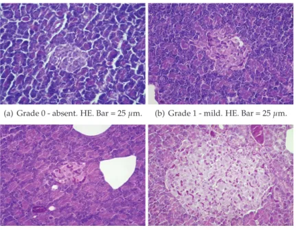
(a) Grade 0 - absent. HE. Bar = 25μm. (b) Grade 1 - mild. HE. Bar = 25μm.
(c) Grade 2 - moderate. HE. Bar = 25μm (d) Grade 3 - severe. HE. Bar = 25 μm
Fig. 5. Semi-quantitative access to congestion
must hereinafter follow one of the following two rules: from the lowest to highest cost or from the most likely diagnosis to the less likely. The goal is that customers do not have to go through the whole plan outlined by the pathologist, and achieve a definitive diagnosis as early as possible, thereby providing savings to owners, who still have to invest heavily in the therapeutic approach/process that follows.
In recent literature (Pena & Andrade-Filho, 2009), the pathologic diagnosis was envisioned as a human task, involving people - the pathologist which can make a diagnosis, based on the strategy called pattern recognition. In other words, pattern recognition is the realization that the histological picture conforms to a previously learned picture of a certain disease, which produces a diagnosis interpretation. Also, other kinds of diagnosis strategies, usually adopted by pathologists, were described in this paper, some leading to pathological difficulties or errors, which need to be compensated with new action plans.
Faced with clinical information that a mass located in the mammary gland was punched, the pathologist will inevitably direct his diagnosis to one of the multiple hypotheses included in the classification of mammary tumours available. He is being driven by location and can hardly be impartial and independent from the information provided. If instead, the clinician punched a herniated organ placed at this location, it is not impossible to get a diagnosis of a mammary adenoma, as the pathologist’s brain was already formatted to diagnose of one of the several types of mammary tumours.
Recent reports showed disagreement between clinical and pathologic diagnoses, except in necropsy, which is the best method to assess overall diagnostic accuracy (Kent et al., 2004; Vos et al., 2005). Also, autopsy is still the most accurate method of determining the cause of death and auditing accuracy of clinical diagnosis, diagnostic tests and death certification
(Roulson et al., 2005). Increased availability of teaching funds may promote efforts to have necropsies performed in veterinary teaching hospitals (Kent et al., 2004). However, in this aspect the problem is getting worse in human medicine, in which the number of autopsies has shown a tendency to decline (Roulson et al., 2005).
The pressure of having to issue a diagnosis that will help save the animal’s life combined with deterioration of the clinical situation increases the responsibility of having to make decisions quickly and can shorten the long and complex process that the pathologist needs to go through to be in possession of the necessary information to make decisions. So, the pathologist is already under sufficient pressure.
(a) Grade 0 - absent. HE. Bar = 25μm (b) Grade 1 - mild. HE. Bar = 25μm
(c) Grade 2 - moderate. HE. Bar = 25μm (d) Grade 3 - severe. HE. Bar = 50 μm
Fig. 6. Semi-quantitative access to fibrosis
More pressure can be caused by the phone ringing continuously in the lab, which will not help to reach an accurate pathological diagnosis. The pathology lab must be a sacred place, where silence and peace must prevail. It is the only way to bring information together, to interpret the observed images correctly. Only then will the images seen on macroscopic exam interact with those observed under the microscope and in the pathologist brain come together to form a conclusion. We must be aware that the pressure clinicians or owners place on the pathologist may precipitate a insufficiently accurate diagnosis.
Nowadays several semi-quantitative and quantitative methods to access macroscopic and microscopic lesions, namely congestion (figure 5), fibrosis (figure 6), inflammatory infiltrate (figure 7), cellular degeneration, mitosis rate, malignancy degrees, among several others, have been developed and published to promote international uniformity, reducing the subjectivity of descriptions depending on the observer, and also to allow statistical studies. Just as a reference, it can be mentioned two: classification and grading of canine mammary tumours (Goldschmidt et al., 2011) and classification of diabetic nephropathy (Tervaert et al., 2010).
(a) Grade 0 - absent. HE. Bar = 25μm (b) Grade 1 - mild. HE. Bar = 25μm
(c) Grade 2 - moderate. HE. Bar = 25μm (d) Grade 3 - severe. HE. Bar = 50 μm
Fig. 7. Semi-quantitative access to inflammatory infiltrate
In research projects two or more observers are required to score lesions independently, in order to enhance the credibility of the results, which is time consuming, expensive and fastidious. Also, some authors defend a standardization of the pathological diagnosis, as a measure to minimize diagnostic variability and error (Foucar, 2000).
The pathologist cannot be reduced to a cognitive instrument for assessing a diagnosis (Pena & Andrade-Filho, 2009) but needs useful instruments to improve his performance. So the urgent need to compile all available information to obtain a diagnosis in a short period of timeframe, which will help to save the animal’s life, is the constant challenge set before the professionals.
A system of rules that allows pathologists to standardize descriptions, easily interpreted by clinicians, will permit an association of descriptions to a conclusive final diagnosis, which would simplify stages in the complex system of diagnosis, thus increasing accuracy. So, the authors propose a model that provides less variability, discrepancies and uncertainties. 3. The re-using knowledge mechanisms
The Artificial Intelligence research community has already proposed some techniques that can be used in several domains to re-use experience. These techniques are described in several bibliographic sources, such as Bratko (2000); Cawsey (1998); Nilsson (1998); Russel & Norvig (2003); Shapiro (1987). In this section, only will be described techniques that have been used in related areas, namely Rule-Based, Pattern and Case-Based Reasoning (CBR).
Rule-Based is the oldest technique. This mechanism emerged from early work on practical and intuitive systems for logical inference (Russel & Norvig, 2003). The rule-based systems have
three properties: locality, detachment and truth-functionality. Proposals by Bachmann et al. (2004); Lloyd-Williams (1994); Tauzovich (1990) are examples of Rule-based systems.
Pattern techniques (Shapiro, 1987), not pattern recognition which is described for example in Duda et al. (2001), are used in many areas. They generally involve the definition of patterns and the use of pattern-matching algorithms. A pattern is a structural sketch of an item with some pieces left undefined. Pattern matching is the process of comparing patterns to see if they are similar. In the Information Systems domain there are several works, such as Batra (2005); Conte et al. (2004); Rieu et al. (2002); Seruca & Loucopoulos (2003), which use pattern techniques.
Finally, the CBR method uses past resolved situations to solve new problems (Aamodt & Plaza, 1994; Kolodner, 1993; Mantaras et al., 2006; Riesbeck & Schank, 1989; Watson, 1999). According to Althoff et al. (1995), CBR is a good method to apply in ill situations, such as modelling tasks. CBR comprises two main elements: Case and Process model.
The case is formed by the problem and its solution (Kolodner, 1993). The objective and the characteristics of the situation are described by the problem. The solution consists of the solution itself, the solution evaluation and reasoning. Identification of case types constitutes the major step forward in the development of the CBR system. The set of cases of the CBR system is called case memory. An important issue related to cases is indexing. The index is a label associated with the case that will allow us to remember it.
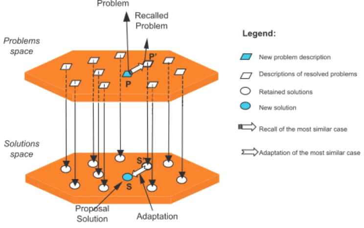
The solving process (or process model), usually called CBR Cycle, begins with the problem description and ends with the solution. The CBR cycle has two main models: 4Rs proposed by Aamodt & Plaza (1994) and the one proposed by Kolodner (1993). The CBR cycle, as illustrated in figure 8, generally involves the following activities:
• case search to find similar cases;
• similarity evaluation to measure the level of similarity between the problem that needs solving and the stored ones;
• adaptation to adjust one or several solutions to the current problem; • case retain to store the newly resolved problem.
Case search is based on problem description. Similarity evaluation is based on similarity functions (Althoff et al., 1995). Consequently, the new solution is built by adapting old solutions to the needs of the current problem. The last task of the CBR cycle is the inclusion of the case in the case memory. Given the fact that a new case is added to the system, the CBR systems could be said to have the ability to learn. It is worth mentioning, that there are a lot of domains where the CBR methodology has been used (Kolodner, 1993; Mántaras & Plaza, 1997; Watson, 1996). CBR application areas consist of software development, health, architectural design (Pearce et al., 1992), meal planning and legal reasoning systems, for example.
4. The framework and the system
It is important to say that this work was developed after a first approach (published in Tomé & Vala (2010)) of applying CBR to a set of 641 with good results. As shown in table 1, 47.1% of the solutions have a similarity greater than 40% of the solution proposed by the veterinary pathologists. Only 16.8% of cases have solutions in the range 0 to 20%. Although
Problem Recalled Problem Proposal Solution Adaptation Problems space Solutions space
New problem description
Retained solutions New solution
Recall of the most similar case
Legend:
Adaptation of the most similar case
P P’
S’ S
Descriptions of resolved problems
Fig. 8. The CBR method2
these results are positive, it is important to mention that the cases are described by regular text which constrains the searching process.
Level of solutions’ similarities [0 , 0.2[ [0.2 , 0,4[ [0.4 , 0.6[ [0.6 , 0.8 [ [0.8 , 1]
108 231 166 80 56
Table 1. Analysis of level of similarity between solutions
As previously mentioned, several authors proposed a diagnosis system (also called a classification schema) based on some animals aspects. These diagnosis systems are used in disease diagnosis by veterinarian pathologists. The WHO proposed a classification schema for animal tumours in twelve, published three books with a classification schema for animal disease based on most affected organs.
The veterinary pathologists, faced with a disease in a particular species, with a certain clinical information, assess which of the body system, and/or specific organ, is most affected. The specialists process a set of likely differential diagnoses, directing the gross and microscopic diagnosis in order to exclude the less likely, until the possession of a set of information consistent with a pathologic diagnosis of a specific aetiology, mostly grouped in bibliography in: congenital and hereditary; abnormalities of development and growth; degenerative; circulatory; traumatic; foreign bodies; metabolic; nutritional; endocrine; toxic; immune-mediated; inflammatory (viral; bacterial; fungal; protozoal; parasitic; miscellaneous); unknown cause (Maxie, 2007).
The veterinary pathologists, faced with a neoplastic disease in a particular species (mostly pets whose lifetime is longer, having a higher predisposition to this pathologic entity) with a certain clinical information, assess in which body system, and/or specific organ, the tumour is primary located, directing the gross and microscopic diagnosis to exclude the less likely diagnosis, using international histopathological classifications. These provided a widely accepted standard nomenclature of domestic animals tumours, promoting advancements in veterinary pathology and facilitating communication between veterinary and medical pathologists, clinicians and researchers. These classifications also pretended to cross geographic and academic boundaries and establish an international consensus pertaining to the nomenclature of domestic animal tumours (Kiupel et al., 2008).
These classifications include a preliminary assessment of the mass, equivalent to gross examination (measurements; superficial and cut section aspects - colour, consistency of the tissues; evaluation of local invasion; attachment to surrounding tissues; evaluation of possible regional lymph node involvement) and then the evaluation of histological criteria which includes cellular phenotype which includes, in turn, nuclear characteristics - shape, colour and staining characteristics, type of chromatin, nucleoli and cytoplasmic characteristics - shape, colour and staining characteristics, intercellular unions); presence or absence of extracellular matrix and its type, when present; cellular pattern organization and cellular differentiation -anaplasia, anysocariosis, anysocytosis, pleomorphism; mitotic rate.
Based on these cellular features and differentiation degree, the tumours are subdivided primarily into benign and malignant groups and then subdivided, based on phenotype and amount of cellular matrix, in mesenchymal or epithelial neoplasias. The third major sub-division consists in the determination of the specific subtype from the classification for the specific target organ. In general, to determine the specific subtype of a given classification of mesenchymal neoplasias, the specialists must take into account the predominant cellular type recognized and the type of cellular matrix produced by the cells (Slayter et al., 1994b). In a given classification of epithelial neoplasias, the veterinary pathologists must take into account the predominant cell type recognized and the cellular organizational pattern. Based on the previous descriptions, it can be concluded that each diagnosis:
• is applied for a specific type of disease; • is based on a set of characteristics;
• is related to a specific type of animal/part..
Based in the previous aspects, it can be concluded that the framework for classification of animal diseases can have the structure illustrated in figure 9.
For instance, the Adenoma disease (defined in (Meuten et al., 2004)) is characterized by: "... rare tumor in domestic animals. ... . Adenonomas are discrete, solitary, tan-white masses confined to the renal cortex. They are small (usually < 2cm) in dogs and cats." Based on the previous description it can be concluded that Adenoma is characterized by the following attributes: Size, Color, Number, Place, Aspect, Shape, Differentiation, Pattern, Cellular
Phenotype, Cytoplasm, Nuclei, Nucleoli, Mitotic Rate and Stroma. The value of each of each
attribute is < 2 cm, Tan-White, Solitary, Renal cortex, Discrete and Masses, Well-differentiated,
Tubular, Cuboidal epithelial, Ample, Single, Single, Rare and Poor. How these attributes and
values are stored in a software application is shown in figure 11.
The way the attributes and values of each type of disease are structured improves the search of previous cases of the animal’s disease. As mentioned at the beginning of this section, searching previous situations with diagnostics described in regular text it is not an easy task because similar situations can be described with the slightest differences.
Fig. 10. Software Application
As previously mentioned, it was developed a software application, of which the main window is shown in figure 10 that can be used for storing knowledge about diseases and cases of animal diseases. The first part of the application - knowledge about diseases - is a library of descriptions of animal diseases described in the bibliography of the domain (such as (Dungworth et al., 1999; Goldschmidt et al., 1998; Head et al., 2003; Hendrick et al., 1998; Kennedy et al., 1998; Kiupel et al., 2008; Koestner et al., 1999; Meuten et al., 2004; Misdorp et al., 1999; Slayter et al., 1994a; Valli et al., 2002; Wilcock et al., 2002)). The second part of the application stores data about cases of animal diseases. In this part it is applied the CBR method described in section 3.
In the CBR part (the structure is shown in figure 12) the four phase cycle of Aamodt and Plaza was implemented (Aamodt & Plaza, 1994). The system implements the retrieve, reuse, revise and retain phases and has a repository with cases and knowledge domain.
As shown in figure 12, the system has components that store data and components that do some of the systems tasks. The components: case memory, vocabulary of domain, adaptation
(a) Definition of Disease types
(b) Adenoma Disease
Fig. 11. Definition of Diseases types
rules, case description, inference rules and metric system, store data; while the components: retrieve, reusing, revision and retaining, implement the system tasks.
Each case is stored as a frame (Minsky, 1974). Each case is divided according the Kolodner (1993) proposal into: objective, characteristics and solution. The characteristics and the solutions are pairs of name x value.
In the domain vocabulary module the most frequent expressions used to describe cases of the domain are stored.
Fig. 12. Structure of the CBR part
The adaptation rules module stores information about how diagnosis can be adapted. In this module if-then rules3can be stored which define how diagnosis can be adapted based on the case’s characteristics.
The Inference rules module is also built out of if-then rules. These rules are based on the knowledge defined in the books of the domain (such as (Dungworth et al., 1999; Goldschmidt et al., 1998; Head et al., 2003; Hendrick et al., 1998; Kennedy et al., 1998; Kiupel et al., 2008; Koestner et al., 1999; Meuten et al., 2004; Misdorp et al., 1999; Slayter et al., 1994a; Valli et al., 2002; Wilcock et al., 2002)).
In figure 13 the process of definition of a new case disease is shown. As can be seen in the figure, the first three elements (dropdowns of the figure) are elements of the Classification Schema illustrated in figure 9. The elements Values usually used and Attributes also used were designed to help the user in the process of defining a new case.
The element Values usually used can be used for retrieving values that are frequently used for a specific attribute. For instance, in the Adenoma situation the attribute size is usually characterized by the value < 2cm. The use of this facility reduces the chance of defining equal values by different ways.
The element Attributes also used aims to help the veterinary pathologist in the definition of the characteristics of the case. Every time the veterinary pathologist defines an attribute, the set of
Attributes also used shows the attributes which were not defined but may defined regarding the
diseases where they appear. For instance, consider the disease and attributes shown in table 2. If the veterinary pathologist already defined the attributes x and y the attributes shown in
Attributes also used are z,w,a and b. The attributes c,d,e,f,g and h do not appear because they are
not in diseases where the attributes x and y appear.
Fig. 13. Definition of Disease types Diseases Attributes A x,y,z,w B x,y,a,b C a,b,c,d D e,f,g,h
Table 2. Example of Diseases and attributes
Finally, it will be explained the functionality of solution finding based on previously resolved/solved cases. This functionality applies CBR to get a case which is the most similar to the current one. The system searches in the Case memory for a case with similarities to the current one. For all the cases in the Case memory the similarity to the current one based on the values of the attributes and on the knowledge of the domain is determined. The case with highest similarity value is selected.
5. Conclusion and future remarks
In this chapter, it is proposed a framework (Conceptual schema) that can be used to structure knowledge about causes of animal death. The framework was developed based on the analysis of the bibliography of the field.
The framework can be used to develop new databases to store data about animal’s death diagnosis and also to improve applications that use previous knowledge of causes of animal death in order to make new diagnoses.
The use of previous knowledge about animal death can improve the task of making diagnoses. First the veterinarian is able to diagnose quickly because he can easily retrieve information about how a similar past situation was diagnosed. Besides that, past knowledge can be considered a library of information where the veterinarian can get useful help or information. Finally, it is important to highlight that the framework is supported (and was tested) by a software application. Through the developing and testing of the software application, it can be guaranteed that the framework fits into aim for which it was developed.
6. References
Aamodt, A. & Plaza, E. (1994). Case-based reasoning: Foundational issues, methodological variations and systems approaches, AI-Communications 7(1): 39–52.
Althoff, K. D., Auriol, E., Barletta, R. & Manago, M. (1995). A review of industrial case-based reasoning tools, Technical report, AI Intelligence.
Bachmann, F., Bass, L., Klein, M. & Shelton, C. (2004). Experience using an expert system to assist an architect in designing for modifiability, Fouth Working IEEE/IFIP Conference
on Software Architecture.
Batra, D. (2005). Conceptual data modeling patterns: Representation and validation, Journal
of Database Management 16(2): 84–106.
Bratko, I. (2000). Prolog Programming for Artificial Intelligence, 3th edn, Addison-Wesley. Cawsey, A. (1998). The Essence of Artificial Intelligence, Prentice-Hall.
Conte, A., Hassine, I., Rieu, D. & Tasted, L. (2004). An information system development tool based on pattern reuse, ICEIS, Porto.
Cotran, R., Kumar, V. & Collins, T. (1999). Robbins Pathologic Basis of Disease (6th ed.), W.B. Saunders, Philadelphia.
Duda, R. O., Hart, P. E. & Stork, D. G. (2001). Pattern Classification, second edn, John Wiley & Sons, Inc.
Dungworth, D. L., Hauser, B., Hahn, F. F., D.W., W. & Haenichen, T. andHarkema, J. (1999). International classification of tumours of domestic animals histological classification
of tumors of the respiratory system of domestic animals, Vol. VI, Armed Forces Institute of
Pathology.
Foucar, E. (2000). Individuality in the specialty of surgical pathology: self-expression or just another source of diagnostic error?, Am J Surg Pathol 24: 1573–1576.
Goldschmidt, M., Dunstan, R., Stannard, A., von Tscharner, C., Walder, E. & Yager, J. (1998). International classification of tumours of domestic animals histological classification
of epithelial and melanocytic tumors of skin of domestic animals, Vol. III, Armed Forces
Institute of Pathology.
Goldschmidt, M., Peña, L., Rasotto, R. & Zappulli, V. (2011). Classification and grading of canine mammary tumors, Vet Pathol 48: 117–131.
Grzybicki, D., Vrbin, C., Reilly, T., Zarbo, R. & Raab, S. (2004). Use of physician extenders in surgical pathology practice, Archives of Pathology & Laboratory Medicine 128(2). Head, K., Cullen, J. M., Dubielzig, R., Else, R.W. andMisdorp, W., Patnaik, A., Tateyama, S.
& Van der Gaag, I. (2003). International classification of tumours of domestic animals
histological classification of tumors of the alimentary system of domestic animals, Vol. X,
Armed Forces Institute of Pathology.
Hendrick, M., Mahaffey, E., Moore, F. & Vos, J.H. andWalder, E. (1998). International classification of tumours of domestic animals. Histological classification of mesenchymal tumors of skin ande soft tissues of domestic animals, Vol. II, Armed Forces Institute of
Pathology.
Kennedy, P., Cullen, J., Edwards, J., Goldschmidt, M. H. andLarsen, S., Munson, L. & Nielsen, S. (1998). International classification of tumours of domestic animals histological
classification of tumors of the genital system of domestic animals, Vol. IV, Armed Forces
Institute of Pathology.
Kent, M., Lucroy, M., Dank, G., Lehenbauer, T. & Madewell, B. (2004). Concurrence between clinical and pathologic diagnoses in a veterinary medical teaching hospital: 623 cases (1989 and 1999), Journal of the American Veterinary Medical Association 224(3): 403–406.
Kiupel, M., Capen, C., Miller, M. & Smedley, R. (2008). International classification of tumours
of domestic animals histological classification of tumors of the Endocrine System of domestic animals, Vol. Second Series; XII, Armed Forces Institute of Pathology.
Koestner, A., Bilzer, T., Fatzer, R., Schulman, F., Summers, B. A. & Van Winkle, T. (1999).
International classification of tumours of domestic animals histological classification of tumors of the nervous system of domestic animals, Vol. V, Armed Forces Institute of
Pathology.
Kolodner, J. (1993). Case-Based Reasoning, Morgan Kaufmann Publishers.
Langone Medical Center, Department of Pathology (2011). For patients: Practice of pathology. Consulted in July of 2011.
URL: http://pathology.med.nyu.edu/patient-care/for-patients/practice-of-pathology
Leake, D. B. (1996). Cbr in context: The present and future, in D. B. Leake (ed.), Case-Based
Reasoning - Experiences, Lessons, & Future Directions, AAAI Press/The MIT Press, pp. 3
– 30. ISBN - 0-262-62110-X.
Lloyd-Williams, M. (1994). Expert system support for object-oriented database design,
International Journal of Applied Expert Systems 1(3).
Mantaras, R. L., Mcsherry, D., Bridge, D., Leake, D., Smyth, B., Craw, S., Faltings, B., Maher, M. L., Cox, M. T., Forbus, K., Keane, M., Aamodt, A. & Watson, I. (2006). Retrieval, reuse, revison and retention in case-based reasoning, The Knowledge Engineering
Review 20(3): 215–240.
Maxie, M. G. (2007). Jubb, Kennedy & Palmer’s Pathology of Domestic Animals, Vol. I, II, III, fifth edn, Elsevier Saunders, Edinburgh.
Meuten, D. (2002). Tumors in domestic animals, 4th edn, Blackwell Publishing Company. Meuten, D. J., Everitt, J., Inskeep, W., Jacobs, R. M., Peleteiro, M. & Thompson, K. (2004).
International classification of tumours of domestic animals histological classification of tumors of the urinary system of domestic animals, Vol. XI, Armed Forces Institute of
Pathology.
Meyer, D. (2001). The acquisition and management of cytology specimens, in R. Raskin, D. Meyer, R. Raskin & D. Meyer (eds), Atlas of canine and feline cytology, W.B. Saunders Company, pp. 1 – 5.
Miller, F. P., Vandome, A. F. & McBrewster, J. (2009). Anatomical Pathology, VDM Publishing House Ltd.
Miller, K. (2002). Immunocytochemical techniques, in J. Bancroft, M. Gamble, J. Bancrofth & M. Gamble (eds), Theory and pratice of histological techniques, 5th edn, Churchill Livingstone, pp. 421–424.
Minsky, M. (1974). A framework for representing knowledge, Technical report, Massachusetts Institute of Technology.
Misdorp, W., Else, R., Hellmén, E. & Lipscomb, T. (1999). International classification of tumours
of domestic animals histological classification of mammary tumors of the dog and cat, Vol.
VII, Armed Forces Institute of Pathology.
Mántaras, R. L. & Plaza, E. (1997). Case-based reasoning: An overview, AI Comunication 10(1): 21–29.
Nilsson, N. J. (1998). Artificial Intelligence: a New Synthesis, Morgan Kaufmann Publishers. Pearce, M., Goel, A. G., Kolodner, J. L., Zimring, C., Sentosa, L. & Billington, R. (1992).
Case-based design support: A case study in architectural design, IEEE Expert October: 14–20.
Pellicciari, C. (2009). Histochemistry through the years, browsing a long-established journal: novelties in traditional subjects, Eur J Histochem 54(4).
Pena, G. & Andrade-Filho, S. (2009). How does a pathologist make a diagnosis?, Arch Pathol
Lab Med 133: 124–132.
Riesbeck, C. K. & Schank, R. C. (1989). Inside Case-Based Reasoning, Lawrence Erlbaum
Associates, Publishers.
Rieu, D., Conte, A. & Giraudin, J. P. (2002). Pattern-based environments for information systems development, The Sciences of Design, Lyon, France.
Roulson, J., Benbow, E. & Hasleton, P. (2005). Discrepancies between clinical and autopsy diagnosis and the value of post mortem histology; a meta-analysis and review,
Histopathology 47: 551–559.
Russel, S. & Norvig, R. (2003). Artificial Intelligence, a Modern Approach, Prentice Hall.
Scott, D., Miller, W. & Griffin, C. (2001). Muller and Kirk’s Small Animal Dermatology, 6th edn, Saunders, Philadelphia.
Seruca, I. & Loucopoulos, P. (2003). Towards a systematic approach to the capture of patterns within a business domain, The Journal of Systems and Software 67: 1–18.
Shapiro, S. C. (1987). Encyclopedia of Artificial Intelligence, Vol. 1 e 2, John Wiley & Sons. Slayter, M. V., Boosinger, T. R., Pool, R. R., Dämmrich, K., Misdorp, W. & Larsen, S. (1994a).
International classification of tumours of domestic animals histological classification of bone and joint tumors of domestic animals, Vol. I, Armed Forces Institute of Pathology.
Slayter, M. V., Boosinger, T. R., Pool, R. R., Dämmrich, K., Misdorp, W. & Larsen, S. (1994b).
International classification of tumours of domestic animals histological classification of bone and joint tumors of domestic animals animals, Vol. Second Series , Vol. I, Armed Forces
Institute of Pathology.
Tauzovich, B. (1990). An expert system for conceptual data modelling, 8th International
Conference on the Entity-Relationship Approach, Toronto, Canada.
Tervaert, T., Mooyaart, A., Amann, K., Cohen, A., Cook, H. & Drachenberg, C. (2010). Pathologic classification of diabetic nephropathy, J Am Soc Nephrol 21: 556–63. Tomé, P. & Vala, H. (2010). Cbr technique in veterinary pathological anatomy, in J. E. Q.
Varajão, M. M. Cruz-Cunha & G. D. Putnik (eds), Enterprise Information Systems, Vol. 2, Springer, Viana do Castelo - Portugal, pp. 401–405.
University of Utah of Spencer S. Eccles (2011). For patients: Practice of pathology. Consulted in March of 2011.
URL: http://library.med.utah.edu/WebPath/HISTHTML/HISTOTCH/HISTOTCH.html Valli, V., Jacobs, R.M. andParodi, A. a. W. & Moore, P. (2002). International classification of
tumours of domestic animals histological classification of hematopoietic tumors of domestic animals, Vol. VIII, Armed Forces Institute of Pathology.
Vos, J., Borst, G., Visser, I., Soethout, K., de Haan, L. & Haffmans, F. (2005). Comparison of clinical and pathological diagnoses in dogs, Vet Q 27(1): 2–10.
Watson, I. (1996). Case-based reasoning tools: an review, 2nd UK Workshop on Case-Based
Reasoning, AI-CBR/SGES Publications, University of Salford, pp. 71–78.
Watson, I. (1999). Cbr is a methodology not a technology, Knowledge Based Systems Journal 12(5-6).
Wilcock, B., Dubielzig, R. R. & Render, J. A. (2002). Histological classification of ocular and otic
tumors ofdomestic animals, Vol. IX, Armed Forces Institute of Pathology.
Williams, D. J., Ansford, A. J. S. D. & Forrest, A. S. (1998). Colour Guide Forensic Pathology, Churchill Livingstone.
Zarbo, R. & Nakhleh, R. (2009). Surgical pathology specimens for gross examination only and exempt from submission, Archives of Pathology & Laboratory Medicine 123(2): 133–139.