Development of new antibiofilm strategies
using plant-based molecules and analogues
Dissertation for Master degree in Bioengineering
Specialization in Biological Engineering
____________________________________________________________________
Faculty of Engineering of the University of Porto (FEUP)
Integrated Master in Bioengineering
João Ricardo Mendes Baptista
Porto, 2016
Supervisor: Anabela Borges (PhD) Co – supervisor: Manuel Simões (PhD)
“Doubt everything. Find your own light”
Acknowledgements
I would like to show my tremendous gratitude to Drª. Anabela Borges for being present all the way through this journey and for being a great professor. Particularly, for all the daily support, to show up whenever I needed help, for all the explanations and guidance as well as for making the great effort to make sure that nothing necessary was missing on the elaboration of this thesis. And also for all the great times we spent together, which was also very important.
I would like to thank to Professor Manuel Simões for all the guidance and support all the way through the elaboration of the project.
I would also like to thank Carla Ferreira, Silvia Faia and Paula Pinheiro from the lab E -103, for all the support and for all the great times we spent together, as well as to the personnel from the lab E 007, specially to Inês Gomes and Joana Malheiro, who always found the time to help me even when they had a lot of work to do.
I also thank to all my colleagues and friends that worked alongside with me for the past months for the friendship, partnership and for always find the time to help each other, and make this journey an easier and more pleasant one. So, I give a special thanks to my partners in lab E -101: Diana Madureira, Joana Russo, Manuel Figueiredo, Rita Costa, Rita Carvalho and Sara Santos.
Finally, I would like to show my gratitude to my parents, brother, uncles and cousins for all the daily support outside the University. As well as to express my gratitude to the financial support by SASUP (Serviços de Ação Social da Universidade do Porto) for the scholarship, because without it, it wouldn’t have been possible for me to finish my master degree in Bioengineering.
Abstract
Bacteria is increasingly acquiring resistance to the conventional antibiotic treatment which is concerning the general public health when considering sustainable and long-term therapeutic strategies to deal with pathogenic bacteria. In addition, the advantageous lifestyle of biofilm forming pathogenic bacteria is known to be related with chronic infections, and significantly increase the resistance to treatment with current antibiotics due to their specific features.
Escherichia coli is related with several infectious diseases, being one of the most common urinary tract
infections (UTIs), associated to catheter devices. Indeed, this bacterium is considered a clinical important opportunistic pathogen that accounts for about 70 to 95% of UTIs, which are typical among nosocomial infections acquired in hospital and other healthcare facilities. The frequency of these infections are particularly related with biofilm development on the indwelling urinary catheters during the period of catheterization. Moreover, the difficulty to eradicate UTIs associated to biofilm formation with the antibiotics available in the market, involve enormous costs for patients and National Health Service. In this sense, the interest in natural plant-based molecules (phytochemicals) for therapeutic purposes have resurged as an alternative and its popularity is increasing in nowadays society.
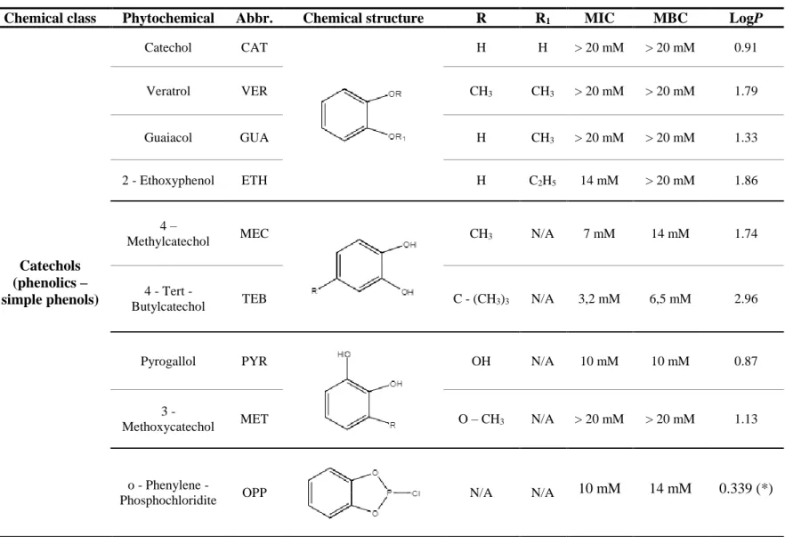
In this study, two main treatment approaches were presented concerning E. coli CECT 434 biofilms, in order to eradicate pre-established ones or to prevent their formation, using twelve selected phytochemicals and analogues (Catechol (CAT), Veratrol (VER), Guaiacol (GUA), 2 – Ethoxyphenol (ETH), 4 – Methylcatechol (MEC), 4 – Tert – Butylcatechol (TEB), Pyrogallol (PYR), 3 – Methoxycatechol (MET), o – Phenylene – Phosphochloridite (OPP), Retinol (RE), Retinyl palmitate (REP) and Retinoic acid (REA). Biofilm control and inhibitions studies were done using a microtiter plate assay followed by mass and metabolic activity quantification using crystal violet and alamar blue staining, respectively, as well as culturability in PCA plates and membrane integrity, using the Live/Dead® BaclightTM viability kit. It was also intended to elucidate the
effect of the selected compounds on some aspects related with biofilm formation, namely alteration of the cells surface hydrophobicity (CSH) and motility (swimming and swarming) inhibition.
Regarding the eradication of pre-formed E. coli biofilms, among the selected compounds, ETH, MEC, TEB, PYR and OPP exhibited the most interesting antibiofilm activity, concerning the reduction of the metabolic activity and culturability of the biofilm cells, as well as membrane disruption. The main additive interaction was observed when these compounds were combined with ciprofloxacin (CIP). A structure activity relationship (SAR) analyze was performed and an increase in the hydrocarbon side chain suggested the improvement of the activity for ETH and TEB on biofilm control. Moreover, ETH, MEC, TEB, PYR and OPP, which exhibited the best antibiofilm activity, can be considered “drug-like compounds”, according to the Lipinski’s rule of five.
Relatively to biofilm prevention, among the tested compounds, TEB and OPP were the most capable of reducing biomass production and E. coli sessile cells metabolic activity. TEB and PYR exhibited the highest
potential to induce membrane disruption and to reduce the culturability. E. coli essential biofilm forming features such as motility and CSH were also affected in the presence of the selected compounds, particularly, CAT, TEB and PYR (swimming); TEB, OPP and REA (swarming); and VER, ETH, MEC, TEB, PYR, MET, OPP, RE, REP and REA (decreased CSH). A global additive interaction of the selected compounds with CIP was also observed.
Overall, the results obtained highlight the potential of these selected plant-based molecules and analogues as an alternative treatment against human pathogenic bacteria, such as E. coli, or a complement to conventional antibiotic treatment. It is noteworthy that this is the first time that the antibiofilm activity of some of the catechols derivatives and retinoids was explored. Moreover, this study reinforces the potential of phytochemical products as a green and sustainable source of new antibiofilm products.
Resumo
As bactérias estão a adquirir resistência a antibióticos de tratamento convencional, o que está a gerar uma problemática relativamente à sua da população, considerando estratégias terapêuticas sustentáveis e duradouras direcionadas a bactérias patogénicas. Para além disso, é consensual que o modo de subsistência vantajoso de bactérias com a capacidade de formar biofilmes está relacionado com infeções crónicas e com o aumento da resistência ao tratamento com antibióticos devido às suas características específicas.
A Escherichia coli está relacionada com diversas doenças infeciosas, sendo uma das mais comuns a infeção do trato urinário (UTIs), associada à utilização de dispositivos médicos como cateteres. Esta bactéria é considerada um patogénico clínico importante relacionado com 70 a 95% das UTIs, que são infeções nosocomiais adquiridas em hospitais e outros estabelecimentos de cuidado de saúde. A frequência deste tipo de infeções estão relacionadas com o desenvolvimento de biofilmes nos cateteres urinários durante o período de cateterização. Para além disso, a dificuldade em erradicar UTIs associadas à formação de biofilme com antibióticos disponíveis no mercado, envolve um enorme custo para os pacientes e para o Serviço Nacional de Saúde. Neste sentido, o interesse em produtos naturais provenientes de plantas (fitoquímicos) para o tratamento clínico ressurgiram como uma alternativa e a sua popularidade tem vinda a aumentar na sociedade.
Neste estudo, duas perspetivas foram analisadas para o tratamento de biofilmes de E. coli CECT 434, de forma a erradicar biofilmes pré-estabelecidos ou prevenir a sua formação, usando doze fitoquímicos selecionados e seus análogos (Catechol (CAT), Veratrol (VER), Guaiacol (GUA), 2 – Ethoxyphenol (ETH), 4 – Methylcatechol (MEC), 4 – Tert – Butylcatechol (TEB), Pyrogallol (PYR), o – Phenylene – Phosphochloridite (OPP), Retinol (RE), Retinyl palmitate (REP) and Retinoic acid (REA). O controlo e inibição de biofilmes foi feito em microplacas, seguido de quantificação de massa e de atividade metabólica, usando coloração de cristal violeta e alamar blue, respectivamente, assim como de culturabilidade em placas de PCA e integridade da membrana celular, usando o kit de Live/Dead® BaclightTM. Foi também avaliado o
efeito dos compostos selecionados na hidrofobicidade da superfície celular (CSH) e a inibição da mobilidade (swimming and swarming).
Considerando a erradicação de biofilmes de E. coli pré formados, entre os compostos selecionados, ETH, MEC, TEB, PYR e o OPP exibiram a mais interessante atividade contra biofilmes, englobando redução da atividade metabólica e culturabilidade, bem como disrupção da membrana celular. Foi observada interação do tipo aditiva quando os compostos selecionados foram combinados com ciprofloxacina (CIP). A relação entre a estrutura e atividade, conhecida com SAR, foi analisada e é sugerido um aumento na cadeia lateral de hidrocarbonetos no caso do ETH e do TEB para um aumento da atividade. Para além disso, o ETH, o MEC, o TEB, o PYR e o OPP, que demostraram melhor atividade anti-biofilme, podem ser considerados “compostos drug-like” de acordo com a regra dos cinco do Lipinski.
No contexto de prevenção de biofilmes, o TEB e o OPP, entre os compostos selecionados, foram os mais capazes de reduzir a produção de biomassa e a atividade metabólica das células sésseis da E. coli. O TEB
e o PYR demonstraram maior capacidade na disrupção da membrana celular e redução na culturabilidade. Algumas características importantes na formação de biofilme da E. coli como a motilidade e a hidrofobicidade da superfície das células (CSH) foram afetadas na presença dos compostos selecionados, particularmente, no caso do CAT, do TEB e do PYR (swimming); do TEB, do OPP e do REA (swarming); e do VER, ETH, MEC, TEB, PYR, MET, OPP, RE, REP e do REA (diminuição da CSH). Do ponto de vista global, os compostos selecionados apresentaram uma interação do tipo aditiva quando combinados com a CIP.
Em geral, os resultados obtidos realçam o potencial destas moléculas com base em plantas e seus análogos como um tratamento alternativo contra bactérias patogénicas humanas, como o caso da E. coli, ou no complemento do tratamento convencional com antibióticos. É de notar que este é o primeiro estudo em que alguns destes catecóis e derivados e retinóis é explorado. Para além disso, este estudo reforça o potencial dos produtos fitoquímicos como uma fonte “verde” e sustentável de novos produtos com propriedades anti-biofilme.
Table of Contents
Acknowledgements ... iv Abstract ... v Resumo ... vii Table of Contents ... ix Tables list ... xiList of figures ... xii
Glossary ... xiv
Abbreviations ... xiv
Indexes ... xv
Chapter 1 | Work outline ... 1
1.1. Background and project presentation ... 1
1.2. Research objectives ... 1
1.3. Thesis organization ... 2
Chapter 2 | Literature review ... 5
2.1. Brief introduction to the biofilm lifestyle ... 5
2.2. Concern of biofilm development from a clinical perspective ... 5
2.3. Biofilm formation ... 8
2.3.1. Stages of biofilm development ... 8
2.4. Biofilm: a strategic microbial lifestyle against antimicrobial agents ... 10
2.5. Strategies to deal with biofilms ... 13
2.5.1. Biofilm prevention ... 13
2.5.2. Biofilm control ... 13
2.6. Phytochemicals as antimicrobials ... 16
Chapter 3 | Biofilm control potential of catechols and retinoids against Escherichia coli ... 21
3.1. Introduction ... 21
3.2. Materials and methods ... 22
3.2.1. Bacterial strain and growth conditions ... 22
3.2.2. Phytochemicals and analogues, EDTA, ciprofloxacin and solution conditions ... 22
3.2.3. Determination of the minimum inhibitory concentration (MIC) and minimum bactericidal concentration (MBC) ... 23
3.2.4. Biofilm formation ... 23
3.2.5. Dual combination of catechols with CIP ... 26
3.2.6. Statistical analysis ... 27
3.4. Results and discussion ... 28
3.4.1. Inhibitory and bactericidal activities of catechols and retinoids ... 28
3.4.3. Effect of the combined application of catechols with CIP on E. coli biofilms ... 40
3.5. Conclusions ... 45
Chapter 4 | Evaluation of the potential of catechols and retinoids on the prevention of Escherichia coli biofilms ... 47
4.1. Introduction ... 47
4.2. Materials and methods ... 48
4.2.1. Bacterial strain and growth conditions ... 48
4.2.2. Catechols, retinoids, EDTA and CIP ... 48
4.2.4. Biofilm prevention analysis ... 48
4.2.5. Motility assay ... 48
4.2.6. Microbial adherence to hydrocarbon (MATH) assay ... 49
4.2.7. Dual combination of catechols or retinoids with CIP ... 49
4.2.7. Statistical analysis ... 50
4.3. Results and discussion ... 51
4.3.1. E. coli biofilm prevention using catechols and retinoids ... 51
4.3.2. Combined effect of the phytochemicals and analogues with CIP... 57
4.4. Conclusions ... 61
Chapter 5 | Concluding remarks and future research perspective ... 63
5.1. General conclusions ... 63
5.2. Future work ... 64
List of tables
Table 1 - Infectious diseases/ site of infection and its associated biofilm pathogenic bacteria. ... 7
Table 2 - Phytochemicals with potential for biofilm prevention and control and their mechanism of action. 18 Table 3 - MIC and MBC values of EDTA and CIP against E. coli. ... 28
Table 4 - MIC and MBC values of CAT derivatives against E. coli. ... 29
Table 5 - MIC and MBC values of retinoids against E. coli. ... 30
Table 6 – Log CFU/cm2 reduction for the selected compounds for 1 and 24 h of exposure. ... 37
Table 7 - ∑ DCI scoring of CAT derivatives (MIC) with CIP (MIC) for biofilm mass reduction, metabolic inactivation culturability and membrane integrity. ... 42
Table 8 - Percentage of Log CFU/cm2 reduction after1 and 24 h of exposure to CIP alone and in dual combination at MIC, 5 × MIC and 10 × MIC with the catechols at MIC (ETH, MEC, TEB, PYR and OPP). ... 43
Table 9 - Percentage of Log CFU/cm2 reduction after 24 h of exposure to the selected phytochemicals and analogues. ... 54
Table 10 – Motility (swimming and swarming) (mm) of E. coli in the absence (control) and in the presence of the selected phytochemicals and analogues. ... 55
Table 11 – Hydrophobicity index (HI (%)) of E. coli cell surface in the absence and in the presence of the selected phytochemicals and analogues. ... 56
Table 12 – Log CFU/cm2 reduction after 24 h exposure to the dual combination of CIP with the selected phytochemicals and analogues. ... 59
Table 13 - ∑ DCI scoring of the selected phytochemicals and analogues (½ × MIC) with CIP (½ × MIC) for biofilm mass reduction, metabolic inactivation and culturability. ... 59
List of figures
Figure 1 - Sequence of the main steps involved in biofilm formation (adapted from Simões et al. 39 ... 9
Figure 2 - Schematic representation of permeability barrier and efflux pump in Gram-negative bacteria (adapted from Nikaido 43) ... 11
Figure 3 - Schematic representation of the differentiation inside a biofilm when treated with antibiotics. Bacteria is represented by oval figures with different colours (fast grower, slow grower, persisters and bacteria with differential gene expression), surrounded by extracellular matrix in a gradient of colours (red - aerobic and high nutrient concentrations; green – anaerobic and low nutrient concentration); dark dots represent the antibiotic molecules which dot density decreases toward the core of the biofilm (adapted from G. G. Anderson (2008) 46)... 12
Figure 4 - Schematic representation of the action of a phage capable of producing dispersin B. The multiplication of the phage and expression of dispersin B along the process are also ilustrated (adapted from Lu 77). ... 16
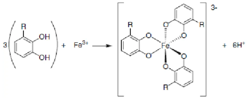
Figure 5 – Percentage of E. coli biofilm mass removal after 1 (■) and 24 h (■) of exposure to the catechols/retinoids and EDTA at MIC, 5 × MIC and 10 × MIC with the exception for CAT, VER, GUA and MET that were tested at 20 mM. Mean values ± standard deviation are illustrated. Bars marked with * are statistically different from the control (P < 0.05). ... 32 Figure 6 - Percentage of E. coli biofilm metabolic inactivation after 1 (■) and 24 h (■) of exposure to the catechols/retinoids and EDTA at MIC, 5 × MIC and 10 × MIC with the exception of CAT, VER, GUA and MET that were tested at 20 mM. Mean values ± standard deviation are illustrated. Bars marked with * are statistically different from the control (P < 0.05). ... 32 Figure 7 – Schematic representation of the expected octahedral geometry of general iron – polyphenol complexes. R=H for the basic structure of catechol. (Adapted from Perron et al. 139) ... 33
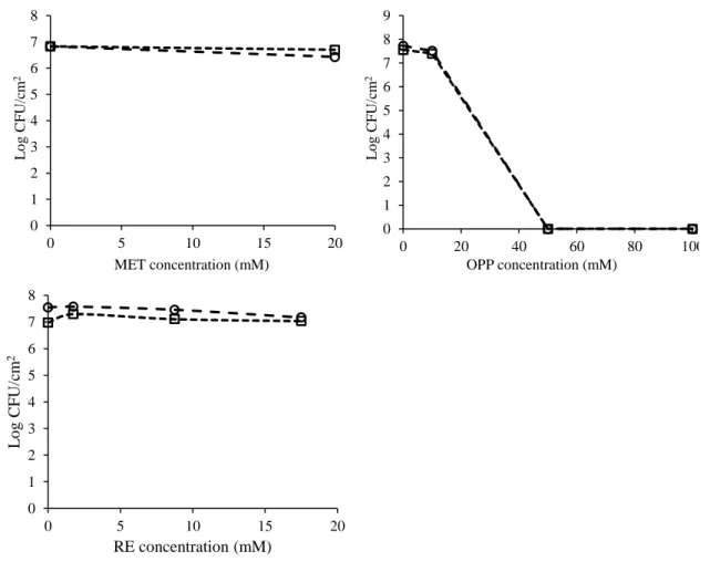
Figure 8 - Effect of the selected compounds (EDTA, CAT, VER, GUA, ETH, MEC, TEB, PYR, MET, OPP and RE) on the culturability of E. coli biofilm cells at different concentrations and times of exposure of 1 ( ) and 24 h ( ). The results are presented as the Log CFU/cm2 as a function of the concentration of the
antimicrobial compounds. Mean values of duplicates are presented. ... 36 Figure 9 – Percentage of E. coli biofilm cells stained with PI (%) after 1 (■) and 24 h (■) of exposure to the selected compounds at MIC, 5 × MIC and 10 × MIC in the case of EDTA, ETH, MEC, TEB, PYR, OPP and RE and at 20 mM for CAT, VER, GUA and MET. Mean values ± standard deviation are illustrated. Bars marked with * are statistically different from the control (P < 0.05). ... 37 Figure 10 – Examples of epifluorescence microscope images of E. coli sessile cells stained with the Live/Dead® BacLightTM viability kit. A – Control; B – Sessile cells exposed to PYR (10 × MIC); C – Sessile
MIC, respectively; G, H and I - Sessile cells exposed to TEB at MIC, 5 × MIC and 10 × MIC, respectively. ... 38 Figure 11 – Percentages of E. coli biofilm mass removal after 1 (■) and 24 h (■) of exposure to CIP alone and in dual combination at MIC, 5 × MIC and 10 × MIC with ETH, MEC, TEB, PYR and OPP at MIC. Mean values ± standard deviation are illustrated. Bars marked with * are statistically different from the control (P < 0.05). ... 41 Figure 12 – Percentages of E. coli biofilm metabolic inactivation after 1 (■) and 24 h (■) of exposure to CIP alone and in dual combination at MIC, 5 × MIC and 10 × MIC with ETH, MEC, TEB, PYR and OPP at MIC. Mean values ± standard deviation are illustrated. Bars marked with * are statistically different from the control (P < 0.05). ... 41 Figure 13 - Examples of epifluorescence microscope images of E. coli sessile cells stained with the Live/Dead® BacLightTM viability kit. A – Control; B – Sessile cells exposed to CIP (10 × MIC); C – Sessile
cells exposed to TEB (10 × MIC). ... 42 Figure 14 – Percentage of E. coli biofilm cells stained with PI (%) after 1 (■) and 24 h (■) of exposure to CIP alone and in dual combination at MIC, 5 × MIC and 10 × MIC with the catechols at MIC (ETH, MEC, TEB, PYR and OPP). Mean values ± standard deviation are illustrated. Bars marked with * are statistically different from the control (P < 0.05). ... 43 Figure 15 - Percentages of E. coli biofilm mass reduction after 24 h (■) of exposure to the selected phytochemicals and analogues at ¼ × MIC and ½ × MIC. Mean values ± standard deviation are illustrated. Bars marked with * are statistically different from the control (P < 0.05). ... 52 Figure 16 - Percentages of E. coli biofilm metabolic inactivation after 24 h (■) of exposure to the selected phytochemicals and analogues at ¼ × MIC and ½ × MIC. Mean values ± standard deviation are illustrated. Bars marked with * are statistically different from the control (P < 0.05). ... 52 Figure 17 - Examples of epifluorescence microscope images of E. coli cells stained with the Live/Dead®
BacLightTM viability kit. A – Control; B – Cells exposed to RE (¼ × MIC); C – Cells exposed to PYR (½ ×
MIC). ... 53 Figure 18 - Percentages of E. coli biofilm cells stained with PI after 24 h (■) of exposure to the selected phytochemicals and analogues at ¼ × MIC and ½ × MIC. Mean values ± standard deviation are illustrated. Bars marked with * are statistically different from the control (P < 0.05). ... 54 Figure 19 - Percentages of E. coli biofilm mass removal after 24 h (■) of exposure to CIP alone and in dual combination at ¼ × MIC and ½ × MIC with the selected phytochemicals and analogues at ½ × MIC. Mean values ± standard deviation are illustrated. Bars marked with * are statistically different from the control (P < 0.05). ... 58 Figure 20 - Percentages of E. coli biofilm metabolic inactivation after 24 h (■) of exposure to CIP alone and in dual combination at ¼ × MIC and ½ × MIC with the selected phytochemicals and analogues. Mean values ± standard deviation are illustrated. Bars marked with * are statistically different from the control (P < 0.05). ... 58
Glossary
Abbreviations
Abbr. Abbreviation
AB Alamar blue
ADME Absorption, distribution, metabolism and excretion
AI Autoinducers
CAT Catechol
CFU Colony forming units
CIP Ciprofloxacin
CRBSI Catheter-related bloodstream infection CSH Cell surface hydrophobicity
CV Crystal violet
DMSO Dimethyl sulfoxide
EDTA Ethylenediamine tetraacetic acid ESBL Extended spectrum beta-lactamases
ETH 2 – Ethoxyphenol
EPS Extracellular polymeric substance
GUA Guaiacol
LBB Luria bertani broth LogP Partition coefficient
LPS Lipopolysaccharides
MATH Microbial adherence to hydrocarbon MBC Minimum bactericidal concentration
MEC 4 – Methylcatechol
MET 3 – Methoxycatechol
MIC Minimum inhibitory concentration
MHA Mueller-Hinton agar
MHB Mueller-Hinton broth
NA No activity
N/A Not applicable.
NG No growth
OD Optical density
OM Outer membrane
OPP o – Phenylene – Phosphochloridite PBS Phosphate buffered saline
PCA Plate count agar
PI Propidium iodide
PS Polystyrene
PYR Pyrogallol
RE Retinol
REA Retinoic acid
REP Retinyl Palmitate
RMA Resistance modifying agents ROS Reactive oxygen species
RT Room temperature
SAR Structure activity relationship TEB 4 – Tert – Butylcatechol USA United States of America
UVC Ultraviolet C
VER Veratrol
Indexes
HI (%) Hydrophobicity index ∑ DCI Dual combination index
C
hapter
1
_____________________________________________________________________________
Work outline
1.1. Background and project presentation
Antibiotic resistance has been increasingly reported in a wide variety of bacteria of clinical importance. 123 A significant factor contributing to the increase of resistance to antibiotics
and host defense mechanisms of many pathogenic bacteria is their ability to form biofilms, which are involved in several device-related and other chronic infections. 4 5 6 Moreover, the present
therapeutic strategies for eradication of acute epidemic bacterial diseases are not effective against biofilm diseases. 5 Therefore, the use of phytochemicals (plant secondary metabolites), is being
increasingly described as an alternative strategy for clinical purposes, by its use alone or combined with antibiotics for drug repurposing as they can potentiate the activity of the currently used antibacterial agents. 789
1.2. Research objectives
The main objective of the present study is to evaluate the activity of plant-based molecules and its analogues on the growth of E. coli in planktonic and biofilm state, which has clinical concern. The plant-based molecules and analogues used in this study belong to the class of phenolics (simple phenols – catechols) and retinoids (vitamin A and analogues). Phenolics have been identified in both higher and edible plants and are commonly related to plants defence against ultraviolet radiation, aggression by pathogens and also to antioxidant properties. 10 11
Retinol can only be found among animal tissues. However, provitamin A carotenoids, retinol precursors that can be converted into retinol, can be found in edible plants such as fruits and vegetables. 12 13 Provitamin A carotenoids are commonly related to its antioxidant activity in
plants. 14 As these compounds are part of human and animal diets, it is important to study their
antimicrobial activity against pathogenic microorganisms and their biofilms.
E. coli is considered a clinical opportunistic pathogen involved in severe infections, namely
those related to urinary catheter devices, and has the capacity to become resistant to multidrugs.
1516 The plant-based molecules and analogues growth inhibitory and bactericidal activities against
E. coli in planktonic state were, firstly, assessed and important information is given by these two
Subsequently, the plant-based molecules and analogues were evaluated on the prevention and control of biofilms and was characterized in terms of produced and removed biomass, sessile cells culturability, metabolic inactivation and membrane integrity. Methods concerning some aspects of the phytochemicals mode of action against E. coli and biofilm forming were evaluated, including motility (swimming and swarming) and cell surface hydrophobicity, due to its importance in preventing cells initial adhesion and biofilm formation.
1.3. Thesis organization
This thesis is divided in 5 Chapters. In Chapter 1, the context, main goals and the guideline of the work described in the next chapters are presented briefly.
Chapter 2 presents a short review of the literature. The biofilm lifestyle, including the stages involved in its development, namely initial adhesion to a substrate, differentiation into micro colony formation, biofilm maturation and the final dispersion and colonization of new niches are discussed. The importance of clinical biofilms and its related pathogenic bacteria is also summarized, regarding biofilm infection in living and non-living tissues such as medical devices. A brief analysis of the intrinsic and extrinsic biofilm related mechanisms of defence to adverse environment conditions in bacteria is provided. An introduction to the large array of phytochemicals, its history evolution in therapeutics and the motivation for seeking new antimicrobial agents in plant-based molecules is also discussed. The mechanism of action of some phytochemicals belonging to different classes, including phenolics as well as their related studied antibacterial and antibiofilm activities against pathogenic bacteria, is also addressed.
The Chapter 3 focuses on the potential of the selected plant-based molecules and analogues in E. coli antibacterial and antibiofilm control. The selected compounds include Catechol (CAT) and its derivatives; Veratrol (VER), Guaiacol (GUA), 2–Ethoxyphenol (ETH), 4–Methylcatechol (MEC), 4–Tert–Butylcatechol (TEB), Pyrogallol (PYR), 3–Methoxycatechol (MET) and o – Phenylene – Phosphochloridite (OPP) and vitamin A or Retinol (RE) and two related compounds; Retinoic acid (REA) and Retinyl palmitate (REP). EDTA was also evaluated as a positive control. The methods applied are briefly described comprising minimum inhibitory and bactericidal concentration, biomass removal (crystal violet staining), metabolic inactivation (Alamar blue assay), biofilm cells culturability in PCA plates and propidium iodide uptake for membrane integrity assessment. The compounds with better performance were selected for dual combination with the antibiotic Ciprofloxacin (CIP).
Chapter 4 focuses on the potential of the selected phytochemicals and analogues in preventing E. coli biofilm formation. The methods used are described briefly. The prevention was analysed in terms of biomass reduction (crystal violet staining), metabolic inactivation (Alamar blue assay), biofilm cells culturability in PCA plates and propidium iodide uptake for membrane
integrity assessment. E. coli motility and cell surface hydrophobicity were also evaluated in the presence of the selected plant-based molecules and analogues, as these properties are important to consider in biofilm initial formation and subsequently development.
At last, Chapter 5 presents an overview of all thesis, with emphasis on the main conclusions and perspectives for further research.
C
hapter
2
_____________________________________________________________________________
Literature review
2.1. Brief introduction to the biofilm lifestyle
Biofilms are communities of sessile cells attached to a surface enclosed in an extracellular matrix in which one or more species can be involved. 1718 These structures are formed when
free-floating bacteria (planktonic bacteria) segregate endogenous signalling molecules in order to coordinate the attachment in a process called quorum sensing. 19 Biofilms can be attached to a
surface, suspended as flocks in fluids or even pellicles in air-liquid interfaces and present a high density of cells that reaches 107 cells/cm2, 20 and take in average 10 days to reach structural
maturity. 21 The polymeric matrix surrounding the cells is mostly composed by extracellular
DNA, proteins and polysaccharides. Polysaccharides are the major component of the matrix and are responsible to mediate most of cell-cell and cell-surface interactions. 22 Furthermore,
non-cellular materials can be part of the extranon-cellular polymeric matrix depending on the surrounding environment such as mineral crystals, corrosion particles, clay or silt particles, or even blood components. 23 Biofilms are able to proliferate in many surfaces such as in solid and liquid
surfaces as well as in soft tissue in living organisms. 22 Moreover, the entire biofilm complex is
usually attached to the surface but that is not necessary. 24
Biofilms usually present great resistance against conventional antibiotics, microbicidal agents and host immune responses 19, as, in sessile state, bacteria have different phenotypes
regarding growth, gene expression and protein production. 18 Some examples of biofilms are
dental plaque, slimy coating in tanks or even algal mats on water. Although biofilms are usually related to infection and disease, they can also be beneficial treating sewage, industrial waste and contaminated soil. Biofilm structure allows single-cell organisms to take part on a group with a multicellular lifestyle which is a great microorganism’s strategy for survival in adverse environments but, as they are getting more resistant to antimicrobial agents, the process of biofilm removal is getting more difficult. Therefore, biofilms are starting to play an important role in many infections as well as a big concern in food industries. 2225
2.2. Concern of biofilm development from a clinical perspective
The development of biofilms is a major concern in therapeutics and there are many ways from which biofilms can become a real issue to human health. For instance, biofilms can develop
in drinking water systems and can be a serious risk to public health if pathogenic forms of microorganisms, such as E. coli, Pseudomonas spp or Aeromonas, are present. When dealing with aquatic environments, bacteria is usually in the form of a biofilm, instead of planktonic, in order to remain attached to a surface, preventing from going with the flow, as long as the environment is suitable for bacterial survival. 26 Biofilms can develop in many different locations which include
industrial or potable water system piping, and even natural aquatic environments. 23
Furthermore, biofilms are becoming a big concern in food industries including brewing, seafood processing, dairy processing and meat processing, since they are able to resist disinfectant treatment and reach the population causing infections. Besides, the microbial control strategies that are used nowadays are not efficient to completely eradicate pathogenic bacteria without compromising the product quality. 25 For instance, the treatment of lettuce with chlorine and
sodium hypochlorite was analysed and it was suggested that procedure wasn’t efficient enough to inactivate E. coli in the vegetable. 27 Some other examples of pathogens which can be present
in food are Salmonella spp. in poultry processing and Listeria monocytogenes in dairy processing.
22
Human societies have been infected by a lot of diseases caused by planktonic cells before the appearance of vaccines and antibiotics, such as the case of Yersinia pestis. 27 According to
that, most of the research that has been done in order to discover the currently available antibiotics, was based on the planktonic state of bacteria. 28 However, 99 % of all bacteria exist in the form
of biofilms, and only 1% in planktonic state. 29 Furthermore, 65 % of microbial infections are
associated with biofilms 29, and around 80% of persistent bacterial infections in the USA are also
associated to biofilm formation 25 such as wound infections due to the skin bacterium
Staphylococcus epidermidis, bacterial prostatitis due to E. coli and other Gram-negative bacteria
that are able to establish biofilm 27, or urinary catheter infections due to E. coli, Gram-negative
rods or Pseudomonas aeruginosa. 30 These biofilm infections can be caused by single species or
by a mixture of species. Biofilm infections develop preferentially on inert surfaces, dead tissue or in medical devices/implants. They can also form in living tissue such as native valve endocarditis by Viridans streptococci. 232627 An example of medical implant associated with biofilm infection
is prosthetic heart valves endocarditis, due to Staphylococcus aureus 23. These types of infection
caused by biofilms tend to be chronic as they persist despite antibiotic treatment, innate and adaptive immune responses mechanisms, and inflammatory responses from the host. 28296 There
are no current drugs that specifically target sessile bacteria in biofilms, but there are some approaches being developed. As said, apart from surgical intervention, antibiotics are the only current option to treat biofilm infections. 28 Antibiotic treatment is able to reverse some symptoms
caused by the released planktonic bacteria, supress growing and spread of the biofilm but cannot eradicate the infection. 272829 The application of disinfectants can be used in accessible areas, like
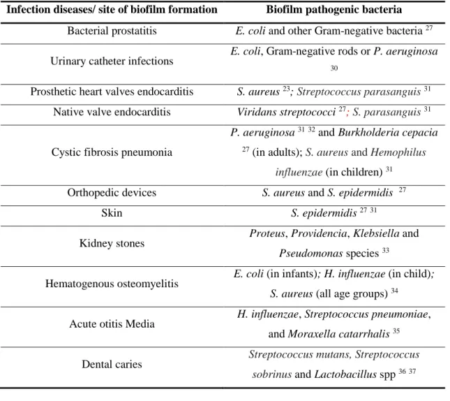
catheters, but there is an urgent need in anti-biofilm drugs for inaccessible areas. 28 In Table 1,
some infectious diseases and their associated biofilm pathogenic bacteria are presented:
Table 1 - Infectious diseases/ site of infection and its associated biofilm pathogenic bacteria.
Infection diseases/ site of biofilm formation Biofilm pathogenic bacteria Bacterial prostatitis E. coli and other Gram-negative bacteria 27
Urinary catheter infections E. coli, Gram-negative rods or P. aeruginosa
30
Prosthetic heart valves endocarditis S. aureus 23; Streptococcus parasanguis 31
Native valve endocarditis Viridans streptococci 27; S. parasanguis31
Cystic fibrosis pneumonia
P. aeruginosa 3132 and Burkholderia cepacia 27 (in adults); S. aureus and Hemophilus
influenzae (in children) 31
Orthopedic devices S. aureus and S. epidermidis 27
Skin S. epidermidis 2731
Kidney stones Proteus, Providencia, Klebsiella and
Pseudomonas species 33
Hematogenous osteomyelitis E. coli (in infants); H. influenzae (in child);
S. aureus (all age groups) 34
Acute otitis Media H. influenzae, Streptococcus pneumoniae, and Moraxella catarrhalis 35
Dental caries Streptococcus mutans, Streptococcus sobrinus and Lactobacillus spp 3637
For example, as described by Parsek 33, 15 to 20 % of kidney stones occur due to urinary
tract infection, where stones are produced by simultaneous action of infecting bacteria, such as
Proteus, Providencia, Klebsiella and Pseudomonas species, and mineral substrates from the
urine. Bacteria was found outside and inside kidney stones in microcolonies encased in a matrix composed by polysaccharides and crystallized minerals (struvite and carbonate-apatite). Furthermore, it was observed that encased bacteria inside the stones would remain viable after prolonged antibiotic treatment, and infection was not eradicated till surgical removal of the stone. Moreover, it was reported by Lauren O. Bakaletz 35 that biofilms may play an important role in
acute otitis media, which affects mostly children under 5 years old. Bacteria such as H. influenzae,
S. pneumoniae and M. catarrhalis, may be the cause of antibiotic failure in treatment and chronic
2.3. Biofilm development
The development of a bacterial biofilm is affected by many factors such as the bacterial strains involved, the material properties of the surface, environment parameters like pH, nutrients, and temperature as well as the hydrodynamic conditions since bacteria respond phenotypically to different shear stress. 2520 Furthermore, biofilms grown in laminar shear or even static conditions
detach and disperse easier than in high shear systems 38 , due to the fact that forces of biofilm
extracellular polymeric substance (EPS) formed during the development are weaker. 20 The main
mechanisms involved in biofilm formation are: rearrangement of attached cells through bacterial motility by pili structures which allows surface aggregation; binary division of attached cells, as new cells spread in different directions to form clusters; and recruitment of planktonic cells. 21
2.3.1. Stages of biofilm development
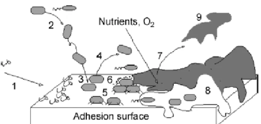
The formation of a biofilm is a coordinated and reversible process and it comprehends an organized sequence of steps (Figure 1) 1839:
1. Preconditioning of adhesion by the presence of some macromolecules suspended on the medium or coated on the surface;
2. Transport of planktonic cells from the medium to the surface; 3. Adsorption of cells to the surface for a limited period of time; 4. Release of reversibly attached cells;
5. Irreversibly attachment of the bacterial cells; 6. Production of cell-cell signalling molecules; 7. Substrate transport to the biofilm;
8. Substrate metabolism by the biofilm cells and consequent products transport out of the biofilm; as well as cellular growth, replication and production of extracellular polymeric compounds;
The initial attachment of the bacteria on the surface can be either active, if the bacteria has motility, or passive, in the case of gravitational transport, sedimentation or liquid flow. 25 This
stage of development depends mostly on the physiochemical properties of the planktonic bacterial surface and its interactions with the surface of the substratum such as: van der Walls forces, hydrophobic, electrostatic and London dispersion forces. 40 The attachment surface properties
also present an important role in biofilm formation such as texture (smooth or rough surface), surface charge, hydrophobicity and conditioning film comprising molecules, as organic substances, that will enhance the attachment of the planktonic bacteria. 25 At this point, the
planktonic bacteria produces a small quantity of EPS 25, and this early step of development usually
requires motility mediated by surface structures as flagella that enables reversible attachment and cell motility relevant for biofilm architecture, for example in E. coli. 41 Flagella may be important
to overcome repulsive forces with the substrate surface. 23 As stated, there is not much
morphological differentiation that leads to biofilm formation at this point.
On the next stage of biofilm formation, the weak interactions turn into a permanent bonding with the increase of EPS, important for strengthening the bond between the bacteria and the surface, stabilizing the colony. 25 At this stage, for irreversible attachment of bacteria, flagella
synthesis is repressed and adhesive organelles are produced, such as curli fimbriae or type I fimbriae in E. coli. 41 As stated by Rodney M. Donlan 23, the rate and degree of attachment is
much dependent on cell surface hydrophobicity, in which fimbriae, that contain many hydrophobic amino acid residues, contribute to this property. Hydrophobicity enables the cell to overcome electrostatic repulsion between cell and substrate surface. Once bacteria irreversibly attach to the surface, they start to form micro colonies. Planktonic bacteria are also recruited from the medium, thanks to cell-cell communication (quorum sensing). Micro colonies allows substrate exchange between different species within the biofilm and mutual product removal. 25 Once the
Figure 1 - Sequence of the main steps involved in biofilm formation (adapted from Simões et
biofilm micro-colony is stabilized, there is a transition to the biofilm maturation in which significant changes occur. In order to reach structural maturity, 10 days are required at least. 20
These changes result in the formation of a 3-dimensional complex with mushroom-shaped or flat and permeate structure, with a network of channels and pores. 25 The network of channels allows
the transport of water, bacterial end-products, nutrients, enzymes, metabolites and oxygen. These channels have gradients of chemicals and ions between micro-zones which provides the power to move the compounds through the biofilm. 22
Once the biofilm gets to their final step of maturation, biofilm cells revert to their first stage, planktonic form. 25 This final stage is very important to the microorganism life cycle
contributing to survival, spread and disease transmission. 18 Biofilms show a very dynamic
environment at this point in which bacterial cells continuously grow and detach; and usually land on surfaces to initiate new colonization. The causes of biofilm detachment can be either external perturbations like fluid shear, or internal processes like endogenous enzymatic degradation or quorum sensing, the release of EPS or other surface-binding protein. 2523 This step of the biofilm
life cycle is very important as a strategy to colonize new niches before space and nutrients become limited. 20 As described by Hall-Stoodley et al. 20, two main types of dispersion can be considered:
dispersal through detachment into the fluid or dispersal over the solid surface. Furthermore, dispersal can be through self-propelled locomotion by swimming, sliding or twitching motility; or through fluid-driven motility by clumping, rippling or rolling. Despite the fact that bacteria with self-propelled locomotion have the ability to choose the direction of movement, they have the disadvantage of being unprotected. On the other hand, cells in clumps disperse along with the fluid but they are protected encased in the biofilm structure. 20
2.4. Biofilm: a strategic microbial lifestyle against antimicrobial agents
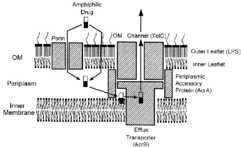
Intrinsic antibiotic resistance of pathogens can be associated to the existence of species-specific bacterial intrinsic resistome (antibiotic resistance genes). 42 Intrinsic antibiotic resistance,in Gram-negative bacteria, is mostly related with two mechanisms: cellular impermeability by the outer membrane (OM) and cell wall; and multidrug efflux pumps that actively pump out drug molecules, such as RND (resistance-nodulation-division) type transporter as AcrB in E. coli, (Figure 2). 43 However, antibiotic resistance can also be obtained through de novo mutations or
acquisition of resistance genes, due to antibiotic exposure and horizontal gene transfer. 42 44
Acquisition of genes by susceptible bacteria from resistant bacteria can happen through conjugation, transformation or transduction. 44 These acquired resistance genes allow bacteria to
produce enzymes that destroy the antibiotic, express efflux systems, modify drug’s target site or to produce alternative metabolic pathways that bypasses the action of the drug. 44 For instance,
some E. coli strains have developed resistance to CIP, trimethoprim, aminoglycosides, amikacin and produce ESBL’s (extended spectrum beta-lactamases). 4445
These mentioned resistance factor are mostly associated to specific genetic factors. However, biofilm formation allows increased resistance to antibiotics and other environment conditions due to its structural nature and the fact that sessile cells produce resistance against antimicrobials. 223929 This allows biofilms to be 100-1000 times less susceptible than equivalent
planktonic bacteria, in terms of minimum inhibitory concentration and minimal bactericidal concentration. 22 39 Furthermore, biofilm infections are tolerant to innate and adaptive immune
responses, tolerant to clinically dosing antibiotics and cause chronic infections (persistent pathoLogy), in contrast to acute planktonic bacterial infections. 29 There are 6 main factors that
confer biofilms their antimicrobial resistance:
1. Biofilm matrix acts as a diffusion barrier which reduces the penetration of antibiotics into the biofilm and prevents them from reaching the target bacterial cells. 22 39 29 Therefore, a
decrease in the level of antibiotics reaching bacteria would increase resistance, by leading to the dead of the outer layer of the biofilm while bacteria within the biofilm would be able to resist and adapt against the stress. 46
2. Microenvironments inside biofilms, as nutrient limitation, which leads to slow or no growth of bacteria that reduces antimicrobial activity. 223929 For example, oxygen limitation has
been investigated and numerous studies have shown the presence of hypoxic zones inside biofilms, which decreases metabolic activity. 46 Slow-growing bacteria are not much susceptible
to antimicrobial agents .27 For instance, β-lactams as cephalosporins and floroquinolones have
shown to kill non growing cells but are much more effective when dealing with fast growing cells;
29
Figure 2 - Schematic representation of permeability barrier and efflux pump in Gram-negative
3. Some bacteria inside biofilm differentiate into persistent cells. 223929 This type of cells
are in a state of dormancy, metabolic inactive, and are usually tolerant to antibiotics, comprising, approximately, 1% of biofilms 47. As stated by Iris Keren et al. 48, there was differentially
overexpression of, approximately, 300 genes in isolated E. coli persistent cells in response to stress such as heat and cold shock. Moreover, there was the expression of several messages coding for proteins that block cellular functions, as RMF (inhibitor of translation) and UmuDC (inhibitor of replication). These facts are consistent with the proposed survival function of persistent cells against harsh conditions including tolerance to antibiotics;
4. Chemical interaction between the antimicrobials and biofilm constituents, such as the polymeric matrix; 2239
5. Induced resistance factors. Mutation frequency in biofilm growing bacteria is much higher when compared to planktonic bacteria and there is an increased horizontal gene transmission. This may explain the multidrug resistant character of sessile bacteria: by producing enzymes that neutralize or degrade antibiotics 22 39 29; antibiotic targets with low affinity and
overexpression of efflux pumps. 29 Furthermore, in stratified biofilms, bacteria on the surface may
be killed by antibiotics but slow growing bacteria may produce differential gene expression induced by lower concentration of antibiotics. 29 As stated by Costerton 27, this induced
heterogeneity is an important strategy for survival. As there is a variety of metabolic states, at least some bacteria would survive to a metabolic directed attack.
6. Programed apoptosis when subjected to antimicrobials, which would allow the community to grow fast after the adverse conditions due to the nutrients release of death cells. 39
Figure 3 shows a schematic representation of the differentiation inside a biofilm when treated with antibiotics:
Figure 3 - Schematic representation of the differentiation inside a biofilm when treated with
antibiotics. Bacteria is represented by oval figures with different colours (fast grower, slow grower, persisters and bacteria with differential gene expression), surrounded by extracellular matrix in a gradient of colours (red - aerobic and high nutrient concentrations; green – anaerobic and low nutrient concentration); dark dots represent the antibiotic molecules which dot density decreases toward the core of the biofilm (adapted from G. G.
2.5. Strategies to deal with unwanted biofilms
2.5.1. Biofilm prevention
One of the strategies applied against pathogenic biofilms is to prevent its formation by killing bacteria in planktonic state before biofilm development. At this point, bacteria is still vulnerable to antibiotics and host defence mechanisms. However, it is very difficult to predict and detect initial biofilm formation due to its little inflammatory responses in a clinical point of view, which makes this a suitable strategy for implants and catheters where antimicrobials can be coated to the surface, but not for current treatment. 28 As described by Hendriks et al. 49, the use of
gentamicin has been proposed in bone cements for total hip arthroplasties fixation, in order to act as a prophylactic drug at the time of surgery and as an agent against infection for the years after. Furthermore, it has been stated by O’Grady et al. 50 that, certain catheters can be coated or
impregnated with antimicrobial/antiseptic agents such as chlorhexidine/ silver sulfadiazine or minocycline/ rifampin, which reduced the potential risk of catheter-related bloodstream infection (CRBSI).
However, the antimicrobial compounds on the surface will eventually run out. In addition, the host-derived glycoproteinaceus film (containing fibronectin, collagen, etc), tissue debris and contaminants formed over the surface can prevent the antibiotic’s release. 28
Accessible biofilms such as in catheters can be treated with ultraviolet C (UVC) light for prevention and killing of bacterial biofilms. In fact, Bak et al. 51 have demonstrated the possibility
of catheter disinfection by radiation with UVC light, and created a device to do so. This device was shown to be effective against E. coli, S. aureus and P. aeruginosa.
2.5.2. Biofilm control
2.5.2.1. Biofilm weakeningAnother approach is to weaken the bacteria within biofilm by neutralizing their virulence factors and their biofilm-forming properties. This is done by interfering with virulence factors, quorum sensing, and iron metabolism. 28
Interfering with virulence factors means to develop specific antibodies or compounds that bind to virulence factors, so that the pathogen lose its virulence and is eradicated by the host defence system or antibiotics. 28 For instance, it was showed by Oana Ciofu et al. 52, that antibodies
(aβab) against the chromosomal β-lactamase of P. aeruginosa showed to improve the treatment with ceftazidime of resistant P. aeruginosa in a rat model with chronic lung infection.
Bacteria communicate to each other through chemical signalling molecules, which are called autoinducers (AI). 53 This process is called quorum sensing and allows bacteria to control gene
signals are produced while bacteria grows until a threshold of concentration, resulting in the activation or repression of specific genes. This concentration is achieved when a sufficient number of cells, nominated as Quorum, is present. 55 There are inducers specific for
Gram-negative bacteria, for Gram-positive bacteria and inducers that can be used in both. For instance, Gram-negative bacteria usually produces acylated homoserine lactone (AHL) as AI. 56
Many features are regulated by quorum sensing including symbiosis, virulence factors, pathogenicity, antibiotic production and biofilm formation. 5357 As bacteria is less virulent, the
immune system of the host is able to kill bacteria, as well as making bacteria more susceptible to antibiotics. 28 Therefore, inhibition of quorum sensing is a great strategy for biofilm weakening.
For instance, as stated by Murugan et al. 58, the methanol extract of Andographis paniculata has
shown to exhibit biofilm inhibitory activity and quorum sensing inhibition against clinical isolates of P. aeruginosa in patients with cystic fibrosis in vitro. Furthermore, curcumin, a phenolic compound from Curcuma longa has shown, by Rudrappa et al. 59, to inhibit virulence factors of
P. aeruginosa such as biofilm formation, pyocyanin biosynthesis, elastase/protease activity and
acyl homoserine lactone (HSL) production, and quorum sensing inhibition which makes curcumin as a supplemental molecule for P. aeruginosa infections treatment. Moreover, Salicylic acid, a signal molecule in plants, has shown to shut down the virulence genes of Agrobacterium
tumefaciens. 60
Iron metabolism is a key vulnerability for pathogenic bacteria since they require Fe for growth and it is critical for pathogenic infections. 61 Therefore, the substitution of Fe for another
metal, such as Ga due to its similarity, has shown to inhibit the growth of P. aeruginosa, its biofilm formation, kill planktonic and sessile bacteria in vitro. 62 There are evidences that many
biological systems are unable to distinguish between gallium and iron. 63
2.5.2.2. Biofilm disruption
Disruption of the biofilm may revert the tolerance of bacterial biofilms to antimicrobial agents since bacteria return to their vulnerable planktonic state. Since the polymeric matrix stabilizes the biofilm, targeting the matrix compounds production or its disruption destabilizes the biofilm with the release of planktonic bacteria, which makes it vulnerable to antibiotics’ action and host immune-system. 28
Disruption of the biofilm can be accomplished by the application of ultrasounds into the pathogenic biofilm. For instance, the use of ultrasounds has been tested in biofilms related to chronic rhinosinusitis by Young 64. In this study, patients demonstrated a significant improvement
in chronic rhinosinusitis symptoms after six sessions of pulsed ultrasound therapy. Therefore, treatment with ultrasound alone or combined with antibiotics should be further analysed as a potential treatment.
Another mechanical technic for biofilm disruption is sonication. According to Bjerkan 65,
sonication was shown to efficiently and reliably dislodge biofilm bacteria from S. aureus,
Enterococcus faecalis and Propionibacterium acnes. Furthermore, sonication can also be used
for improving microbioLogical diagnosis of periprosthetic joint infection, by sonication of the implant and analysing the remaining fluid by multiple PCR. This technic has proven to be useful especially in patients receiving antibiotics, as cultures have limited sensitivity. 66
The use of enzymes is another strategy that can be applied to degrade the biofilm polymeric matrix, by targeting matrix components such as polysaccharide, eDNA and proteins, with the release of antibiotic susceptible planktonic bacteria. 17 For example, Dispersin B is able to degrade
poly-N-acetylglucosamine, biofilm matrix polysaccharide that mediates attachment of
A. actinomycetemcomitans cells, intercellular adhesion and resistance against antimicrobials and
host immune system. 67 Furthermore, it was suggested by Chaignon 68, that a treatment with
Dispersin B capable of degrading poly-N-acetylglucosamine in biofilm matrix followed by a protease (proteinase K or trypsin) would eradicate staphylococcal strains biofilms. Dispersin B has also shown to degrade the biofilm matrix in several bacterial species such as E. coli and
Pseudomonas fluorescens. 28
Alginate lyase can also be applied in biofilms of alginate-producing strains of
P. aeruginosa as it would disrupt the biofilm matrix and therefore, make the bacteria more
susceptible to antibiotic treatment such as gentamicin or ceftazidime. 69
Another interesting technic for biofilm disruption is based on the natural dispersal of antibiotic vulnerable planktonic bacteria from mature biofilms in the last stage of biofilm formation. This occurs in response to carbon availability, when bacteria needs to search for new sources of nutrients. 28 For instance, it was demonstrated that nitric oxide (NO), a signalling
molecule in biological systems, causes dispersal of P. aeruginosa biofilm bacteria, and was suggested that combined exposure to NO and antimicrobial agents would be a new strategy for persistent P. aeruginosa biofilm infections. 70 NO was also shown to cause dispersal in several
strains of Gram-negative and Gram-positive bacteria, such as Vibrio cholerae, E. coli,
S. epidermidis. 71 Moreover, cis-2-decenoic acid, a short chain fatty-acid messenger produced
during growth, was demonstrated to induce dispersion of a mature biofilm and inhibiting biofilm development at a nanomolar concentration. It was shown to disperse biofilms of a multitude of microorganisms such as E. coli, P. aeruginosa, S. aureus and Candida albicans. 72 In addition, it
is possible to induce biofilm dispersal by interfering with c-di-GMP level. As stated by Qun Ma
73, it is possible to use proteins that bind and block c-di-GMP and induce biofilm dispersal. For
instance, the protein BdcA is able to directly bind to c-di-GMP, and is able to increase the motility and eDNA production, and to reduce exopolysaccharides, cell-length and aggregation in E. coli. In fact, high levels of c-di-GMP in E. coli have shown to regulate genes that induce biofilm formation and decrease bacterial motility. 74
2.5.2.3. Bacterial biofilm direct killing
It is possible to kill bacteria inside biofilm with antibiotics as a single-agent strategy. For instance, colistin and imipenem showed to eradicate P. aeruginosa biofilms. However, higher concentrations and time of exposure to this antibiotics were required for bacteria eradication when compared to planktonic bacteria. 75 So, this technic is not quiet suitable for in vivo due to the
toxicity to the host.
Another strategy for direct killing is the use of bacteriophages, which are viruses that can infect bacteria and replicate on the site of infection. This technic has showed to kill bacteria inside biofilms. However, it is necessary to consider the susceptibility of the bacteria to the bacteriophage, since they are specific for each strain; and to consider the ability of the phage to degrade the EPS matrix and reach bacteria. 76 However, this problem can be overcome by
combining two technics. Bacteriophages expressing dispersin B have shown to be effective against E. coli biofilms, by Lu 77, which explored the possibility of using biofilm dispersion and
microbial direct killing at the same time as illustrated in Figure 4.
Another strategy to deal with pathogenic biofilms is to kill persistent cells that are one of the main causes for chronic infections. 47 As described by Allison 78, this can be done by adding
glycolysis intermediates, such as pyruvate, in order to stimulate metabolic activity and generate a proton-motive force (PMF) facilitating aminoglycosides uptake. This strategy has shown to kill
E. coli and S. aureus persisters.
2.6. Phytochemicals as antimicrobials and antibiofilm agents
Plants have been used for thousands of years as a natural source for healing purposes by people from all over the world. Since the appearance of antibiotics in 1950’s, the use of natural
Figure 4 - Schematic representation of the action of a phage capable of producing dispersin B. The
multiplication of the phage and expression of dispersin B along the process are also illustrated (adapted from
plant derivative compounds as antimicrobials was inexistent. 79 As an uncontrolled use of
conventional antibiotics lead to multidrug resistance bacteria, there is an urgent need for new antibacterial products and plants resurged as an alternative for antibacterial sources. 80
Furthermore, only six antibiotics were approved in the last decade, due to bacterial resistance to conventional antibiotics. 80. Moreover, the population is getting aware of the over prescription,
misuse and the toxicity of conventional drugs, and botanical medicines and plant extracts are getting popular as an alternative. 79
Plants produce a large array of natural products including phytochemicals. Most of them confer antimicrobial properties as a plant defence mechanism against potential pathogens including bacteria, fungi, nematodes, and insects, which can be used for clinical purposes and biofilm control 8182
As described by Staskawicz 83, bacterial pathogens share common strategies to infect and
colonize plants and animal hosts, as delivering effector proteins into the host cells in order to mimic, supress and modulate host defence signalling pathways. In opposition, plant and animal host evolved surveillance mechanisms to detect bacterial pathogens. So, it’s expected that pathogenic bacteria in animals are vulnerable to phytochemicals as pathogenic bacteria in plants.
39 There are some main reasons to seek for new antibacterial agents in plant products:
1. Uncontrolled use of conventional antibiotics lead to the development of multidrug resistance by pathogenic microorganisms; 80
2. Popularity of natural products as an alternative for health care. 79
3. Plants are a major source of chemicals diversity; 81
4. Phytochemicals show antibacterial activity, alone and as synergists of less effective products, against a wide range of pathogenic bacteria; 8485
5. Phytochemicals can be used as resistance modifying agents (RMAs) in order to reduce the spread of bacterial drug resistance, facilitating the recycling of ineffective antibiotics that are cheaper and less toxic than new antimicrobials. 8687
6. Lack of development of new antibiotics. 80
There are many classes of phytochemicals such as phenolics, terpenoids (essential oils), sulphur containing compounds, coumarins, quinones and alkaloids. 79 88 89 90 Many studies
regarding the use of phytochemicals as antimicrobial agents against a wide range of bacteria have been made as well as their use as antibiofilm agents.
Some phytochemicals with antibiofilm activity and their mechanisms of action on different bacterial strains are presented on Table 2.
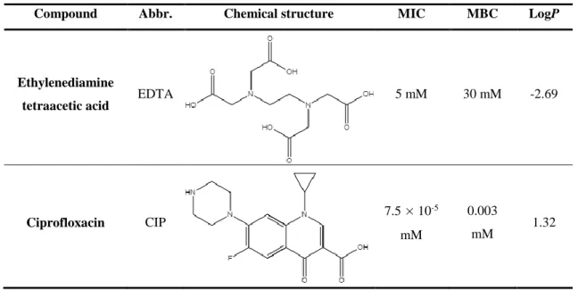
Table 2 - Phytochemicals with potential for biofilm prevention and control and their mechanism of action.
Chemical class Phytochemical Bacterial strains Mechanism of action
Phenolics
Gallic acid L. monocytogenes;
E. coli Inhibition of swimming (L. monocytogenes) and swarming
(L. monocytogenes and E. coli) 91; Reducing swimming
motility 92
Ferulic acid L. monocytogenes;
E. coli 91; B. cereus; P. fluorescens 92
Salycilic acid B. cereus and P. fluorecens
92; E.coli
and S. aureus 7
Reducing swimming motility; quorum sensing inhibition 92;
quorum sensing inhibition; reducing swimming and sliding motility 7
Resveratrol and piceatannol E. coli Inhibition of ATPase activity and ATP synthesis 93
Quercetin; quercetrin;
quercetin-3-_-d glucoside E. coli Inhibition of ATPase activity
93
Quercetin; Tannic acid (A.
japonica) S. aureus
Repressing genes related to biofilm formation; quorum sensing and virulence inhibition; hemolysis activity inhibition
(only quercetin) 94
Naringenin; quercetin,
sinensetin and apigenin E. coli; Vibrio harveyi
Antagonistic activity against autoinducer-mediated cell–cell signalling 95
Curcumin (Curcuma longa)
P. aeruginosa 59; Burkholderia
pseudomallei 96; Vibrio
parahaemolyticus, Vibrio vulnificus
and V. harveyi 97; Gram-negative
bacteria 98
Quorum sensing inhibition 59; Inhibition of biofilm formation 96; quorum sensing inhibition ,reduction of bioluminescence,
inhibition of alginate production, motility and biofilm development 97; quorum sensing inhibition, inhibited the
expression of C4 and C6 homoserine lactones (HSLs)-mediated phenotypes 98
Proanthocyanidins
(Cranberry fruit) P. aeruginosa Inhibition of swarming motility 99
Punicalagin (Pomegranate)
Phloretin (Apple) E. coli Reducing biofilm formation; Repression of autoinducer-2