Universidade de Lisboa
Faculdade de Farmácia
Staphylococcus aureus escapes killing from extracellular
histones through plasminogen binding and activation by
staphylokinase or tissue plasminogen activator
Vanessa da Silva Lopes
Mestrado integrado em Ciências Farmacêuticas
2
Universidade de Lisboa
Faculdade de Farmácia
Staphylococcus aureus escapes killing from extracellular
histones through plasminogen binding and activation by
staphylokinase or tissue plasminogen activator
Vanessa da Silva Lopes
Monografia de Mestrado integrado em Ciências Farmacêuticas apresentada à
Universidade de Lisboa através da Faculdade de Farmácia
Orientador: Doutora Isabel Antolin Rivera
3
Università degli studi di Pavia
Dipartamento di Medicina Moleculare
Supervisor: Doctor Giampiero Pietrocola
4
Abbreviations
MRSA - Methicillin Resistant Staphylococcus aureus
CA-MRSA - Community-Associated Methicillin Resistant Staphylococcus aureus MSSA - Methicillin Susceptible Staphylococcus aureus
MSCRAMM - Microbial Surface Components Recognizing Adhesive Matrix Molecules ECM - Extracellular Matrix
OD - Optical Density
BSA - Bovine Serum Albumin PLG - Plasminogen
uPA - Urokinase Plasminogen Activator tPA - Tissue-type Plasminogen Activator SDS - Sodium Dodecyl Sulfate
PBS - Phosphate Buffered Saline
ELISA - Enzyme-Linked Immunosorbent Assay
SDS-PAGE - Sodium Dodecyl Sulfate - Polyacrylamide Gel Electrophoresis FnBPA - Fibronectin-Binding Protein A
FnBPB - Fibronectin-Binding Protein B FBG - Fibrinogen
Sort A - Sortase A
CFU - Colony Forming Unit AU - Absorbance Units
5
Resumo
Staphylococcus aureus é uma bactéria comensal que coloniza mais de 20% da
população humana. Encontra-se sobretudo na mucosa nasal, mas na maioria dos casos não origina doença. Contudo em caso de cortes ou em situações cirúrgicas, a nível hospitalar, S. aureus poderá entrar nos tecidos e provocar diversas lesões, podendo mesmo pôr em risco a vida do doente.
Atualmente, cada vez é maior a quantidade existente de estirpes resistentes à antibioterapia. Em Portugal, a prevalência de S. aureus resistente à meticilina (MRSA) encontra-se por volta dos 50%, sendo uma das maiores taxas a nível europeu. Portanto, urge a necessidade de novas formas de tratamento, de modo a conseguir impedir cenários desastrosos de resistências por parte desta bactéria.
O sistema imunitário desempenha um papel bastante importante como primeira linha de defesa contra diversos microrganismos. A ativação dos neutrófilos poderá em ultima instância levar à ocorrência de um processo designado NETosis. Este processo origina a libertação de NETs, estas são redes de filamentos contendo histonas e outros componentes. As histonas extracelulares são conhecidas por ter grande capacidade bactericida. Deste modo, a formação de NETs permite aprisionar e matar estas bactérias. Contudo, S. aureus terá arranjado uma forma de conseguir escapar a este efeito bactericida.
Com base no artigo: “Streptococcus pyogenes exhaust killing from extracellular histones through plasminogen binding and activation by streptokinase” tentámos transpor esta proposta para o caso do Staphylococcus aureus. Sendo assim, neste trabalho, foi mostrado que tal como a bactéria deste artigo, também S. aureus consegue ligar plasminogénio nas suas proteínas de superfície. Foi também analisado se a ativação do plasminogénio por estafilocinase (SAK - fator de virulência produzido pela bactéria) ou pelo ativador tecidular do plasminogénio (tPA - produzido por células endoteliais) consegue degradar as histonas e deste modo esta bactéria consegue escapar ao efeito bactericida das mesmas.
Apesar das histonas terem demonstrado uma grande capacidade bactericida, ainda tem que ser desenvolvida uma forma de evitar que estas sejam degradadas pela plasmina.
Palavras-chave:
Staphylococcus aureus; Histonas; Plasminogénio; Estafilocinase6
Abstract
Staphylococcus aureus is a commensal-bacterium that colonizes more than 20% of the human population. It is found mostly in the nasal mucosa, but in most cases it does not cause disease. However, in case of cuts or at hospital level in surgical situations, S. aureus may spread into the tissues and cause various lesions and may even lead to a potentially life-threatening-situation.
Currently, there is an increasing number of strains resistant to antibiotic therapy. In Portugal, the prevalence of MRSA is around 50%, being one of the highest rates in Europe. Therefore, it is urgent the need for new forms of treatment, in order to prevent worst scenarios of antibiotic resistances.
The immune system plays a very important role as the first line of defense against microorganisms. Activation of neutrophils may ultimately lead to the occurrence of a process called NETosis. This process leads to the release of NETs, these are composed by filaments networks containing histones and other components. Extracellular histones are known to have high bactericidal capacity. In this way, the formation of NETs allows to trap and kill these bacteria. However, S. aureus will have managed a way to escape this bactericidal-effect.
Based on the article: "Streptococcus pyogenes exhaust killing from extracellular histones through plasminogen binding and activation by streptokinase", we tried to transpose this proposal to the particular case of Staphylococcus aureus. Thus, in this work, we show that as the bacteria of this article, S. aureus can also bind plasminogen to its surface proteins. It is also analyzed whether the activation of plasminogen by staphylokinase or tissue plasminogen activator can degrade the histones and thus this bacterium can escape from their bactericidal-effect.
Although histones have demonstrated a high bactericidal-capacity, a way to prevent them from being degraded by plasmin has to be developed in future projects.
Keywords:
Staphylococcus aureus; Histones; Plasminogen; Staphylokinase (SAK);7
INDEX
Abbreviations ... 4 Resumo ... 5 Abstract ... 6 1. Introduction ... 10 1.1. Staphylococcus aureus... 10 1.1.1. History ... 101.1.2. Classification and Morphology ... 10
1.1.3. Habitat ... 11
1.1.4. Clinical manifestations ... 11
1.1.5. Epidemiology ... 12
1.1.5.1. Strains – Methicillin-resistant Staphylococcus aureus ... 13
1.1.5.2. The spread of USA300 internationally ... 14
1.1.6. Pathogenesis ... 14
1.1.6.1. Virulence factors ... 15
1.1.6.1.1. MSCRAMMs ... 15
1.1.6.1.3. Staphylokinase ... 17
2. Plasminogen ... 18
3. The innate immune system ... 20
3.1. Histones ... 20 4. Objectives ... 22 5. Materials e methods ... 23 5.1. Materials ... 23 5.1.1. Equipment ... 23 5.1.2. Biological material ... 23 5.1.3. Chemical material ... 24
8
5.2. Methods ... 26
5.2.1. ELISA ... 26
5.2.2. Bactericidal assay with S. aureus USA300 LAC ... 27
5.2.3. SDS-PAGE ... 28
6. Results and discussion ... 31
6.1. Plasminogen binding to the bacteria cell wall proteins: FnBPA and FnBPB ... 31
6.2. Histones effect on the bacteria survival ... 33
6.3. Histones degradation ... 34
6.3.1. Effect of SAK in the histones degradation ... 35
6.3.2. Effect of tPA in the histones degradation ... 37
7. Conclusion ... 39
8. Bibliography ... 40
Appendix ... 45
Figure Index
Figure 1. Scanning electron micrograph (SEM) of Staphylococcus aureus. Under a high magnification of 10,000x ... 10Figure 2. Diagram of the body showing the common sites of infection and diseases that S. aureus causes ... 12
Figure 3. Prevalence of MRSA and MSSA community-associated clones in Europe .. 14
Figure 4. Panel A shows the surface and secreted proteins. Panels B and C show cross sections of the cell envelope... 15
Figure 5. Basic fibrinolytic system ... 18
Figure 6. Schematic illustration of plasminogen ... 19
Figure 7. Chromosome constitution ... 20
Figure 8. A- Microtiter; B- Schematic representation of an indirect ELISA ... 27
9
Figure 10. SDS-PAGE: principal steps of the sample preparation ... 29 Figure 11. ELISA - Determination of PLG bound to the bacteria cell wall proteins ... 31 Figure 12. Schematic representation of FnBPA and FnBPB. ... 32 Figure 13. A- Number of bacteria colonies grown in plates with different histones concentrations. B- Bacterial survival rate ... 33
Figure 14. SDS-PAGE of control samples ... 34
Figure 15. SDS-PAGE of histones with plasminogen and staphylokinase ... 35
Figure 16. Bactericidal effect of SAK and plasminogen in the bacterial survival of the
histones ... 36
Figure 17. SDS-PAGE of histones with plasminogen and tissue plasminogen activator
... 37
Figure 18. Bactericidal effect of tPA and plasminogen in the bacterial survival of the
histones ... 38
Table Index
10
1. Introduction
1.1. Staphylococcus aureus
1.1.1. History
In the 1500s, the Italian physician Girolamo Fracastoro suggested that germs could carry disease. Long after this revolutionary proposal, Dutch naturalist Antoni van Leeuwenhoel observed bacteria in 1674 with his roughly made microscope. The spherical colonies that he called “animalcules” were probably some species of
Staphylococcus aureus [1].
In 1878, Robert Koch was the first to describe staphylococci in human pus. Two years later, Pasteur cultivated the microorganism in a liquid medium. The following year Ogston demonstrated that staphylococci were pathogenic to mice and guinea pigs, causing acute and chronic abscesses. The German physician Anton Rosenbach, in 1884, described the two most common species, S. aureus and S. epidermidis. These two species are also known by the test most commonly used to differentiate them, being
S. aureus often referred to as "coagulase-positive" [2].
Although it has been recognized and described more than 100 years ago,
Staphylococcus aureus still remains a versatile and dangerous pathogen in humans [1].
1.1.2. Classification and Morphology
Staphylococcus aureus is a member of the Micrococcaceae family [3]. This family
includes the genera Micrococcus, Staphylococcus, and Planococcus [4]. It is one of the about 32 species in the Staphylococcus genus, but most of the others species are found in other mammals and do not infect humans. S. aureus is a facultative anaerobic gram-positive coccus, 1μm in diameter that is microscopically observed as individual organisms, in pairs, and in irregular, grapelike clusters (Fig.1), and it is a nonmotile (not
11 able to have movement) and non-spore forming bacterium [1]. Individual cocci of pathogenic strains, such as S. aureus, tend to be slightly smaller than nonpathogenic strains. Size also depends partly on the age of the culture and the medium in which it is grown [4].
In fact, staphyle is the Greek name for “bunch of grapes”, and cocci, which means “spherical bacteria”, once the bacteria are perfectly spherical in shape. The name
aureus, which is a Latin word for “gold”, was given to the bacteria because it grows in
large yellow colonies [1,5].
S. aureus is distinguished from other staphylococcal species on the basis of the
gold pigmentation of colonies and coagulase positive, mannitol fermentation, and deoxyribonuclease tests and often have hemolytic capacity [1,3]. Colonies are usually large (6-8 mm in diameter), smooth, and translucent [4].
1.1.3. Habitat
S. aureus lives on the skin and mucous membranes of warm-blooded animals,
normally inhabits the body surfaces of many species of mammals and birds. About 20-60% of humans carry relatively large populations of this organism in their noses, because the nasal membranes provide a perfect habitat for S. aureus colonies since they are warm and moist; many of these people are also skin carriers [1,4].
It is also found in a variety of other habitats, such as water, decaying matter, and on just about any surface [1,4]. S. aureus is extremely resistant. It is among the hardiest of all nonspore-forming bacteria and is relatively resistant to drying or moderate temperature changes [4]. It grows in a wide temperature range of 15 - 45C, and one reason why S. aureus is so strong is that its cell wall is extremely thick compared with the cell walls of other bacteria [1].
1.1.4. Clinical manifestations
Usually, S. aureus colonizes the nasal passage, vagina, pharynx and axillae and sometimes this commensal bacteria can cause both superficial and invasive infections, and lead to a potentially life-threatening situation [5,6]. Although this bacterium colonizes the skin of many healthy individuals, most people who are carriers do not become infected, because the skin provides a substantial barrier to the entry of bacteria into the body. If a break in the skin happens due to injury or surgery, it will provide a point of entry for S. aureus. Thus, it can produce many superficial skin lesions, such as infections of
12 hair follicles, acne, boils1, sties2 and also cause infections in other areas of the body, and
serious internal infections such as osteomyelitis, meningitis, endocarditis and sepsis [1,4,5]. In Fig. 2, it is possible to observe the wide range of sites in the body where S.
aureus can cause infection and consequently disease. In community-acquired infections,
usually the skin infections are predominant, whereas infections of the lung dominate among nosocomial S. aureus infections [7]. - - -
1.1.5. Epidemiology
A major cause of hospital acquired (nosocomial) infection of surgical wounds is
S. aureus. Nosocomial infections are predominantly associated with catheterization or
implants [8]. People colonized with this bacterium has increased risk for subsequent infections. S. aureus may also cause more serious infections, particularly in people debilitated by chronic illness, traumatic injury, burns or immunosuppression [5]. Rates of staphylococcal colonization are high among patients with type 1 diabetes, intravenous drug users, patients undergoing hemodialysis, surgical patients, and patients with acquired immunodeficiency syndrome. Patients with qualitative or quantitative defects in leukocyte function are also more prone to suffer staphylococcal disease [3]. - - - - - - - -- - -
1 Boil is a deeper pus filled abscesses of the skin 2 sty is an inflammation of a gland in the eyelid
13 Even today S. aureus infections result in substantial morbidity and mortality, due to the ineffective antibiotic therapy [9]. This can be explained by the increased incidence of staphylococcal diseases, that include methicillin-resistant S. aureus (MRSA) strains. Therefore, efforts to develop alternative options and vaccine able to prevent these life threatening infections are being made in response to this increases [8].
1.1.5.1.
Strains – Methicillin-resistant Staphylococcus aureus
Chambers and Deleo identified serial “waves of resistance” in the history of 20th-century S. aureus epidemiology. They described the emergence of penicillin-resistant S.
aureus in the 1940s and rapid spread during the 1950s and 1960s, initially in the health
care setting and then in the community, as the first wave of resistance. In 1959 the first wave of resistance happened due to the introduction of semisynthetic penicillin (methicillin). Lately, methicillin started another wave of resistance with methicillin-resistant S. aureus (MRSA) which was described in hospitalized populations [10].
MRSA infections are increasingly common in the community being a serious threat to public health worldwide [11,12]. Antibiotic treatment is often ineffective due to the development of antibiotic-resistant strains, since the resistance to methicillin confers resistance to all penicillinase-resistant penicillins and cephalosporins [6,11]. Methicillin resistance is not caused by inactivation of the antibiotic by an enzyme such as beta-lactamase, but instead seems to be related to a difference in cell wall permeability that prevents entry of the antibiotics [2]. MRSA is prevalent in hospitals, and hypervirulent MRSA strains have recently spread throughout the community [6].
The community-associated MRSA (CA-MRSA) strain USA300 was first reported in the USA as a cause of skin and soft tissue infection among college football players in Pennsylvania and among prisoners in Missouri in 2000. USA300 became the predominant community-associated MRSA strain circulating in the United States by 2011 [10,13].
Although USA300 isolates were initially resistant only to semi-synthetic penicillins and macrolides, they have widened their resistance profiles considerably over the last 5 years. This includes the addition of clindamycin resistance and tetracycline resistance. Several USA300 isolates have developed reduced susceptibility to vancomycin and, in some cases, to daptomycin, in addition to occasional resistance to gentamicin and trimethoprim–sulfamethoxazole [13].
14
1.1.5.2.
The spread of USA300 internationally
USA300 isolates have been recognized in all Europe. [8] MRSA is prevalent in hospitals, and this strain has recently spread throughout the community [5]. The prevalence of MRSA in Portuguese hospitals has been close to 50% for over 10 years and remains one of the highest in Europe. In Europe, the epidemiology of nosocomial MRSA is well understood, but the epidemiology of CA-MRSA is incompletely documented due to poor geographic coverage and outdated data [9]. However, some studies have been made to observe the distribution of the most prevalent clones in Europe (Fig. 3) [14].
1.1.6. Pathogenesis
S. aureus is responsible for the majority of skin and soft tissue infections [15].
Infections are initiated when a breach of the skin or mucosal barrier allows staphylococci access to contiguous tissues or to bloodstream. When S. aureus invades the bloodstream, it can be devastating. Whether an infection is contained or spreads depends on a complex interplay between S. aureus virulence determinants and host defense mechanisms [3]. It carries a risk of systemic spread and life-threatening bacteremia, so it is a great deal discovering about the contribution of bacterial factors to the development of infection [3,15]. For the majority of diseases caused by this organism,
15 pathogenesis is multifactorial [5].
S. aureus has a diverse arsenal of components and products that contribute to
the pathogenesis of infection [3]: factors that probably inhibit phagocytosis and toxins that damage host tissues and cause disease symptoms [5].
1.1.6.1.
Virulence factors
Staphylococcus aureus has developed many mechanisms to escape from human
immune responses [16]. Therefore, it possesses over 50 virulence factors that allows the bacterium to adapt to a variety of host niches and to cause a diverse spectrum of diseases. These factors include biofilm formation, a number of “microbial surface components recognizing adhesive matrix molecules” (MSCRAMMs), capsular polysaccharides (CPs), staphylokinase (SAK), toxins and even more factors that decrease bacterial clearance by the host [17,18].
1.1.6.1.1. MSCRAMMs
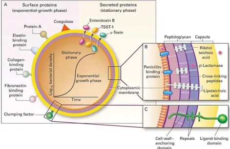
The major group of proteins involved in virulence are the surface-attached proteins, which are known as cell wall anchored (CWA) proteins, which significantly enhance staphylococcal virulence by helping in immune evasion and binding to host tissues [6,15].
The range of CWA proteins on the surface varies among strains, and the expression of CWA proteins can be different according to growth conditions or phase of growth; for example, some proteins are found predominantly on cells in the exponential
Figure 4. Panel A shows the surface and secreted proteins. The synthesis of many of these proteins is dependent on the growth phase, as shown by the graph. Panels B and C show cross sections of the cell envelope [3].
16 or stationary phases (Fig.4) and in some cases they are only expressed under iron-restricted conditions [6,19].
The adherence between S. aureus and the nasal epithelium is a multifactorial process. The primary bacterial adherence is thought to be mediated by wall teichoic acid (WTA), whereas MSCRAMMs have critical roles at later stages of colonization of the human nose [20]. Over 20 different of these related proteins bind extracellular matrix molecules and have been designated microbial surface components recognizing adhesive matrix molecules (MSCRAMM) [3,10]. They recognize and bind to human extracellular matrix components such as fibrinogen (FBG) or fibronectin [21]. The name of the proteins may have been defined according to the function with which it was first associated [6]. Others were given generic names.
MSCRAMM
Function
Clumping factors A (ClfA) Immune evasion
Clumping factor B (ClfB)
Expressed during nasal colonization and contributes to nasal colonization in humans
Iron-regulated surface determinant A (IsdA)
Interact with harvested human desquamated epithelial cells
Serine–aspartate repeat protein C (SdrC) Homophilic SdrC–SdrC interaction
results in biofilm formation
Serine–aspartate repeat protein D (SdrD) Promote adhesion of bacteria to
desquamated nasal epithelial cells
Serine–aspartate repeat protein E (SdrE) Immune evasion
Fibronectin binding proteins A (FnBPA)
Adhesion to ECM
Fibronectin binding proteins B (FnBPB)
Collagen - binding protein (Cna) Adhesion to collagen-rich tissue
Table 1. Principal Staphylococcal MSCRAMM proteins and their function [6].
1.1.6.1.2. Capsular polysaccharides
In order to resist phagocytose, S. aureus expresses a polysaccharide capsule which masks the bacterial surface and surface-associated proteins [16].
17
1.1.6.1.3. Staphylokinase
Staphylokinase (SAK) is considered one of the virulence factors produced by Staphylococcus aureus. Strains of S. aureus express this bacteriophage encoded protein during the late exponential phase of the growth cycle [21,22]. The SAK gene is carried by certain prophages and its synthesis is positively regulated by the accessory gene regulator Agr and is negatively regulated by the staphylococcal accessory regulator SarA [23,24]. SAK is a 15.5 kDa protein that is constituted by 136 amino acids comprising a single chain and does not contain disulfide bonds [25].
The role of staphylokinase during bacterial infection is based on its interaction with the host proteins, such as plasminogen (PLG) [21,24]. It has been shown that SAK is a highly specific activator of human PLG [24]. Due to this binding with SAK, plasminogen changes its conformation and it is activated into plasmin molecules at the bacteria surface [9,26]. It is important to refer that staphylokinase belongs to indirect plasminogen activators, so it does not possess enzymatic activity, therefore its activity is gained after the formation of stoichiometric complexes (1:1) with plasminogen [25].
Activation of plasminogen promotes penetration of S. aureus through physiological barriers via plasmin-induced proteolysis, since it digests fibrin blood clots and many components of extracellular matrix and basal membranes, thus allowing the bacterial invasion into host tissues [24,27,28]. Despite this negative aspects, SAK is a promising antithrombotic agent factor, because of its fibrin-selective plasminogen activity [29].
Apart from its ability to activate plasminogen, staphylokinase induces extracellular release of α-defensins from polymorphonuclear cells. Furthermore,a binding between them and staphylokinase leads to a complex formation. Consequently, this interaction inhibits the bactericidal effect of the defensins [23]. Defensins are short cationic, amphiphilic, cysteine-rich peptides that play important roles in innate immune defense against infectious pathogens, since it forms structured nanonets3 to entrap bacterial
pathogens and disorganize bacterial cell membranes [30,31].
SAK also plays a role in the innate immune defenses, due to its anti-opsonic capacity. By activating human plasminogen into plasmin at the bacteria surface it creates bacterium-bound serine protease activity that cleaves two host opsonins: human immunoglobulin G (IgG) and human C3b [26]. So, it protects the bacteria from phagocytosis [24].
Even though the importance of fibrin generation for S. aureus virulence has been established, the role of SAK remains uncertain [18].
3 Nanonets result from oligomerize α-defensins, that prevent intimate contact with epithelial cells and
18
2.
Plasminogen
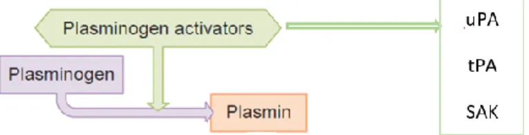
As previously referred, the zymogen form of plasmin, plasminogen, is an enzyme that plays an important role in fibrinolysis, and in the degradation of extracellular matrix and other key proteins involved in immunity and tissue repair [32,33]. In the skin, PLG is present mostly in the basal layer of the epidermis and in the walls of hair follicles, and it can be also found in significant quantities in extravascular fluids [27,33]. The coagulation system and the innate immune defense have the same evolutionary origin. So, for that reason, tissue damage as well as bacterial infection trigger activation of the coagulation system, that leads to thrombin generation and fibrin deposition. Fibrin not only has its role in preventing leaking blood vessels, but it is also part of the first barrier against bacterial spread [34].
Physiologically, the plasminogen conversion is catalyzed by the activators urokinase plasminogen activator (uPA) or tissue-type plasminogen activator (tPA) secreted by endothelial cells [35,36]. Other proteases, such as factors XIa and XIIa, are also described as being capable of functioning as plasminogen activators [33].
Tissue-type plasminogen activator (tPA) is a serine protease, which is synthesized as a single-chain protein, but can be converted by plasmin into a two-chain form in which the chains are connected by a disulfide bond [37].
Plasminogen is mainly synthesized by the liver [38]. However, other extrahepatic tissues, such as cortex and medulla of the thymus, splenic white, adrenal glands, kidney, brain, testis, heart, lung, uterus, and cornea have the capacity to provide local sources of plasminogen [32,39]. PLG is synthesized as an 810 amino acid polypeptide protein, the mature form of this protein is cleaved during secretion forming a single polypeptide chain that contains 791 amino acids [40].
PLG (Glu-plasminogen) is a single-chain glycoprotein of approximately 90kDa that is composed of a pre-activation peptide (region AP represented in Fig. 6), followed by five kringle4 domains (triple-disulfide-linked peptide regions) and an active serine protease
4 The name kringle comes from the resemblance to a Scandinavian pastry [72].
Figure 5. Basic fibrinolytic system. Different plasminogen activators can initiating this system. This one of the functions of the plasmin activation. [36]
19 site [34,41,42]. The kringle domains contain lysine binding sites, which facilitate the binding to large substrates (fibrinogen, plasminogen and others), bacterial proteins, and mammalian cell surfaces, small molecule ligands (Cl-) and amino acids, interactions that
also regulate the activation of this protein [40]. Glu-plasminogen can circulate in two ways, as closed conformation that cannot be instantly activated by tPA or uPA, or can assume an open conformation when it is bound to fibrin or at the cell surface [33].
The plasminogen activation can occur by a variety of mechanisms, but all of them lead to the cleavage Arg 561 and Val 562 bond, resulting in the formation of the two-chain
called plasmin, constituted by a heavy chain and a light chain, being these chains covalently linked by two disulfide bonds [34,41]. If plasmin cleaves peptide bonds after Arg67 , or Lys76,77, it leads to the formation of a truncated form of plasminogen (85 kDa,
Lys-PLG), called Lys-plasminogen, which assumes an open conformation, that has a higher affinity for fibrin and is faster activated by tPA or uPA than the original molecule [43]. Both Glu- and Lys-plasminogen in the open conformation assume a flexible formation where the activation loop is exposed for cleavage by this plasminogen activators [33].
Figure 6. Schematic illustration of plasminogen where the seven domains are shown: plasmin preactivation site; tissue plasminogen activator (tPA) and urokinase plasminogen activator (uPA) cleavage sites [34]; and five kringle domains
20
3.
The innate immune system
As the first line of human defense, it responds in order to non-self and danger signals from microbial invasion or tissue injury [44]. Innate immune system recognize microbial infections both to elicit immediate defense and to generate long-lasting adaptive immunity [45]. The immediate defense is composed by many components. It includes soluble recognition molecules, such as natural antibodies, pentraxins (for example C-reactive protein), and the complement system. There are also cellular components in the innate immune system, which are composed by phagocytic cells (neutrophils), antigen presenting cells (dendritic cells), and killing cells (natural killer cells) [44].
3.1. Histones
Histones were discovered by Albrecht Kossel in 1884. They are highly conserved, alkaline, small and positively charged proteins. Histones are mostly known as nuclear proteins that wind up the double-stranded DNA to form chromatin and for regulate expression of DNA [46–48].
There are five classes of histones:
- H2A, H2B, H3, and H4 also called “core histones”; - while H1/H5 represents “linker histone" [49].
The core histones have similar structures with a conserved central motif domain (designated histone fold) and an unstructured amino-terminal tail. Histone fold is composed of a long central helix with a loop or a short helix flanked on either side [46]. Histone fold exhibits high hydrophobic interactions to form dimers and tetramers within the core histones. The nucleosome, organizational unit of chromatin (Fig. 7), is composed by two each of the histones H2A, H2B, H3, and H4, which is referred to as
Figure 7. Chromosome constitution: DNA is wrapped twice around a histone octamer to make a nucleosome and associated with H1 maintain the chromosome structure [74,75].
21 histone octamer [49].
All histones have their N-terminal tails rich in lysine and arginine residues. These amino-terminal sites are flexible and undergo numerous posttranslational modifications [49].
Histones are important structural elements of nuclear chromatin and play a key role in regulation of gene transcription. In the nucleus, histones are completely inert [48,49]. On the other hand, when histones are unchained and released extracellularly upon cell death or from activated neutrophils, as part of neutrophil extracellular traps (NETs), they assume pro-coagulant effects, cytotoxicity and immune stimulatory effects [50]. The formation of these extracellular traps has been recognized as an important mechanism of the host innate immune response against infections [51].
The release of histones requires rupture of the nuclear and plasma cell membrane, for example during cell death. Regulated form of necrosis (NETosis/ETosis) is restricted to immune cells like neutrophils (NETosis) and other granulocytes or macrophages (ETosis) [49]. NETosis is a defense mechanism that plays an important part in host innate defense as it catapults chromatin out of the cell to trap bacteria [49,50]. However, the knowledge about the NETosis function in the early immune response to an invading pathogen is little [52]. NETs were discovered in 2004 and since this milestone they have been described as a potential bactericidal [53]. As a result, the studies on the antimicrobial capacity of histones have grown. NETs consist of a framework of nuclear DNA (chromatin) with histones, proteases and several antibacterial components that are expelled from activated neutrophils, providing a matrix to entrap and kill various microbes [54,55].
Activated neutrophils undergo dramatic morphological changes, to release NETs. After their activation, neutrophils flatten and firmly attach to the substratum [56]. The NET formation is described as beginning by the global chromatin decondensation and then the disintegration of the nuclear membrane occurs concomitant with cytoplasmic granule dissolution, allowing the NET components to mix in the cytoplasm prior to extracellular release [51]. (It can be seen a schematic representation of NETosis in the appendix A1)
As antimicrobial peptides, histones play an important role in skin defense, but are also found in other tissues, such as stomach or intestine, reproductive tissue, as well as in blood [48].
22
4.
Objectives
This work was based on the article "Streptococcus pyogenes exhaust killing from extracellular histones through plasminogen binding and activation by streptokinase" [57].
This thesis aims to verify if, as the bacterium studied in this article, S. aureus could have the same ability to escape from extracellular histones, which are part of the innate immune system, by fixing and activating PLG on its cell wall. In order to attain this milestone, the project proposed intends to:
- confirm if the components present on the bacteria surface (FnBPA and FnBPB) can bind plasminogen by enzyme-linked immunosorbent assay (ELISA);
- analyze the effect of the histones on the bacterial survival by Bactericidal assay with S. aureus USA300 LAC;
- observe the consequences of adding plasminogen, tPA or SAK to histones by SDS-PAGE.
23
5.
Materials e methods
5.1. Materials
5.1.1.
Equipment
Refrigerated Incubator shaker – C24KC
Autoclave – Vapour-line VWR
Vortex machine – Super mixer
Balance
UV-Vis Spectrophotometer – JASCO V-630
Wide Mini-Sub Cell GT electrophoresis cell with PowerPac Basic power supply Fume hood – VBH 4B PP/99 Cuvettes Plates pH meter Microplates
Microplate reader Bio-Rad laboratories, Model 680
5.1.2.
Biological material
Bacterial strain and culture conditions
Staphylococcus aureus USA300 LAC strain (bacteria were kindly donated by the
research groups of Prof. Timoty J. Foster and Prof. Joan Geoghegan (Microbiology Department, Moyne Institute of Preventive Medicine, Trinity College, Dublin 2, Ireland) was let to grow in Brain Heart Infusion (BHI) at 37C with constant shaking (195 rpm) and overnight in order to obtain the stationary phase. In the next morning, the overnight bacterial culture was diluted (1:40) in fresh BHI and incubated at 37C with shaking to obtain the exponential phase, the incubation being stopped when it was observed the desired OD600nm.
For our proposal, the staphylococcal cells were harvested from cultural medium by centrifugation (10 minutes at 4000 rpm) and the supernatant was discarded. Then, the bacterial cells were washed with 5mL of 10mM Tris-HCl buffer (pH 7.4) and after that they were centrifuged (10 minutes at 4000 rpm), the supernatant was removed and finally the bacteria were re-suspended at the appropriate density.
24
Plasminogen
Plasminogen was produced in the laboratory using a purification method called “Lysine-sepharose affinity chromatography” by Sanderson-Smith [58]. In this method plasminogen was purified from human plasma.
Concentration - 1µg/µL
Histones and tPA
They were purchased from SIGMA.
Mixture of histones concentration – 7.5µg/10µL; tPA concentration - 100µg/mL
SAK protein
It was generously provided by Dr. S.H.M. Rooijakkers, University Medicinal Center Utrecht, The Netherlands.
Concentration – 1µg/10µL
FnBPA and FnBPB
The recombinant proteins were kindly donated by Prof. Timoty J Foster and Prof. Joan Geoghegan. FnBPA concentration – 1.69 mg/mL FnBPA concentration – 1.65 mg/mL
5.1.3.
Chemical material
Name
Distributor OPD (o-Phenylenediaminedihydrochloride) tablets 2mg Sigma
Prestained Protein SHARPMASS V
plus Protein MW Marker Euroclone
Sample buffer, Laemmli 2x concentrate Sigma
5.1.4.
Solutions and buffers
Pure Water – Millipore, Q-Gard 1 Tris-HCl 10mM pH7.4
25
Electrophoresis Buffer (1x)
It was prepared by the following formula a 10x concentrated buffer:
Tris – 30g/L (0.025M)
Glycine – 140.4g/L (0.19M)
SDS – 10.0g/L (0.0035M)
After everything weighted and mixed, the pH was adjusted to the value 8.3 and then this solution was filtrated and stored at 4°C until further use. Subsequently, every time that it was needed the electrophoresis buffer, it was diluted in order to obtain a solution with a concentration 1x.
ELISA Buffer
60mL NaCl 5M
2mL Tween 20
BHI medium
To prepare this medium, brain heart infusion powder was dissolved in sterile water, in a concentration of 37g/L. Then this solution was sterilized by autoclaving (121C for 20min) and stored at 4C.
Brain Heart infusion (BHI) agar plates
This medium is recommended for the cultivation and isolation of a variety of microorganisms, including bacteria. In order to prepare plates without contamination, it was carried out several steps.
1. The BHI (37.8g/l) and agar (1.8%) powders were weighted; previously the right quantity was calculated according to the number of plates needed (6mL / plate).
2. Then, the water was added into the recipient with the BHI and agar. 3. The BHI agar solution was autoclaved (121C for 20min).
4. When the sterilization was finished, the solution was put inside the plates, still warm. This step took place into the fume hood to avoid contamination. 5. Finally, the plates were stored at 4ºC until further use.
26
5.2. Methods
5.2.1.
ELISA
ELISA (enzyme-linked immunosorbent assay) is a micro-well plate-based assay technique designed for detecting and quantifying substances such as peptides, proteins, antibodies and hormones. [59] This technique combines the specificity of antibodies with the sensitivity of simple enzyme assays, by using antibodies or antigens coupled to an easily-assayed enzyme. ELISA can provide a useful measurement of antigen or antibody concentration [60].
There are different types of ELISA:
Direct ELISA
Indirect ELISA
Sandwich ELISA
Competitive ELISA
ELISpot
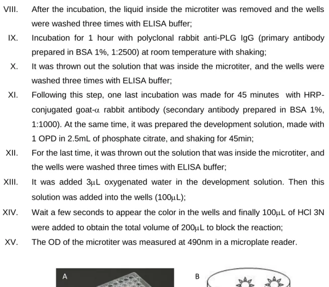
The ELISA type used in this experience was the indirect ELISA (Fig. 8). This method is a two-step ELISA which involves two binding process of primary antibody and labeled secondary antibody [61]. To screen bacteria surface proteins (FnBPA and FnBPB) binding to plasminogen (PLG), an ELISA assay was performed. All the experience is described below.
I. Firstly, solutions with a concentration of 5g/mL for each protein - FnBPA, FnBPB and BSA 1% (control) were prepared. These solutions were prepared in 50mM Na2CO3 pH 9.5;
II. Microtiter wells were coated with this solutions, two wells for each protein (100L).
III. Incubation at 4C overnight (in fridge);
IV. In the next morning, the solutions inside the microtiter were discarded and the wells were washed three times with ELISA buffer;
V. Incubation with BSA 2% (200L) for 1 hour at room temperature with shaking to block unspecific binding sites;
VI. The solutions inside the microtiter were discarded again without the washing step;
VII. Incubation with 0.5g/well of human PLG dissolved in PBS (100L) in each well for 1h at room temperature with shaking;
27 VIII. After the incubation, the liquid inside the microtiter was removed and the wells
were washed three times with ELISA buffer;
IX. Incubation for 1 hour with polyclonal rabbit anti-PLG IgG (primary antibody prepared in BSA 1%, 1:2500) at room temperature with shaking;
X. It was thrown out the solution that was inside the microtiter, and the wells were washed three times with ELISA buffer;
XI. Following this step, one last incubation was made for 45 minutes with HRP-conjugated goat- rabbit antibody (secondary antibody prepared in BSA 1%, 1:1000). At the same time, it was prepared the development solution, made with 1 OPD in 2.5mL of phosphate citrate, and shaking for 45min;
XII. For the last time, it was thrown out the solution that was inside the microtiter, and the wells were washed three times with ELISA buffer;
XIII. It was added 3L oxygenated water in the development solution. Then this solution was added into the wells (100L);
XIV. Wait a few seconds to appear the color in the wells and finally 100L of HCl 3N were added to obtain the total volume of 200L to block the reaction;
XV. The OD of the microtiter was measured at 490nm in a microplate reader.
5.2.2.
Bactericidal assay with S. aureus USA300 LAC
Exponential growing cultures of S. aureus USA300 LAC, after the wash with 10Mm tris-HCl buffer, were suspended in 15mL of Tris-HCl buffer (OD600 = 0.4). Then
500L of bacterial suspensions were mixed with 500L histones solutions of different concentrations and incubated for 2 hours at 37C.
Figure 8. A- Microtiter; B- Schematic representation of an indirect ELISA (1-PLG; 2-anti-PLG; 3- anti-rabbit)
A B
1
2 3
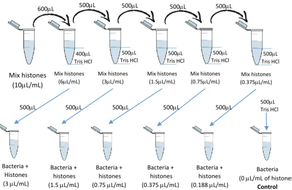
28 To prepare the different histone concentrations, it was used a solution of mix histones (10L/mL), and diluted with Tris-HCl buffer (Fig. 9).
After incubation (2h at 37C with shaking), sample (bacteria with different concentrations of histones) serial dilutions were plated on BHI agar5 and left in the
incubator (37C at 120rpm) overnight. Bacterial counts were determined the next day. All results were related to a non-treated control.
5.2.3.
SDS-PAGE
SDS-PAGE (sodium dodecyl sulfate polyacrylamide gel electrophoresis) is a technique that allows the resolution of proteins based on their different molecular weights. In polyacrylamide gel electrophoresis, proteins migrate in response to an electrical field through pores in a polyacrylamide gel matrix; pore size decreases with increasing acrylamide concentration. The combination of pore size and protein charge, size, and shape determines the migration rate of the protein [62]. The standard Laemmli method is used for discontinuous gel electrophoresis under denaturing conditions, that
5 It was prepared with BHI 37.8g/L and Agar 1.8%
Mix histones (10L/mL) Bacteria + Histones (3 L/mL) Bacteria + histones (1.5 L/mL) Bacteria + histones (0.75 L/mL) Bacteria + histones (0.375 L/mL) Bacteria + histones (0.188 L/mL) Bacteria (0 L/mL of histones) Control 400L Tris HCl 500L 500L 500L 600L 500L Tris HCl 500L Tris HCl 500L Tris HCl 500L Tris HCl 500L 500L 500L 500L 500L 500L 500Tris HCl L Mix histones (6L/mL) Mix histones (3L/mL) Mix histones (1.5L/mL) Mix histones (0.75L/mL) Mix histones (0.375L/mL)
Figure 9. Sample preparation in the bactericidal assay. In this process, between which dilution the solutions were vortexed, to have homogeneous solutions
29 is, in the presence of sodium dodecyl sulfate (SDS).
To evaluate the degradation of histones, a SDS-PAGE was made by casting the stacking gel (4.8% polyacrylamide: see below the composition) which is where the sample is loaded, over the 15% polyacrylamide (see below the composition) running (or separating) gel, ideal for the resolution of 14-65 KDa proteins, and allowing them to polymerize between two glass plates separated by spacers.
Stacking gel 4.8%: 4.8% acrylamide 16% 0.5 M Tris-HCl pH 6.8; 0.13% ammonium
persulfate (APS 10%, SIGMA); 0.13% N-N-N-N-Tetramethylethylenediamine (TEMED, SIGMA).
Running gel 15%: 15% acrylamide; 25% 1.5 M Tris-HCl pH 8.8; 0.08% ammonium
persulfate (APS 10%, SIGMA); 0.08% N-N-N-N-Tetramethylethylenediamine (TEMED, SIGMA).
At first, the running gel was prepared and put between two glasses, then propanol was added (this alcohol can straighten up the gel surface) and after 15' it was removed and the stacking gel was added. Once the gel polymerized, it was placed in a vertical migration electrophoresis tank (Mini Protean IITM BIORAD) full with the running
buffer (20mM tris-HCl pH 8.3, 0.2 M glycine, 0.1% SDS).
Histones (CTH) were used to observe the time dependent degradation by SAK (staphylokinase) and tPA (tissue plasminogen activator). In one microtube, 60L of histones, 6.2L of plasminogen and 24L of SAK were mixed. (Fig.10) This mix was vortexed and 10L of this solution was removed to other microtube at each time (t=0’,
10L 60L histones 6.2L plasminogen 24L SAK vortex
Time= 0min Time= 5min Time= 15min Time= 30min Time= 1hour Time= 2 hours Time= 4 hours 10L 10L 10L 10L 10L 10L
Figure 10. SDS-PAGE: principal steps of the sample preparation. In this picture the volumes inside the microtubes are not in scale
30 5’, 15’, 30’, 1h, 2h, 4h). Between each time, the sample was incubated at 37C with shaking (100 rpm).
The protein samples were treated by adding an equal volume (10L) of SDS-PAGE loading buffer (Laemmli 2x - 0,1% (w/v) bromophenol blue, 20% (v/v) glycerol, 2% SDS in 50 mM Tris-HCl pH 6.8) containing 20mM di-thio-threitol (DTT) (reducing conditions), then they were put in the freezer till the end of the 4 hours, finally they were defrost and boiled (T=95°C) for 5'. The high temperature, as well as the presence of SDS, denatures the proteins preventing them from forming aggregates and facilitating their passage through the acrylamide pores.
This process was repeated with tPA. Firstly, it was prepared the initial solution with plasminogen (6.2µL), histones (60µL), tPA (1.2µL) and Tris HCl (22.8 µL) and then the following steps were exactly the same as with SAK.
The controls were the following solutions:
- 7.5L histones (1mg/mL) + 1L plasminogen - 7.5L histones (1mg/mL) + 2.5L Tris-HCl - 7.5L histones (1mg/mL) + 0.2L tPA - 7.5L histones (1mg/mL) + 3.1L SAK
Furthermore, prestained protein SHARPMASS TM V PLUS (protein MW marker
10-250kDa, EuroClone) was also loaded. Initially, the equipment was turned on with a voltage of 90 V, to allow the sample to migrate through the stacking gel, after 30' the voltage was raised to 120 V once the protein reached the separating gel. As soon as the proteins were separated, the gel was recovered from the glass plates and immersed in staining solution (Coomassie Brilliant blue 0.2% (BioRad, Hercules, CA) in 30% isopropanol, 20% acetic acid) and left with shaking for 30' before being destained in SDS-PAGE destaining solution (10% acetic acid, 45% methanol) for 1-2 hours (in this moment the protein bands become visible).
31
6.
Results and discussion
6.1. Plasminogen binding to the bacteria cell wall proteins:
FnBPA and FnBPB
In order to confirm the capacity of S. aureus to bind plasminogen on its cell surface, it was performed an ELISA. The exploitation of plasminogen can be used as a shield against the histones and to escape from innate immunity. As observed in the figure below (Fig. 11), these bacteria surface proteins can immobilize plasminogen strongly. FnBPA has an absorbance of 1.949AU and FnBPB has 1.926AU. The blank (BSA) has 0.2AU, which is nearly zero. These results show that FnBPA and FnBPB were capable to bind PLG efficiently. These results are discussed below.
The fibronectin binding proteins (FnBPs) were one of the first proteins of Gram-positive bacteria to be characterized [63]. The C-terminal domains of FnBPs include a sorting signal (LPETG), a hydrophobic membrane-spanning domain, and positively charged residues that are required for covalent attachment of the proteins to cell wall peptidoglycan by Sortase A6. The N termini of the mature proteins (A domains) comprise
related amino acid sequences and are expected to fold into three subdomains, N1, N2,
6 Sort A is a membrane enzyme responsible for the anchoring of surface-exposed proteins to the cell
wall. BSA FnB PA FnB PB 0.0 0.5 1.0 1.5 2.0 2.5 Abs 4 9 0 n m
Figure 11. ELISA - Determination of PLG bound to the bacteria cell wall proteins. The assay was performed as described. All data was collected after the reaction was blocked with HCl and the measurement was developed with a wavelength of 490nm, at 28,3ºC. PLG solution used in the microtiter had a concentration of 5μg/mL (0.5μg/well). The first antibody was anti-PLG (α-PLG) prepared with a proportion of 1:2500 and the second antibody was anti-rabbit with a smaller proportion of 1:1000.
32 and N3. The subdomains (N2 and N3) of FnBPA and FnBPB have undergone substantial amino acid sequence divergence. They differ from each other by the A domains having only about 40 % of equal amino acid sequence and the fibronectin binding motifs of FnBPB comprising 10 repeats compared to the 11 repeats in FnBPA.
There are approximately seven different isotypes of FnBPA and FnBPB and despite considerable antigenic differences, they retain ligand-binding functions with similar affinities [64,65]. A hydrophobic trench located between the N2 and N3 subdomains forms the plasminogen-binding site [66]. Despite the differences, both surface proteins have the domain to bind PLG, as it can be observed in the following figure (Fig. 12 – was edited from the original article). This corroborates the absorbance values, once that in the results there are no significant differences between them, meaning that the binding with PLG is nearly the same on this two surface proteins. Once, FnBPA and FnBPB can bind the host plasma protein plasminogen, S. aureus is capable of fixing plasminogen to its surface [63].
The A domain of FnBPB and FnBPA is responsible for binding PLG. The kringle 4 (part of the PLG, is represented in Fig. 6) probably comprises the only binding domain within PLG for FnBPB, that binds to its subdomain N2/N3 (163–480). PLG also bind to the subdomain N2/N3 (194-511) of the FnBPA [67].
The presence of active plasmin on the surface of it most likely contributes to the pathogenesis of different infections. For example, degradation of fibrin clots could help bacteria to spread through the tissues [68]. The PLG system constitutes a potent proteolytic potential that S. aureus uses to promote its own spreading through tissue barriers and to escape from the innate immune system (confirmed in the next results)
Figure 12. Schematic representation of FnBPA and FnBPB. In this scheme is indicated the position of the signal sequence (S), the plasminogen binding A domain (N1, N2, N3), and the fibronectin binding motifs (numbered). The wall -spanning region (M) and sorting signal (W) and LPETG motif at the C terminus [64].
Plasminogen
Plasminogen
33 [34]. (In the appendix 1 it can be seen all the effects of the association and activation of PLG to the bacteria surface.)
Confirming that S. aureus expresses a set of plasminogen adhesin proteins, the recruitment of the plasminogen to the bacterial surface might be an actual strategy by the surface proteins to protect themselves from killing by the bactericidal effect of the histones.
6.2. Histones effect on the bacteria survival
The aim of this work was to observe if S. aureus could protect itself from histones by the acquisition and activation of plasminogen. Therefore, the most important step of this investigation was made the bactericidal essay to analyze and confirm the impact of histones on the bacterial survival. Many studies have shown that histones kill bacteria at very low concentrations, compared to other antimicrobial peptides [57]. Considering this, solutions with different histones concentrations were prepared to observe their ability to kill bacteria and examine the Staphylococcus aureus susceptibility. The effect of each single type of histone is not analyzed. Mixtures of all histones with concentrations ranging from 0.18 to 3.0μg/ml were used instead.
In the graphics above, it is possible to observe the high capacity of histones to kill S. aureus. A correlation between both variables is shown: as CTH concentration (0
Figure 13. Bactericidal effect of histones with S. aureus. A- Number of bacteria colonies and B- Bacterial survival rate. Exponentially growing (OD600 =0.3-0.4) S. aureus USA300 Lac WT were washed once with 10 mM tris buffer and
suspended in 10 mL of fresh buffer to have 1x10⁸/ml; the bacteria were incubated with CTH at different concentrations.
Serial dilutions of the samples were plated on BHI agar plates to determine CFU after incubation overnight at 37˚C. Bacterial were counted the next day. Survival was calculated by equating buffer-incubated bacteria with 100%. All results were related to a nontreated control.
34 to 3 µg/ml) increases, the bacteria survival decreases, as expected. With 3μg/ml of histones, the number of bacteria surviving was significantly decreased. These results prove that intact histones are potent bactericidal proteins that kill S. aureus USA300 at concentrations in the nanomolar range.
Especially dealing with extracellular histones function in NETs, their role in an innate immune mechanism has recently been supported by several studies. The release of extracellular histones triggers the recruitment of immune cells to the site of infection, that leads to the induction of systemic inflammatory reactions [53,54]. Though, their functions in the early immune response to an invading pathogen is not very well known and how this bactericidal effect happens still remains unknown [52]. It has been suggested that this effect results of their basic charge and their capacity to bind strongly to the bacterial cell wall, that causes loss of its osmotic barrier [49].
6.3. Histones degradation
Once proved that plasminogen can bind to the bacteria surface and that histones have the ability to kill the S. aureus at low concentrations, the next step was to perform a SDS-PAGE to observe the consequences of adding SAK and tPA to the activation of plasminogen and this way observe if it causes the histones degradation.
First of all, the molecular weight of histones is not an exact value, because all the single histones have different weights, but seems to be between a range of 11-27 kDa, as shown in the Fig. 14 B, where we can clearly observe two bands. The upper band is composed by H1, and the other histones: H2A, H2B, H3 and H4 belong to the lower band. [57,69] In Fig. 14 A, it can be observed plasminogen at the top of the gel (at ≈ 90kDa), and histones at the bottom. As it can be observed, histones were not degraded by plasminogen alone. In order to represent the proportion used in the experience
Figure 14. SDS-PAGE of control samples. A: PLG + Histones; B: Histones; C: tPA + Histones; D: SAK + Histones. They were prepared in the same conditions as the experience samples
A
B
C D
35 25 15 10
35 samples, the control with tPA (Fig.14 C) was made with a small amount of this protease, so it is not possible to observe the correspondent band. According to bibliography, the single chain form has a molecular weight of approximately 70 kDa, which is in agreement with the obtained results, once it was not converted by plasmin to two-chain form, since there is no plasminogen in the sample. [37,70] The molecular weight of SAK is 15.5 kDa, so in Fig. 14 D, it is possible to observe (at ≈15kDa) a longer band compared to the other control with only histones.
6.3.1.
Effect of SAK on the histones degradation
As previously stated, this study aims to observe if SAK could neutralize histones, and this way protect the bacteria from killing. Thus, it was realized a SDS-PAGE, in which the mix of histones were incubated with PLG and SAK, and samples were collected at different times in order to analyze the degradation over time. In the results represented above (Fig.15), it is possible to observe that histones were degraded when SAK was added to the sample. Therefore, SAK could activate plasminogen and plasmin caused the histones degradation. The band corresponding to histone 1 completely disappeared, and the other band lost its color intensity and the size decreased over time, meaning that part of the histones were degraded but some still remain intact. Besides, they were not completely degraded, it was found that plasmin is supposed to hydrolyze effectively all types of histones and can degrade several extracellular matrix components [69].
The results are not exactly as expected, since there should be further histone degradation, which is not observed. It can be caused by several reasons. One reason behind this result can be related to the type of plasminogen activation. Since SAK do not
5’ 15’ 30’ 60’ 120’ 240’ 0 Weight marker 35 25 15 10
Figure 15. SDS-PAGE of histones with plasminogen and staphylokinase. This assay was performed as described. Exponentially growing cultures (OD600 =0.3-0.4) of USA 300 LAC WT (1x10⁸) were immobilized and then incubated
36 possess enzymatic activity, it gains activating activity after the formation of equimolar complexes with PLG. Therefore, its participation in this activation process must be indirect [25]. The exact mechanism that allows plasminogen to be activated by SAK is not completely known and, compared to streptokinase SAK, it is a weaker plasminogen activator in solution. In previous studies, it had been shown that in the presence of soluble fibrin the plasminogen activation by SAK increases by 2–3 times, so the fact that there is not fibrin in the sample can be responsible for reduced the effectiveness of plasmin to degrade histones[25].
There might be some errors associated with the experience related to the operator, such as incorrect use of the pipette that would originate an incorrect measured volume. Since we are working with small volumes, a small deviation can lead to a huge difference in the final results. Although there is an error associated with the obtained result, it proves that, in fact, SAK can activate plasminogen and destroy part of the histones.
In the other hand, the temperature might have influenced the experience results, since the experiment was not carried out under the perfect conditions for the activity of the SAK (37ºC) but at room temperature. For this reason, in order to check the SAK capacity, it was performed another bactericidal assay, where the incubation temperature was at 37ºC. This experience was not performed by me, but was made by my laboratory colleague, Giulia Nobile of the Università degli studi di Pavia, who kindly gave me her results to complete this work.
In the figure above (Fig. 16), there are two types of samples. Samples with bacteria incubated only with histones (1.5 μg/mL), and in this case it was similar to the experience realized in 6.2, but instead of different concentrations, the same
5 15 30 60 120 240 0 10 20 30 40 50 60 70
USA300 + PLG + SAK + histones USA300 + Histones time (min) Ba c te ri a l S u rv iv a l (% )
Figure 16. Bactericidal effect of SAK and plasminogen in the bacterial survival of the histones. Exponentially growing cultures (OD600 =0.3-0.4) of USA 300 LAC WT bacteria (1x10⁸) were incubated with PLG (1 mM) for 1 hour, unbound
PLG were removed by an additional washing step, then was added SAK (10 nM) and histones (1.5 μg/mL). After incubation of 5’, 15’,30’, 1h,2h,4h serial dilution were plated on BHI agar and incubated at 37˚C. Bacterial counts were determinated the next day. All results were related to a non treated control.
37 concentration at different times was used. In the other sample, S. aureus was incubated with histones plus PLG and SAK. Only the PLG that was bound to the bacteria surface is present in the samples, because unbound PLG was washed out. We examined whether the addition of SAK could protect S. aureus against histone-induced killing.
In these results, it is clearly visible that in the presence of PLG and SAK, the bacteria can survive to the histones bactericidal effect, showing a survival rate of approximately 50%. On the other hand, without PLG and SAK, the survival rate is almost zero. Indeed, the survival rates of the bacteria increased significantly when bacteria were allowed to bind plasminogen and were subsequently exposed to SAK and histones. It is certain that S. aureus exploits human plasminogen by activating it through its virulence factor staphylokinase.
SDS-PAGE shows that not all histones were degraded. Nevertheless, this assay showed that plasminogen and SAK increase the bacteria rate survival. Therefore, it may be concluded that binding PLG and posterior activation can have a protecting effect against killing by histones.
6.3.2. Effect of tPA in the histones degradation
The key of this thesis is to confirm if, as Streptococcus pyogenes, S. aureus can neutralize histones by proteolytic degradation by plasmin and if it could be a relevant process to escape from this innate immune mechanism. As previously mentioned, humans have two distinct PLG activators, tissue-type plasminogen activator (tPA) and urokinase (uPA). The tPA is produced mainly by endothelial cell and it was used in this experience to analyze its effect upon histones degradation [34]. As can be seen, in Figure 17, the histones were completely degraded when tPA and PLG were added. Even
5’ 15’ 30’ 60’ 120’ 240’ 0 Weight marker 35 25 15 10
Figure 17. SDS-PAGE of histones with plasminogen and tissue plasminogen activator. This assay was performed as described. Exponentially growing cultures (OD600 =0.3-0.4) of USA 300 LAC WT (1x10⁸) were immobilized and then
38 after 5 minutes the bands corresponding to the histones disappear. The effect of tPA is quite evident, it can activate plasminogen that protects the bacteria from the bactericidal effect of histones, proving that activation to plasmin is necessary for the degradation process. Contrarily to the previous results with SAK, none type of histones remain intact. Moreover, this experiment is corroborated by the figure below (Fig.18) gently provided Giulia Nobile, where it is clear the tPA power to activate plasminogen, leading to the histones degradation and this way the bacteria were capable to survival. It must be emphasized that the bacteria incubation was made with Tris-HCl buffer, so it is not expected bacteria multiplication since this medium is not ideal for reproduction. The objective of this experience is to observe the killing effect of histones or the protecting effect of the activated plasminogen, so we do not want its reproduction, it would affect the values.
Histones can almost achieve killing rates of 100%, but when plasminogen and its respective activator are added, their capacity to kill decreases abruptly. At the different times bacterial survival rates are similar, so time does not have a significant impact on the protecting effect of plasmin.
Taken together, these results show that intact histones are potent bactericidal proteins that kill bacteria, since without plasminogen and its activators the survival rate is very low. On the other hand, these results show that bacteria have found a survival mechanism to escape killing from extracellular histones.
Figure 18. Bactericidal effect of tPA and plasminogen in the bacterial survival of the histones. Exponentially growing cultures (OD600 =0.3-0.4) of USA 300 LAC WT bacteria (1x10⁸) were incubated with PLG (1 μM), unbound PLG were
removed by an additional washing step, then was added tPA (27nM) and histones (1,5 μg/mL). After incubation of 5’, 15’,30’, 1h, 2h, 4h serial dilution were plated on BHI agar and incubated at 37˚C. Bacterial counts were determinated the next day. All results were related to a non treated control.
5 15 30 60 120 240 0 10 20 30 40 50 60 70
USA 300+ PLG+ tPA + Histones USA 300+ PLG + Histones time (min) Ba c te ri a l S u rv iv a l (% )
39
7.
Conclusion
Antibiotic treatment is often ineffective due to the development of antibiotic-resistant strains [6]. Therefore new forms of treatment are increasingly needed. The innate immune system plays an important role as a potent antimicrobial mechanism. The white blood cells called neutrophils release NETs, maybe as a last resort, to control microbial infections [56]. NETs are constituted by many components, such as filaments and histones, which have the ability to trap and kill pathogens extracellularly. As it was proved on the performed bactericidal assay, histones are a potent antimicrobial. The results of this experience are clear, with a nanomolar quantity of histones, the bacteria survival decreased exponentially.
However, S. aureus has been developing many forms to escape from the innate immune system; for this reason, in this thesis we analyzed the way that S. aureus escapes from the histones degradation. Our results show that this bacterium has the ability to exploit plasminogen by activating it, on its surface cell wall, by SAK or by the endogenous plasminogen activator, tPA. This process leads to the degradation of extracellular histones and contributes to the invasion capacity of S. aureus from the host innate immune mechanism. It was performed an ELISA, where it was observed the high capacity of FnBPA and FnBPB to bound plasminogen. This interaction with PLG, not only provides protection when it is activated into plasmin, but it can also work as a shield against phagocytosis.
There are five types of histones and every single one can show different antimicrobial effects. Unfortunately, in this work, the effect of the different types of histones was not studied. We only wanted to analyze the bactericidal effect of the histones mixture, once that in NETs all types of histones are present. Therefore, it is an important aspect that can be studied in the future, since the antimicrobial effect can be provided for one specific histone, and can be used against the bacteria as a potential antimicrobial treatment. More studies have to be done, to find a way to prevent the plasminogen bounding to the cell surface proteins FnBPA and FnBPB. Thus, histones capacity to kill will be useless, because they will be degraded by the activated plasminogen bound to the bacteria.
![Figure 1. Scanning electron micrograph (SEM) of Staphylococcus aureus. Under a high magnification of 10,000x [73].](https://thumb-eu.123doks.com/thumbv2/123dok_br/15561952.1046921/10.892.262.625.893.1133/figure-scanning-electron-micrograph-sem-staphylococcus-aureus-magnification.webp)

![Figure 3. Prevalence of MRSA and MSSA community-associated clones in Europe [14]. A –MRSA; B –MSSA](https://thumb-eu.123doks.com/thumbv2/123dok_br/15561952.1046921/14.892.118.768.380.795/figure-prevalence-mrsa-mssa-community-associated-clones-europe.webp)
![Table 1. Principal Staphylococcal MSCRAMM proteins and their function [6].](https://thumb-eu.123doks.com/thumbv2/123dok_br/15561952.1046921/16.892.132.778.464.957/table-principal-staphylococcal-mscramm-proteins-function.webp)
![Figure 6. Schematic illustration of plasminogen where the seven domains are shown: plasmin preactivation site; tissue plasminogen activator (tPA) and urokinase plasminogen activator (uPA) cleavage sites [34]; and five kringle domains [33]](https://thumb-eu.123doks.com/thumbv2/123dok_br/15561952.1046921/19.892.254.615.299.511/schematic-illustration-plasminogen-preactivation-plasminogen-activator-urokinase-plasminogen.webp)
![Figure 7. Chromosome constitution: DNA is wrapped twice around a histone octamer to make a nucleosome and associated with H1 maintain the chromosome structure [74,75]](https://thumb-eu.123doks.com/thumbv2/123dok_br/15561952.1046921/20.892.151.741.914.1095/figure-chromosome-constitution-nucleosome-associated-maintain-chromosome-structure.webp)