Universidade de Aveiro Ano 2016/2017
Departamento de Química
Eliana Janine
de Paiva Soares
Glicoperfil do CD44: Estabelecimento Molecular de alvos
terapêuticos no Cancro da bexiga
CD44-glycoprofilling: Establishing the molecular basis for
targeted therapeutics in bladder cancer
DECLARAÇÃO
Declaro que esta dissertação é integralmente da minha autoria, estando devidamente referenciadas as fontes e obras consultadas, bem como as citações estão identificadas de modo claro. Não contém, por isso, qualquer tipo de plágio quer de textos publicados, qualquer que seja o meio dessa publicação, incluindo meios eletrónicos, quer de trabalhos académicos.
Universidade de Aveiro Ano 2016/2017
Departamento de Química
Eliana Janine
de Paiva Soares
Glicoperfil do CD44: Estabelecimento Molecular de alvos
terapêuticos no Cancro da bexiga
Dissertação apresentada à Universidade de Aveiro para cumprimento dos requisitos necessários à obtenção do grau de Mestre em Bioquímica Clínica, realizada sob a orientação científica do Doutor José Alexandre Ferreira, Investigador do grupo Patologia e Terapêutica Experimental do Centro de Investigação do Instituto de Oncologia do Porto, coorientação do Doutor Luís Carlos Lima, Investigador do grupo Patologia e Terapêutica Experimental do Centro de Investigação do Instituto de Oncologia do Porto e do Professor Doutor Francisco Manuel Amado, Professor Associado com Agregação do Departamento de Química da Universidade de Aveiro.
“Everything you do has some effect, some impact”
Dalai Lama
o júri
presidente Professora Doutora Rita Maria Pinho Ferreira
Professora auxiliar da Universidade de Aveiro
Doutor André Moreira Neto da Silva
Investigador externo da Faculdade de Ciências da Universidade do Porto
Doutor José Alexandre Ribeiro de Castro Ferreira
Investigador do grupo Patologia e Terapêutica Experimental do Centro de Investigação do Instituto de Oncologia do Porto
O autor declara que participou na conceção e na execução dos seguintes trabalhos publicados e em publicação durante o ano de desenvolvimento da Dissertação de Mestrado.
I. Lima L, Neves M, Oliveira MI, Dieguez L, Freitas R, Azevedo R, et al. Sialyl-Tn identifies muscle-invasive bladder cancer basal and luminal subtypes facing decreased survival, being expressed by circulating tumour cells and metastases. Urologic Oncology: Seminars and Original Investigations. 2017 Dec 13;35(12):675.e1-675.e8.
II. Azevedo R, Soares J, Gaiteiro C, Peixoto A, Lima L, Ferreira D, et al. Glycan affinity magnetic nanoplatforms for urinary glycobiomarkers discovery in bladder cancer. (submitted to Talanta)
agradecimentos Ao Doutor José Alexandre Ferreira, orientador deste trabalho, pela sua dedicação, paciência, ensinamento, por ter confiado e acreditado nas minhas capacidades e em especial por toda a ajuda, motivação e amizade.
Ao Doutor Luís Carlos Lima, coorientador deste trabalho, por permitir a realização do estágio, por me ter inserido no seu grupo de trabalho e por me ter dado esta grande oportunidade.
Ao Doutor Francisco Manuel Amado pelo apoio dado no desenvolvimento deste trabalho
Aos meus colegas do Grupo de Patologia e Terapêutica Experimental por todos os bons momentos passados e pela grande ajuda, que sem ela não teria conseguido estar aqui hoje.
Ao meu pai e à minha mãe pelo esfoço que fazem para eu puder estar aqui hoje, por sempre acreditarem em mim e nos meus sonhos, por me aturarem e me darem força e mesmo quando há desilusões me motivarem a fazer melhor. Sem eles não seria a pessoa que sou hoje.
palavras-chave Cancro da bexiga, isoformas de CD44, glicosilação em proteínas, quimioresistência,
resumo O cancro da bexiga (BC) apresenta uma das maiores taxas de recorrência entre os tumoures sólidos e é a segunda causa de morte, relativamente a doenças do trato geniturinário. A introdução de modelos moleculares para um melhor prognóstico e desenvolvimento de terapias dirigidas efetivas, continua a ser um aspeto desafiador devido à significativa heterogeneidade molecular inter e intratumoral. No entanto, a CD44, uma proteína de membrana fortemente O-glicosilada e envolvida nas interações célula-célula, adesão celular e migração, parece desempenhar um papel crítico na progressão e disseminação do cancro da bexiga, abrindo portas para potenciais terapêuticas dirigidas. No entanto, o gene que codifica esta proteína geralmente sofre splicing alterativo, o que resulta em diversas isoformas funcionalmente distintas, de pesos moleculares variáveis e com vários locais de glicosilação. No entanto, a natureza dessas isoformas no contexto do cancro da bexiga ainda não está bem esclarecida. Com base nestas ideias, este trabalho tem como objetivo determinar as isoformas da CD44 mais clinicamente relevantes e com potencial de direcionar para clones mais agressivos. É dado particular ênfase à identificação de O-glicanos associados ao cancro, que visam aumentar o entendimento molecular para o desenho de ligandos altamente específicos. Consequentemente observou-se que a CD44 está aumentada na urina de doentes com cancro de bexiga, comparativamente com urinas controlo de indivíduos saudáveis. Esse efeito é mais pronunciado em estadios avançados da doença, particularmente após a invasão muscular, o mesmo se verifica com expressão da CD44 nos tumores de bexiga.
Além disso, uma abordagem direcionada por RT-PCR demonstrou que o modelo celular de tumores superficiais de cancro da bexiga, a linha celular 5637 e os tumores de bexiga não invasivos sobre-expressam isoformas da CD44 de alto peso molecular (CD44v3-10high, CD44v8-10high, CD44slow). Por
outro lado, as linhas celulares T24 e HT1376 derivadas de tumores musculo-invasivos e estes mesmos tumores sobre-expressam predominantemente CD44s, uma isoforma de menor peso molecular (CD44v3-10low, CD44v8-10low,
CD44shigh). Além disso, os clones quimiorresistentes das células T24, tratadas
com cisplatina, também sobre-expressam CD44s. Da mesma forma, os tumores invasores apresentaram um fenótipo semelhante, apoiando a associação da CD44 com fenótipos mais agressivos. Os estudos de glicómica e glicoproteómica envolvendo a linha celular T24 demonstraram ainda a expressão de CD44 glicosilado com antigénios sialil-Tn (CD44-STn) e di-sialil-T (dSdi-sialil-T), anteriormente associados a um pior prognóstico. Em paralelo, ensaios de imuno-histoquímica e de ligação de proximidade in situ confirmaram a
keywords Bladder cancer, CD44 isoforms, glycosylation, protein glycosylation
abstract Bladder Cancer (BC) presents one of the highest recurrence rates amongst solid tumours, and constitutes the second deadliest disease of the genitourinary track. The introduction of molecular models for disease management and effective targeted therapeutics remains a challenging aspect due to significant inter and intra-tumour molecular heterogeneity. Nevertheless, CD44, a heavily O-glycosylated membrane protein involved in cell-cell interactions, cell adhesion and migration has been suggested to play a critical role in bladder cancer progression and dissemination, holding potential for targeted therapeutics. However, the gene encoding for CD44 generally undergoes significant alterative splicing, which results in many functionally distinct isoforms of variable molecular weights and glycosylation sites. Nevertheless, the nature of these isoforms in bladder cancer are yet to be fully disclosed. Building on these insights, this work aims to highlight clinically relevant CD44 isoforms with potential for targeting more aggressive clones. Particular emphasis is also given to the identification of cancer-associated O-glycans envisaging the molecular rational for designing highly specific cancer ligands. Accordingly, it was observed that CD44 is increased in the urine of bladder cancer patients in relation to healthy controls. This effect is more pronounced for advanced stages of the disease, particularly upon muscle invasion, mimicking CD44 expression in bladder tumours.
Moreover, a targeted approach by RT-PCR demonstrated that superficial bladder cancer cell model 5637 and non-invasive bladder tumours overexpress high molecular weight CD44 isoforms (CD44v3-10high, CD44v8-10high, CD44slow
phenotype). Conversely, T24 and HT1376 cell lines derived from muscle invasive tumours and invasive lesions predominantly overexpress lower molecular weight isoform CD44s (CD44v3-10low, CD44v8-10low, CD44shigh
phenotype). In addition, chemoresistant clones from T24 cells challenged with cisplatin also overexpressed CD44s. Likewise, bladder tumours from patients with invasive tumours presented a similar phenotype, supporting CD44s association with more aggressive phenotypes. Glycomics and glycoproteomics studies involving T24 cell line further demonstrated the expression of CD44 glycosylated with sialyl-Tn (CD44-STn) and di-sialyl-T (dST) antigens, previously associated with poor prognosis. In parallel, immunohistochemistry and in situ proximity ligation assays confirmed the existence of CD44-STn and CD44-dST in muscle invasive tumours.
In conclusion, CD44s, possibly modified with cancer-associated STn and dST glycans, holds potential to selectively target more aggressive bladder cancer lesions and chemoresistant clones, setting the molecular rational for ligands design. Future studies should now focus on disclosing the functional impact of
CD44-glycoprofilling: Establishing the molecular basis for targeted therapeutics in bladder cancer| Eliana Soares
Index
LIST OF FIGURES
III
LIST OF TABLES
VI
ABBREVIATIONS
VIII
CHAPTER 1
1
1.
INTRODUCTION
3
1.1. Bladder cancer 3 1.1.1. Epidemiology 31.1.2. Pathophysiology and disease progression 4
1.1.2.1. Diagnosis and therapeutics of bladder cancer 5
1.2. Glycoprotein CD44: potential biomarker of poor prognosis in bladder cancer 6
1.2.1. Molecular structure and biological role 6
1.2.2. Clinical relevance in solid tumours 9
1.2.3. CD44 in bladder cancer 10
1.3. Protein glycosylation and cancer 13
1.3.1. Patterns of glycosylation 13
1.3.2. Altered glycosylation in bladder cancer 15
1.3.3. CD44 glycosylation in solid tumours 19
CHAPTER 2
21
2.
AIMS AND SCOPES
23
CHAPTER 3
25
3.
MATERIAL AND METHODS
27
3.1. Patient and sampling 27
3.2. Cell lines and cell culture conditions 27
3.3. Isolation of Urine Proteins 28
3.4. CD44 analysis by Slot-Blot 28
3.5. RNA extraction and mRNA expression analysis 29
3.6. CD44 analysis by Western Blot 30
3.7. Cellular O-glycome analysis by MALDI-TOF-MS 31
3.8. Isolation of CD44 isoforms by immunoprecipitation 32
3.9. CD44 isoforms identification by nanoLC-MS/MS 33
3.10. Immunohistochemistry 34
3.11. In situ proximity ligation assays on tissue sections 35
3.12. Statistical analysis 36
CD44-glycoprofilling: Establishing the molecular basis for targeted therapeutics in bladder cancer| Eliana Soares
CHAPTER 5
53
5.
DISCUSSION, CONCLUDING REMARKS AND FUTURE PERSPECTIVES
55
REFERENCES
59
CD44-glycoprofilling: Establishing the molecular basis for targeted therapeutics in bladder cancer| Eliana Soares
List of figures
Page Figure 1. Schematic representation of bladder cancer staging and grading. 5
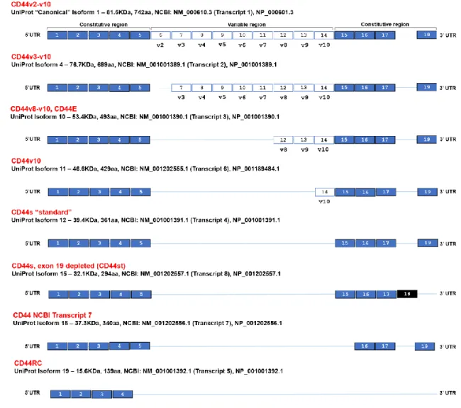
Figure 2. Human CD44 structure and examples of alternatively spliced mRNA transcripts.
9
Figure 2. Schematic representation of protein-associated glycan structures relevant in bladder cancer.
14
Figure 4. Schematic representation of PCR probes recognition zones. 30
Figure 5. Validation of CD44 expression in urine samples and immunohistochemistry of bladder cancer patients.
40
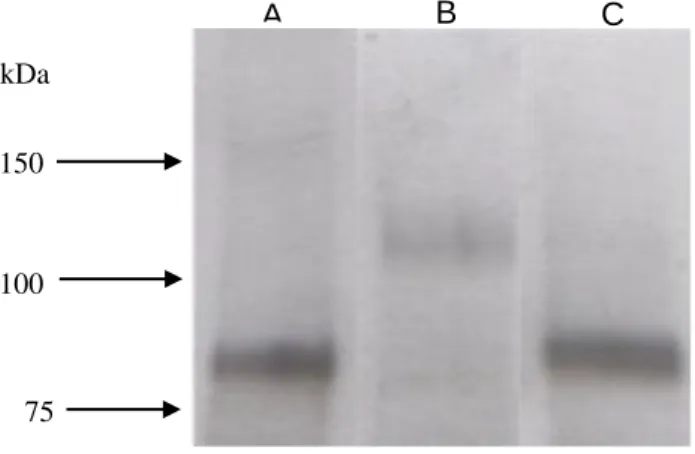
Figure 6. Western blot of T24, 5637 and HT1376 for CD44. 41
Figure 7. Graphical representation of CD44 isoforms expression in BC cell lines.
42
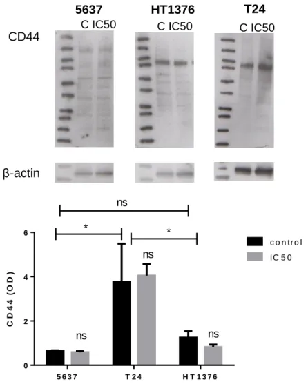
Figure 8. CD44 expression in superficial tumour derived cell line 5637 and muscle-invasive bladder cancer derived T24 and HT1376 cell lines.
42
Figure 9. Mass spectrometry spectra of identified O-glycoforms in T24 cells. 43
Figure 10. Identification of CD44 isoforms by mass spectrometry. 45
Figure 11. In situ proximity ligand assay and immunohistochemistry for the simultaneous detection of CD44/ST and dST as well as CD44/STn glycoforms.
47
Figure 12. Western blot analysis of bladder cancer tumours.
Figure 13. Graphic schematization of CD44 isoforms expression in NMIBC and MIBC.
48 49
Figure 13. Graphic schematization of CD44 expression in pre- and post-chemotherapy tumour samples.
CD44-glycoprofilling: Establishing the molecular basis for targeted therapeutics in bladder cancer| Eliana Soares
List of tables
Page
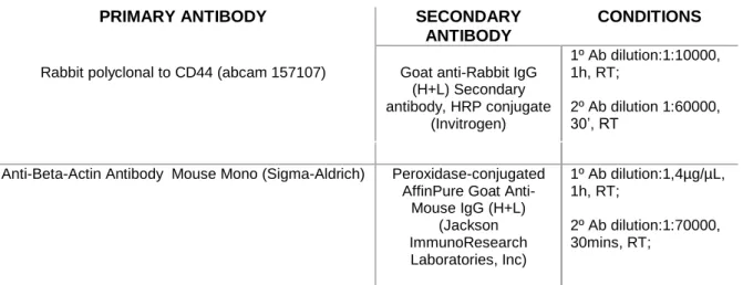
Table 1. Specification of primary and secondary antibodies conditions Table 2 - Representation of CD44 isoforms analysed compared to decribed and predicted isoforms
31 45
Abbreviations
BC Bladder cancer
BCG Bacillus Calmette-Guérin BCICs Bladder cancer initiating cells
CAMs Cell adhesion molecules
CIS Carcinoma in situ
C1GalT-1 Core 1, β1-3 galactosyltransferase COX-2 Cyclooxygenase-2
CSC Cancer stem cell
Ct Cycle threshold
dST Disialyl-T antigen
ECM Extracellular matrix
EMA Epithelial membrane antigen
ER Endoplasmic reticulum
FFPE Formaline-fixed paraffin embedded
FGFR3 Fibroblast Growth Factor 3
GA Golgi apparatus
GAG Glycosaminoglycan
Gal Galactose
GalNAc N-Acetylgalactosamine
GC Gemcitabine and cisplatin chemotherapy
GCH Glucocorticoid hormones
GlcNAc N-Acetylglucosamine
HA Hyaluronic acid
HABD Hyaluronan binding domain
HRAS Harvey rat sarcoma viral oncogene homolog
IL-1β Interleukin 1β
MIBC Muscle invasive bladder cancer
MVAC Methotrexate, vinblastine, adriamycin, and cisplatin chemotherapy
MoAbs Monoclonal antibodies
NMIBC Non-muscle invasive bladder cancer
OSTase Oligosacharide transferase
CD44-glycoprofilling: Establishing the molecular basis for targeted therapeutics in bladder cancer| Eliana Soares
PGE-2 Prostaglandin E2
PLA in situ Proximity ligantion assay
ppGalNAc UDPGalNAc-polypeptide N- acetylgalactosaminyl- transferases
PTEN Phosphatase and tensin homolog
PTM Post-translational modification
RB Retinoblastoma
Rg Receptor globulins
Ser Serine
S6T Sialyl-6-T antigen
SLea Sialyl Lewis a
SLex Sialyl Lewis x
ST Sialyl-T antigen
STn Sialyl-Tn antigen
T antigen Thomsen-Friedenreich antigen
TCC Transitional cell carcinoma
Thr Threonine
T-ICs Tumour initiating cells
Tis Tumour in situ
TUR Transurethral resection
TNF-α Tumour necrosis factor alpha
Chapter 1
CD44-glycoprofilling: Establishing the molecular basis for targeted therapeutics in bladder cancer| Eliana Soares
1. Introduction
1.1.
Bladder cancer
1.1.1. Epidemiology
Bladder cancer (BC) is the most common malignancy of the urinary tract, the seventh most common cancer in men, and the seventeenth in women (1). Also, it is the ninth most common cancer worldwide, with 429,000 new cases and 165,000 deaths in 2012 (2). In Portugal, BC is the eighth most common cancer, with 1829 new estimated cases and 900 deaths in 2010 (3). The disease is three times more iterated in men than in women and its incidence increases with age, reaching a peak between 50 and 70 years (4). Moreover, at diagnosis, women present more advanced stages of the disease and have less favourable prognosis (5).
Environmental risk factors have a significant role in BC initiation. Chemical or environmental exposures and chronic inflammation are well known risk factors for the development of BC and may lead to genetic and molecular changes (6). Critical exposures include aromatic amines, aniline dyes, nitrites and nitrates, acrolein, coal, and arsenic. Other casual factors include Schistosoma spp (Schistosoma haematobium) infection, and pelvic irradiation (7). However, tobacco smoke is the predominant risk factor associated with BC, accounting for 50% of BC cases (1). Genetic predisposition also has a significant influence on BC development, especially on the susceptibility to other risk factors (8).
Haematuria is the most frequent BC symptom, occurring in approximately 80% of patients. Other symptoms include urgency and dysuria, or, in more advanced tumours, pelvic pain, and symptoms related to urinary tract obstruction (1).
CD44-glycoprofilling: Establishing the molecular basis for targeted therapeutics in bladder cancer| Eliana Soares
1.1.2. Pathophysiology and disease progression
Urothelial cancers arise from two distinct but overlapping pathways: papillary and non-papillary. Approximately, 80% to 85% of urothelial cancers are papillary lesions, stemming from hyperplastic epithelium or minimal dysplasia of a preneoplastic clone. The continuous growth of this clone results in the development of low-grade superficial papillary tumours that present loss of heterozygosity of chromosome 9, frequent mutations in the Fibroblast Growth Factor 3 (FGFR3), Harvey rat sarcoma viral oncogene homolog (HRAS) and phosphatidylinositol 3-kinase (PI3KCA) genes (9). Non-papillary and invasive tumours are thought to arise from severe dysplasia or carcinoma in situ (CIS). In a non-papillary pathway, cells from the initial hyperplasia develop genetic instability with frequent loss of tumour suppressor genes, such as Retinoblastoma (RB) and p53 (10,11). Interestingly, most muscle-invasive lesions emerge without a prior history of disease (primary invasive bladder cancer) (12,13).
Urothelial carcinoma (transitional cell carcinoma or TCC), squamous cell carcinoma, and adenocarcinoma comprise the three main bladder cancerous lesions, with TCC accounting for approximately 90% of cases (14,15). At diagnosis, approximately 75% of patients present superficial lesions and low grade tumours, (stages Ta, T1, or tumours in situ [Tis]), 10% of which progress to recurrent muscle-invasive and metastatic disease (Figure 1) (12,13). Consequently, non-muscle invasive bladder cancer (NMIBC) is a chronic disease with varying oncologic outcomes requiring frequent follow-up and repeated treatments, making the cost per patient (from diagnosis to death) one of the highest of all cancers (16,17).
Figure 3 Schematic representation of bladder cancer staging and grading (18). The stage of the primary tumour (T) is based on the extent of penetration or invasion into the bladder wall.Tis, Tumour in situ: ‘‘flat tumour’’; Ta, Non-invasive papillary carcinoma; T1, Tumour invades subepithelial connective tissue; T2, Tumour invades muscle; T2a, Tumour invades superficial muscle (inner half); T2b, Tumour invades deep muscle (outer half); T3, Tumour invades perivesical tissue; T4, Tumour invades any of the following: prostate, uterus, vagina, pelvic or abdominal wall. Regarding tumour grading, bladder lesions can be classified as urothelial papilloma (a benign lesion), papillary urothelial neoplasm of low malignant potential (PUNLMP), low-grade papillary urothelial carcinoma and high-grade papillary urothelial carcinoma.
CD44-glycoprofilling: Establishing the molecular basis for targeted therapeutics in bladder cancer| Eliana Soares
1.1.2.1. Diagnosis and therapeutics of bladder cancer
The most common clinical presentation of BC is asymptomatic hematuria, which prompts evaluation with cystoscopy, renal function testing, and upper urinary tract imaging in adults over 35 years with irritative voiding symptoms, and risk factors for bladder cancer (1,14). Moreover, tremendous efforts have been put in the development of biomarker panels for early diagnosis, with promising results regarding several glycans, lectins and proteoglycans (18)
The standard therapy for low-grade NMIBC is transurethral resection (TUR) of the tumour, allowing staging and primary treatment design. Further management is based upon risk factors, stage, grade, multimodality and recurrence history. Accordingly, a dose of perioperative chemotherapy can be used to reduce the risk of recurrence, and adjuvant chemotherapy is reserved for patients with multiple or multifocal recurrence. Moreover, there are subgroups of high-risk NMIBC patients which endure TUR and perioperative chemotherapy, followed by intravesical Bacillus Calmette-Guérin (BCG) therapy (19). Of note, intravesical BCG therapy is highly effective in comparison to chemotherapy alone (20). However, only two thirds of patients respond to BCG and one third of those responders will have recurrent disease (21), urging biomarkers able to predict therapy response and stratify patients who benefit the most from treatment.
Upon tumour invasion of the bladder wall (T2), perivesical tissue (T3), or adjacent pelvic organs (T4), endoscopic tumour resection and intravesical therapy become insufficient and the standard approach is radical cystectomy with pelvic lymphadenectomy and adjuvant or neoadjuvant chemotherapy (cisplatin-based combinations: methotrexate, vinblastine, cisplatin and doxorubicin - MVAC, or gemcitabine and cisplatin - GC) (14,22). Radical cystectomy may achieve a good local control; however, the relapse rate after radical cystectomy is as high as 50%, depending on the pathological stage of the primary tumour and the presence of loco-regional or distant metastasis (23). Importantly, the introduction of cisplatin-based chemotherapy in recent therapeutic schemes has allowed improving survival in comparison to cystectomy alone (24). Moreover, cisplatin-based chemotherapy results in nearly 15.2 and 14.0 months overall survival for MVAC and GC, respectively, compared to monotherapy and other combinations (25). Data from retrospective surgical series show that despite the early use of such aggressive approaches, many patients with muscle invasive bladder cancer (MIBC) still have high risk of recurrence and death from bladder cancer. The majority of recurrences occur within 3 years, with approximately 75% of patients already presenting distant metastasis (23).
CD44-glycoprofilling: Establishing the molecular basis for targeted therapeutics in bladder cancer| Eliana Soares
Currently, there is a lack of specific biomarkers for targeting aggressive cell phenotypes. Additionally, the high molecular heterogeneity of MIBC is responsible for significant variations in disease course, as well as elevated recurrence and progression
rates, hampering patient stratification regarding treatment response (BCG,
chemotherapy). As such, to improve current clinical practices, bladder tumours of different stages have been widely screened for disease-specific glycosylation biomarkers capable of improving diagnose, surveillance, prognosis and offer novel therapeutic options.
Lately, there have been some evidences linking the expression of CD44 and the isolation of bladder tumour-initiating cells. Chan K, et al., (26) isolated tumour-initiating cells (T-ICs) from bladder tumours based on the expression of basal cell markers. Particularly, CD44+, CK5+, CK20- (T-ICs) cells could be successively transplanted up to 3
passages to induce tumour formation in vivo, with the tumours retaining the heterogeneity of the primary tumour. CD44+, CK5+, CK20- cells could yield either CD44+, CK5+, CK20- or
CD44-, CK5-, CK20+ (differentiated progeny) tumours. In contrast to T-ICs, the
differentiated subpopulation could not be successively transplanted, revealing its limited self-renewal and/or proliferative capacities. Later, the same authors stratified bladder cancer into subtypes regarding cellular differentiation states based on keratin profiles, demonstrating that undifferentiated (basal-like) cells were CD44+ (27). These results
suggest that CD44 expression characterizes undifferentiated BC cells with sustained self-renewal, thereby being a cancer stem cell (CSC) marker.
1.2.
Glycoprotein CD44: potential biomarker of poor
prognosis in bladder cancer
1.2.1. Molecular structure and biological role
The CD44 antigen is a multifunctional and densely glycosylated transmembrane glycoprotein that together with selectins, integrins and cadherins composes the cell adhesion molecules (CAMs) family, mediating cell-matrix and cell-cell interactions, cell adhesion, migration, and tissue integrity (28,29).
The CD44 gene comprises 19 exons (1-19), of which only exons 1-5 and 15-19 are conserved, while the remaining 9 exons (6-14 exons also known as v2-v10) undergo
CD44-glycoprofilling: Establishing the molecular basis for targeted therapeutics in bladder cancer| Eliana Soares
variable exons (v2-v10) without the conserved exon 18, constituting the bulkiest CD44 isoform (250 kDa) (30,31). In turn, the standard CD44 (CD44s or CD44h) constitutes a much shorter CD44 isoform by losing all variable exons and exon 18, yielding an 85 kDa protein (32,33) (Figure 2). Moreover, the CD44v3-v10 isoform loses both v2 and 18 exons, being primarily found in keratinocytes, while the isoform CD44v8-v10 (CD44E) loses both v2-v7 and 18 exons and is preferentially expressed in epithelial cells (34). In addition, CD44v10 isoform loses all variable exons except v10, as well as the constitutive exon 18. Finally, the CD44st isoform, also known as short-tail CD44, loses all variable exons and exon 19. More recently, two more CD44 isoforms, namely CD44 NCBI Transcript 7 and CD44RC were described in UniProt and NCBI databases (35,36). Particularly, CD44 NCBI transcript 7 loses all variable exons, exon 15 and exon 18, while CD44RC is solely constituted by the first four conserved exons (Figure 2) (35,36). Of note, CD44 has several predicted isoforms that remain to be described in research models or humans, urging more in-depth studies regarding these markers.
Moreover, CD44 presents multiple post-translational modification sites, with at least five N-glycosylation sites in its ectodomain and several potential O-glycosylation sites in the membrane proximal extracellular region (37,38); thereby affecting its ligand binding characteristics, modulating its functions and further diversifying its structure (39). CD44 can also be considered a “part-time” proteoglycan, since some of its alternative splicing variants present glycosaminoglycan (GAG)-initiation sites (40). Further modifications can occur through tyrosine sulphation (41).
Hyaluronic acid (HA), an important component of the extracellular matrix (ECM), is the most common, but not the only, ligand of CD44. HA is a linear polymeric GAG and there are at least three sites for its binding on the CD44 molecule, one of which is the “link” domain encoded by exon 2 (42), and the other two overlap in the region encoded by exon 5 (43). Although all CD44 isoforms contain HA recognition sites, not all cells expressing CD44 constitutively bind the HA ligand. Cells can express CD44 in an active, an inducible, or an inactive state with respect to HA binding. The differences in the HA binding state of CD44 are cell specific and have been related to post-translational modification patterns (44). This interaction between CD44 and HA has been reported in chondrocytes, being an important process to direct the assembly of pericellular matrices (45) and chondrosarcoma cell lines adhesion (46). In addition, cells undergoing repair appear to upregulate both CD44 and HA. Mukesh Jain, et al., demonstrated that CD44 and HA are expressed in minimal amounts on smooth muscle cells in normal arteries, in
CD44-glycoprofilling: Establishing the molecular basis for targeted therapeutics in bladder cancer| Eliana Soares
opposition to injured arteries where the expression of these two molecules was much higher (47).
Several lymphocyte functions also appear to be CD44-dependent. Namely, there is an increase in cell surface levels of CD44 in activated T-cells (48). Additionally, CD44 expression seems to mediate the adhesion of lymphocytes to vascular endothelial cells via its binding to HA, and this interaction seems to be associated to T-cell extravasation into inflammation sites in mice (49) and humans (50). The homing of lymphocytes to inflammatory sites through CD44–HA binding is enhanced by the induction of HA synthesis in vascular endothelium by the proinflammatory cytokines tumour necrosis factor α (TNF-α) and interleukin 1β (IL-1β) (51). Accordingly, the presence of CD44 splicing variants appears to be obligatory for the migration and function of Langerhans and dendritic cells from peripheral organs to lymph nodes for antigen presentation (52). As it was previously discussed CD44 presents different ligands beside hyaluronic acid, such as osteopontin. Osteopontin (OPN) is highly expressed in bone. It is also known as, early T-lymphocyte activation (ETA-I) (53). This protein has revealed to be associated with regulation of immune cells, namely, promoting cell-mediated immune responses, playing a role in chronic inflammatory diseases, infiltration of macrophages during inflammation and induction of pro-inflammatory cytokines expression (54,55).
As it was described above, CD44 is involved in cell-cell and cell-matrix interactions, proliferation, migration and adhesion, which are directly involved in tumour progression and metastasis (56,57). As such, the clinical relevance of CD44 will be discussed in the next sections.
CD44-glycoprofilling: Establishing the molecular basis for targeted therapeutics in bladder cancer| Eliana Soares
Figure 2 Human CD44 structure and examples of alternatively spliced mRNA transcripts. Blue filled boxes represent constitutive exons, white filled boxes represent alternative exons. The blue line represents the alternative region that is missing in each CD44 isoform. The exon box width is not proportional to the bp number. Exon 18, filled black, is not coding and contains early 3′UTR. Its inclusion gives rise to a short cytoplasmic tail mRNA that translates into CD44st. Seventy amino acids of cytoplasmic tail are encoded by exon 19. Cytoplasmic tail of CD44 contains intracellular signalling motifs and mediates interaction with cytoskeleton. Variable region of CD44 is a site of heavy O-glycosylation. The globular amino terminal domain of CD44 contains 3 disulphide bonds and two hyaluronic binding motifs.
1.2.2. Clinical relevance in solid tumours
The expression of CD44 isoforms and its binding to HA is associated with tumour growth and development (58). Moreover, some studies describe unusual CD44 transcripts not found in the corresponding normal tissues (30,59). Furthermore, it was demonstrated that in vivo tumour formation by human lymphoma Namalwa cells, stably transfected with CD44s, can be suppressed by a soluble human CD44s-immunoglobulin fusion protein disrupting the interaction between CD44s and its physiologic ligands. Moreover, no
CD44-glycoprofilling: Establishing the molecular basis for targeted therapeutics in bladder cancer| Eliana Soares
tumour growth was detected even 100 days after the initial tumour cell injection. These results may provide new insides for tumour growth control in vivo (60). Both CD44s and its isoform CD44v10 expression are highly associated with malignant melanoma, and CD44v10 appears to facilitate local invasion. Accordingly, treatment of B16F10-bearing C57BL/6 mice either with a CD44s-/ CD44v10-specific antibody, or with receptor globulins (Rg) containing the extracellular part of CD44s or CD44v10 linked to the constant region of the immunoglobulin kappa light chain resulted in a reduction of lung metastasis. However, only CD44 Rgs prevented spread and settlement of melanoma cells in distant organs. These findings confirm the involvement of both CD44s and CD44v10 in melanoma progression, and is suggestive for the use of Rgs as therapeutic reagents (61). A similar study in malignant melanoma also demonstrated that CD44 blockage by specific antibodies inhibits tumour growth and metastisation in vivo by disrupting CD44-HA interactions (62). In line with these findings, tumour cell lines with high levels of CD44 protein were shown to be capable of forming more aggressive tumours in animal models (63,64).
CD44 also seems to be involved in lymphocytes preservation. Particularly, CD44 appears to inhibit DNA fragmentation and apoptosis of immature lymphocytes and peripheral T lymphocytes induced by anti-CD3 monoclonal antibodies (MoAbs) and glucocorticoid hormones (GCH), therefore modulating T-cell survival (65). In addition, CD44 provided resistance to anti-integrin antibody induced apoptosis of colon carcinoma cell lines, demonstrating the possibility of this molecule involvement in poor prognosis of colon carcinoma and metastasis development (66).
Furthermore, osteopontin and CD44 intereractions have been associated to tumour dissemination and metastization in brain tumours (67). Through the interaction with integrin receptors on macrophages and CD44, OPN seems to promote a delayed immune system response, inducing cellular immunity and an anti-oxidant effect preventing cell damage (67). Moreover, tumour dissemination is associated to neovascularization. CD44 isoforms and integrin aVβ3 are highly associated with angiogenesis process in tumours. Expression of CD44, osteopontin and integrin aVβ3 have been demonstrated to be involved in endothelial cell migration by vascular permeability factor/vascular endothelial growth factor (68,69).
CD44-glycoprofilling: Establishing the molecular basis for targeted therapeutics in bladder cancer| Eliana Soares
There have been some studies relating the expression of CD44 proteins and several biologic and clinical roles in bladder cancer. Kuncová J, et al. (70) evaluated the expression of CD44s and CD44v6 in 5637 and HT1197 BC cell lines, reporting that expression of both isoforms was dependent on the differentiation state of cells. Particularly, undifferentiated 5637 cells presented a more regular positive staining for CD44 proteins, while the more differentiated HT1197 displayed variations in CD44 staining. Also, CD44 proteins expression was correlated with higher proliferative activity and stem-like phenotypes of HT1197 and 5637, as well as with cellular atypia in HT1197. Moreover, it was suggested that variations in CD44 expression might be connected to the derangement of differentiation in tumour cells, manifested by prominent cytological atypia in high-grade tumours.
It is known that CSCs can be isolated from several solid tumours, such as breast (71) and brain tumours (72); however the lack of bladder cancer stem (initial) cells (BCICs) markers has hampered the same progress in bladder cancer. Yang Y, et al. (73) reported that BCICs might be among the epithelial membrane antigen (EMA)- CD44v6+ subset isolated from bladder tumours by magnetic cell sorting, since both EMA- cells and CD44v6+ cells possess the ability for colony-forming, self-renewal and proliferation. Importantly, these cells are usually located in the basal layer of normal urothelium, the potential location of BCICs, and would infiltrate from basal layer towards superficial layer in tumourous urothelium.
Inflammation events in normal and CSCs might be associated to prostaglandin E2 (PGE2) signalling (74), which has been reported to regulate hematopoietic stem cells homeostasis (75). Cyclooxygenase-2 (COX-2), the PGE2-generating enzyme, is determinant to inflammation-related carcinogenesis (76) and COX inhibitors present antitumour activity (77). Having the above in consideration, Thanan R, et al. (78) suggested that COX-2 may play an important role in tumour initiation, regulation of stem cell proliferation and differentiation in inflammation-related bladder cancer carcinogenesis. Particularly, S. haematobium-induced BC was correlated with stemness marker Oct3/4 expression, while BC without associated infection correlates with CD44v6 expression. Moreover, the expression of Oct 3/4 and CD44v6 was associated with nuclear localization of COX-2, suggesting that inflammation could mediate stem cell proliferation and differentiation in bladder cancer.
In a clinical approach, CD44v9 was reported to be an important prognostic biomarker for disease progression and cancer-specific death in MIBC and high-grade NMIBC (79). Its localization in invasion fronts demonstrated the clinical value of CD44v9
CD44-glycoprofilling: Establishing the molecular basis for targeted therapeutics in bladder cancer| Eliana Soares
expression, which could be used as stratification marker to select patients who might progress to muscle-invasive bladder cancer. In turn, it was demonstrated that CD44s and CD44v6 correlate with tumour stage and grade (80). Particularly, low-grade papillary TCC exhibited staining of basal layer cells, with a decreasing intensity of staining towards the urothelium surface, while high-grade TCC displayed strong staining of CD44s and CD44v6 throughout the entire neoplastic urothelium. Tumour grade and depth of invasion also correlated with positive staining of CD44s and CD44v6, confirming the diagnostic and prognostic value of both markers. It was also evidenced that CD44v6 is an indicator of metastatic potential with an increased expression in higher stages and in tumours with infiltrating borders (80). Another study also associates CD44s expression with higher grade, stage, and density of tumour infiltrating lymphocytes, as well as shorter overall survival, while CD44v6 expression was associated with better prognosis, lower grade and increased overall survival (81). Contrastingly, another study reported that the loss of CD44v6 expression is an independent adverse predictor of recurrence and overall survival, further suggesting that routine evaluation of CD44v6 could be important in identifying high-risk patients which would benefit from more aggressive therapeutic approaches. Also it was hypothesised that CD44v6 expression could be a much better predictor of recurrence than BC grade (82).
The knowledge regarding the clinical significance of CD44 in BC is still scares, which allied to conflicting reports has hampered the true development of targeted therapies involving this marker. These discrepancies can be explained by the lack of standard immunohistochemical assays, the use of antibodies with different specificities, and differences in the clinicopathological status of bladder tumours used in the different studies. Therefore, integrative and standardized studies are necessary to elucidate the role of CD44 in BC as it holds an important biological and clinical value and may serve as therapeutic target. Moreover, the structural complexity of this glycoprotein, including the coexistence of different splicing variants in the same tumour (83) and significant heterogeneity in glycosylation patterns (39) also may explain the existence of conflicting data. Therefore, it is crucial to determine the nature of the splicing variants expressed by bladder tumour of different histopathological natures, as well as their biological and clinical significances. Moreover, since CD44 is a highly glycosylated protein, exploiting its O-glycosylation patterns may allow narrowing down its isoforms to clinically relevant glycospecies capable of improve BC management (detection, prognosis, follow-up, and therapy selection).
CD44-glycoprofilling: Establishing the molecular basis for targeted therapeutics in bladder cancer| Eliana Soares
1.3.
Protein glycosylation and cancer
1.3.1. Patterns of glycosylation
Protein glycosylation is the most frequent post-translational modification (PTM) of membrane-bound and secreted proteins. This non-templated process involves the highly coordinated action of several glycosyltransferases, glycosidases and nucleotide sugar transporters in the endoplasmic reticulum (ER) and Golgi apparatus (GA) to generate carbohydrate-associated proteins. Glycosylation increases proteome complexity due to the diversity in sugar compositions, glycosidic linkages (N- , O- and C-linked glycosylation, glypiation (GPI anchor attachment) and phosphoglycosylation), chain length and substitution patterns (84–88). Given its structural diversity, glycans are important mediators of a plethora of biological events, such as cell-cell adhesion, cell differentiation, migration, signalling transduction, immune recognition, host-pathogen interactions, protein folding, traffic and stability (89–91).
Two main classes of glycans can be found at the cell surface, namely N- and O-glycans (Figure 3). Although glycosylation has been characterized as a post-translational modification, N-glycosylation often occurs during the translation and transport of proteins into the ER, promoting the proper folding of newly synthesized polypeptides. Precursor N-glycan synthesis begins on the cytosolic face of the ER and is further elongated after the initial structure is flipped into the ER lumen (91). During N-glycan synthesis, a 14-sacharide “core” unit (Glc3Man9GlcNAc2-) is transferred by oligosaccharide transferase (OSTase) to asparagine (Asn) residues in an Asn-X-Ser/Thr sequence of the nascent protein, with X being any amino acid except proline. The diversification of the glycans occurs in the GA to yield either partially unprocessed oligomannose antenna or, more frequently, complex or hybrid type structures (92). Mature N-glycans often yield highly relevant terminal structures, such as Lewis (Le) blood group related antigens and ABO(H) blood group determinants (93). Further glycome complexity is added by sugar phosphorylation, O-acetylation of sialic acids, and O-sulfation of galactose and N-acetylgalactosamines (94).
N-glycosylation does not prevent glycosylation from happening, as
O-glycosylation commonly takes place on glycoproteins previously N-glycosylated in the ER.
O-glycosylation occurs post-translationally by covalently α-linking a
CD44-glycoprofilling: Establishing the molecular basis for targeted therapeutics in bladder cancer| Eliana Soares
by UDPGalNAc-polypeptide N- acetylgalactosaminyl- transferases (ppGalNAc-Ts), forming the simplest O-glycan Tn antigen. Subsequently, T antigen (core 1 or Thomsen-Friedenreich antigen) results from the attachment of a galactose (Gal) to GalNAc by T synthase (β1-3 galactosyltransferase or C1GalT-1) (95). Moreover, T antigen can be the precursor of several more complex core structures (from core 2 to 8) bearing similar terminal structures than mature N-glycans (96). In addition, Tn and T antigens can be sialylated by sialyltransferases, forming the sialyl-Tn (STn), sialyl-T (ST) and disialyl-T (dST) antigens. Upon formation of the STn antigen, any further processing of the oligosaccharide chain stops (97).
In cancer tissues, glycosylation patterns are profoundly altered, being characterized by the expression of highly branched and heavily sialylated/fucosylated glycans, alterations in glycan terminal structures, and overexpression of truncated glycans (18,98). Glycome alterations at the cell surface derive from the synergism of different events that go beyond altered glycosyltransferases and/or glycosidases expression. These may include alterations in peptide backbone or nascent glycan structures (99), glycosidases or glycosyltransferases activity (100,101), chaperone functions (102), glycosyltransferases mislocation in secretory organelles (103), and the bioavailability of sugar nucleotide donors and cofactors (104). Importantly, many glycoepitopes constitute tumour-associated antigens. Those found at the cell surface are easily accessible to antibodies and lectins to selectively target specific tumour cells, while those secreted or shed into bodily fluids can be explored for non-invasive cancer diagnosis through serological assays. As such, cancer specific alterations to protein glycosylation provide a unique opportunity for clinical intervention.
CD44-glycoprofilling: Establishing the molecular basis for targeted therapeutics in bladder cancer| Eliana Soares
Figure 4 Schematic representation of protein-associated glycan structures relevant in bladder cancer (18). The figure represents specific N-linked and O-linked glycan structures, as well as terminal Lewis and sialylated Lewis structures that have biological significance in bladder cancer. Key enzymes mediating the addition of specific sugars are also shown. Protein N-glycan alterations include the β1-6 branching of N-glycans in result of GlcNAcT-V (GnT-V) overexpression, and the addition of bisecting GlcNAc branches by GlcNAcT-III (GnT-III) glycosyltransferases. Alterations in O-glycosylation pathways are also a common hallmark of malignant transformations of the bladder. Herein, we represent the overexpression of simple mucin-type O-glycans and their sialylated counterparts, T, sialyl T (ST), Tn and sialyl Tn (STn) antigens. Altered expression of terminal structures is also a common feature of bladder tumours. Namely, the abnormally low or absent expression of ABO(H) blood group determinants is frequently found in high grade and invasive disease. Carbohydrate terminal Lewis antigens are significantly under-expressed in healthy urothelium when compared to bladder tumours and are
also highlighted here. Lewis type 1 antigens includes Lewisa (Lea), and sialyl Lewisa (SLea), while the type 2 group includes
Lewisx (Lex) and sialyl Lewisx (Slex).
1.3.2. Altered glycosylation in bladder cancer
It has been long known that advanced stage bladder tumours present severe dysregulations in glycosylation pathways, translated by the loss of terminal ABO blood group determinants at the cell-surface of ABH secretor individuals (105,106), over- and/or de novo expression of short-chained O-GalNAc glycans (107), Lewis blood group related antigens, and their sialylated counterparts (108,109), as well as oversialylation and fucosylation of glycan chains (110). Particularly, ABO(H) antigens in the initial biopsy of bladder carcinomas is predictive of a much higher chance of subsequent invasion than in those tumours in which the ABO(H) antigens are detectable (111). However, a significant number of patients whose initial tumours were reported as blood group antigen negative failed to develop an invasive tumour (111). It is possible that these conflicting results may, at least in part, be explained by differences in methodology, interpretation, or both. These antigens are present on normal bladder epithelium but not on some low-grade and early-
CD44-glycoprofilling: Establishing the molecular basis for targeted therapeutics in bladder cancer| Eliana Soares
stage papillary transitional cell carcinomas of the bladder (112). In bladder urothelium the most studied change has been the deletion of blood group A antigens from A individuals and H antigen from O individuals. Moreover, the loss of activity of the A and B gene-encoded transferases (112), and ABO(H) gene and/or its promoter hypermethylation (105) have been suggested to be amongst the events driving these, explaining the deletion of these antigens in bladder tumours.
The A, B, H antigens have biosynthetic and structural similarities with the Lewis antigens, including the type 1 Lewisa and type 2 Lewisx antigens and their sialylated
counterparts, namely sialyl-Lewis a (SLea) and x (SLex). Several authors have associated
Lewisa expression patterns with malignant transformations, reporting significantly lower
expression of this antigen in healthy urothelium when compared to invasive tumours (113,114). As such, the expression of Lewisa can be associated with worse bladder
cancer phenotypes. Moreover, Lewisa antigen expression patterns change at an early
neoplastic stage, suggesting that Lewisa determination might be useful in the diagnosis of
very early premalignant changes in the urothelium (115). In addition, the employment of Lewisa staining scores allows the sub-classification of histologically identical tumours into
prognostically different groups, pointing to a relationship between the pathological grade and stage of the evaluated tumours and a morphological and functional de-differentiation (115). Given this, Lewisa antigen is a valuable functional marker of the malignant potential
in superficial bladder cancer. In turn, the Lewisx antigen is not expressed in normal
urothelium, except for occasional umbrella cells, but is demonstrated in most invasive tumours, regardless of blood type and secretor status of the individuals studied (106). In cancer cells, both SLea and SLex mimic their normal expression on blood cells, allowing
cancer cell binding to endothelial selectins and extravasation of cancer cells (116). Minimal structural alterations in SLe terminal motifs may significantly alter their biological behaviour, allowing them to be explored has biomarkers of disease. Namely, a glycan epitope that is very similar to the selectin ligand SLex, the sialyl-6-sulfo Lewisx, is
preferentially expressed in normal epithelial cells compared to cancer cells (117). Essentially all genes involved in the synthesis of SLex are predicted to be the same as
those governing the synthesis of sialyl-6-sulfo Lewisx, except for the genes engaged in its
sulfation. This finding supports the hypothesis that reduced expression of sialyl-6-sulfo Lewisx induces SLex expression in cancers(118). Sialyl-6-sulfo Lewisx antigens are
expressed in bladder urothelial carcinomas playing divergent roles in its progression, since it promotes E-selectin-mediated tumour cell adhesion to vascular endothelial cells,
CD44-glycoprofilling: Establishing the molecular basis for targeted therapeutics in bladder cancer| Eliana Soares
enhance anti-tumour immune responses (119). A structure very similar to SLea termed
disialyl-Lewisa is preferentially expressed in non-malignant cells, and is useful as a marker
for tissue injuries occurring in benign diseases (120). The structural difference between SLea and disialyl-Lewisa is the presence of one extra sialic acid residue attached to the
C-6 position of βGlcNAc in the carbohydrate structure of the latter glycan epitope. This implies that the α-2-6 sialylation of the GlcNAc moiety is impaired in cancer cells compared to non-malignant cells. Moreover, this event has been associated with the epigenetic silencing of the ST6GalNAcIV gene responsible for the α2-6 sialylation of the βGlcNAc moiety in cancer cells (120,121). The sialylated forms of Lewis antigens have also been associated with BC malignant potential. Particularly, it has been demonstrated that loss/reduction of SLea expression was associated with higher atypia grade (122),
while SLex has been closely linked to invasive and metastatic potential of primary bladder
tumours (123). Yet, another study demonstrated no associations between SLex, grade or
stage in urothelial carcinoma of the renal pelvis, ureter, and urinary bladder (122). Notwithstanding, the overall increase in cell-surface sialic acid content was shown to reduce the attachment of metastatic tumour cells to the extracellular matrix, possibly protecting them from recognition by the alternative pathway of complement activation and favouring metastatic spread (124).
ABO(H) loss may also stem from premature stop in protein O-GalNAc glycosylation, ultimately translating in the accumulation of Tn, T and their sialylated counterparts STn and ST antigens.In particular, the overexpression of ST6GalNAc-I has been found to promote the premature sialylation of the Tn antigen and consequent formation of the STn antigen in bladder cancer (125,126). Specifically, the STn antigen is absent in the healthy urothelium, while being present in more than 70% of high-grade NMIBC and MIBC, denoting a cancer specific nature (125). This posttranslational modification of cell surface proteins is mostly expressed in non-proliferative tumour areas, known for their high resistance to cytostatic agents currently used to improve the overall survival of advanced stage bladder cancer patients (125). Recently, a novel STn-dependent mechanism for chemotherapeutic resistance of gastric cancer cells to cisplatin has been described, in which STn protects cancer cells against chemotherapeutic-induced cell death by decreasing the interaction of cell surface glycan receptors with galectin-3 and increasing its intracellular accumulation (113). The relationship between chemoresistance and STn overexpression remains to be fully explored in bladder cancer. Furthermore, STn expression is significantly higher in MIBC when compared to NMIBC, denoting its association with muscle invasion and poor prognosis (127). Studies in vitro
CD44-glycoprofilling: Establishing the molecular basis for targeted therapeutics in bladder cancer| Eliana Soares
have further demonstrated that this antigen plays an important role in bladder cancer cell migration and invasion through mechanisms so far unexplored (125,128). However, glycoproteomics studies of bladder cancer cell models highlighted that STn was mainly present in integrins and cadherins, further reinforcing a possible role for this glycan in adhesion, cell motility and invasion (128). Also, recent work from our group has demonstrated the presence of STn in lymph node and distant metastasis, strengthening the notion that STn expression may influence cancer cell motility and matastization capability (129). Moreover, STn inclusion improved the predictive capacity of a molecular model proposed for stratification and prognostication of bladder tumours based on keratin (KRT14, 5, and 20) expressions (129). Accordingly, the STn antigen has been associated with basal-like phenotypes (KRT14+ and/or KRT5+, KRT20-) facing worst prognosis (129). Furthermore, STn-expressing BC cells have shown the ability to induce a tolerogenic microenvironment by impairing dendritic cells maturation, allowing cancer cells to evade innate and adaptive immune system responses (130). Interestingly, the tolerogenic effect of short-chained O-glycans has also been correlated with bladder tumour metastasis through a mechanism in which MUC1 carrying core 2 O-glycans functions as a molecular shield against NK cells attack, thereby promoting metastization (131). In addition, STn expression in bladder cancer tissues has been used in combination with other surrogate markers of tumour aggressiveness envisaging patient stratification regarding disease stage and therapeutic benefit. Specifically, STn and sialyl-6-T (S6T), an STn-related antigen, expression are independent predictive markers of BCG treatment response and were found useful in the identification of patients who could benefit more from this immunotherapy (132). Moreover, STn was found to be a marker of poor prognosis in bladder cancer and, in combination with PI3K/Akt/mTOR pathway evaluation, holds potential to improve disease stage stratification (127).
In turn, several reports associated the presence of Tn and T antigens with recurrence and metastization in bladder cancer, suggesting that these antigens may be surrogate markers of profound cellular alterations (133,134). Also, there is growing evidences linking the overexpression of the sialyl-T antigen and ST3Gal.I, the enzyme responsible by T antigen sialylation, with bladder cancer aggressiveness, recurrence (135), poor prognosis, and tumour grade (133,136). Moreover, the expression of T antigen is significantly associated with higher risk for subsequent recurrences with deep muscle invasion and metastatic involvement of regional lymph nodes (133,136,137). The fact that these simple glycans are absent, significantly under-expressed or restricted to some cell
CD44-glycoprofilling: Establishing the molecular basis for targeted therapeutics in bladder cancer| Eliana Soares
types in healthy tissues, makes them ideal diagnostic and therapeutic targets for bladder cancer therapy (138).
1.3.3. CD44 glycosylation in solid tumours
As previously described, CD44 undergoes extensive post-translational
modification, including N- and O-linked glycosylation and substitution with high molecular weight glycosaminoglycans (139), thereby modulating its biological functions. Particularly, Dasgupta et al. demonstrated that CD44s O-linked glycosylation decreased CD44s-mediated adhesion to hyaluronate (HA), while N-linked glycosylation had minimal influence on CD44 function. These findings suggest that O-linked glycosylation may be as important as alternative splicing in the regulation of CD44 function and the broad spectrum of biological processes attributed to it, including normal development, tumour metastases, and lymphocyte function (38). Contrastingly, Bartolazzi et al. showed that treatment of a panel of human cell lines which constitutively express CD44 with the inhibitor of N-linked glycosylation tunicamycin results in the loss of attachment of these cells to HA-coated substrate (140) The authors further demonstrated that treatment of the same cells with deoxymannojirimycin, which inhibits the conversion of high mannose oligosaccharides to complex N-linked carbohydrates, results in either no change or an increase in CD44-mediated adhesion to hyaluronate, suggesting that complex N-linked oligosaccharides may not be required or even inhibit CD44-HA interaction. Using human melanoma cells stably transfected with CD44 N-linked glycosylation site-specific mutants it was shown that integrity of five potential N-linked glycosylation sites within the HA recognition domain of CD44 is critical for HA binding. Mutation of any one of these potential N-linked glycosylation sites abrogates CD44-mediated melanoma cell attachment to HA-coated surfaces, suggesting that all five sites are necessary to maintain the HA-recognition domain in the appropriate conformation. They also demonstrated that mutation of serine residues, which constitute the four Ser-Gly motifs in the membrane proximal domain and provide potential sites for glycosaminoglycan side chain attachment, impairs HA binding (140). These observations suggest that the relevance of N-glycosylation sites for CD44 functions is site specific. Olgun Guvench developed molecular dynamics simulations that provide atomic-resolution mechanistic understanding of CD44-HA interaction to help bridge gaps between existing experimental binding and structural biology data.Findings from these simulations included that Tyr42 may function
CD44-glycoprofilling: Establishing the molecular basis for targeted therapeutics in bladder cancer| Eliana Soares
as a molecular switch that converts the HA-binding site from a low affinity to a high affinity state; in the partially disordered form of hyaluronan binding domain (HABD), basic amino acids in the C-terminal region can gain sufficient mobility to form direct contacts with bound HA to further stabilize binding; and terminal sialic acids on covalently attached N-glycans can form charge-paired hydrogen bonding interactions with basic amino acids that could otherwise bind to HA, thereby blocking HA binding to glycosylated CD44 HABD (141). Glycosylation modulates CD44 biological role not only by affecting its interaction with ligands. Namely, Hu et al. suggest that the Lewis y antigen, as an important component of the molecular structure of CD44, promotes proliferation and inhibits apoptosis of ovarian cancer cells, leading to drug resistance via activation of PI3K/AKT signalling pathways. Moreover, it is proposed that the Lewis y antigen, as a structural component of CD44, integrins a5b1 and avb3, as well as EGFR, play a role in various cell adhesion processes that mediate both cell adhesion and drug resistance (142).These results suggested that fucosylation of CD44 is related to drug resistance in ovarian cancer. Campos et al. uncovered CD44-STn glycoforms in gastric cancer patients serum, further validating its expression in gastric cancer tissue, suggesting that aberrantly expressed CD44 as potential biomarkers in gastric cancer (143). Particularly, the CD44v6 expression levels were associated with pre-malignant and malignant lesions of the stomach, providing a potential biomarker for gastric mucosa transformation (143). Singh et al. also reported the coexpression of oncofetal carbohydrate antigens T and STn on CD44 splice variants providing a link between cancer-associated changes in glycosylation and CD44 splicing, both of which correlate with increased metastatic potential of colon cancer (144).
Regarding BC, Carrascal et al. reported that STn+-CD44 cancer cells impair
dendritic cells (DC) maturation and endow DCs with a tolerogenic function, limiting their capacity to trigger protective anti-tumour T cell responses, suggesting that STn antigens and, in particular, STn+ glycoproteins, such as CD44, are potential targets for
circumventing tumour-induced tolerogenic mechanisms (145). More recently,
glycoproteomic analyses of advanced bladder tumours based on enzymatic treatments, Vicia villosa lectin-affinity chromatography enrichment and nanoLC-ESI-MS/MS analysis resulted in the identification of several key cancer-associated glycoproteins carrying altered glycosylation, including STn+-CD44 (146). These observations suggest that
exploring CD44 glycosylation may improve its biomarker potential. Nevertheless, the comprehensive glycomics mapping of CD44, including N- and O-glycan patterns, in
Chapter 2
2. Aims and Scopes
Bladder Cancer presents one of the highest recurrence rates amongst solid tumours, and constitutes the second deadliest disease of the genitourinary track. Moreover, the only therapeutic approach for managing advanced patients are cisplatin-based regimens that often fail to prevent disease relapse and dissemination. The high molecular heterogeneity presented by advanced bladder tumours and the lack of specific biomarkers for targeting more aggressive cancer cells remain challenging topics in bladder cancer management.
According to the current state-of-the art, CD44 presents an opportunity to address more aggressive bladder cancer cells and consequently improve these patients management. However, the gene encoding for CD44 generally undergoes significant alterative splicing, which results in many functionally distinct isoforms of variable molecular weights and glycosylation sites. Nevertheless, at the moment there are no consensus regarding the nature of CD44 isoforms associated to muscle invasion, which would be crucial for designing targeted therapeutics.
Having as starting point preliminary associations between increased CD44 levels in urine of cancer patients with more aggressive forms of the disease (Appendix 1), the first part of this work will be devoted to confirming these observations. The second part of the work will focus in disclosing the nature of CD44 isoforms associated with muscle invasion and ultimately chemoresistance using molecular models and patient samples, envisaging the molecular rationale for intervention. Emphasis will also be set on CD44 O-glycosylation patterns with the objective of narrowing down the isoforms to clinical relevant species.
Based on these observations, this work comprehends the following specific aims:
i. Identification of clinically relevant CD44 isoforms for targeted therapeutics;
ii. O-glycome characterization of CD44 isoforms envisaging highly cancer-specific
target;