Mestre em Bioquímica Estrutural e Funcional
Characterization of Nitric Oxide Reductase
(NOR) from
Pseudomonas nautica
, a Study
on Biologic Nitric Oxide Reduction
Dissertação para obtenção do Grau de Doutor em Bioquímica
Orientador: Isabel Moura, Professora Catedrática, Faculdade de Ciências e Tecnologia da Universidade Nova de Lisboa
Presidente: Prof. Doutor António Manuel Dias de Sá Nunes dos Santos
Arguentes: Prof. Doutora Margarida Maria Portela Correia dos Santos Romão
Prof. Doutor José João Galhardas de Moura
Vogais: Prof. Doutora Maria de Lurdes Afonso Barreira Alves de Mira
Prof. Doutora Maria Conceição Santos Silva Rangel Gonçalves
Prof. Doutor Manuel Aureliano Pereira Martins Alves
Prof. Doutora Maria Margarida Catalão Almiro e Castro
DoutoraCristina Maria Grade Couto da Silva Cordas
nº arquivo
Characterization of Nitric oxide reductase
(NOR) from
Pseudomonas nautica
, a study on
biologic nitric oxide reduction.
Lisboa 2011
Dissertação apresentada para a obtenção do grau
de doutor em Bioquímica, especialidade em
Bioquímica-Fisica, pela Faculdade de Ciências e
Tecnologia da Universidade Nova de Lisboa.
Dissertation submitted to obtain the phD degree
in Biochemistry, specialty in
Physical-Biochemistry, by the Faculdade de Ciências e
Tecnologia from the Universidade Nova de
“Saber interpor-se constantemente entre si próprio e as coisas
é o mais alto grau de sabedoria e prudência”
Em primeiro lugar gostaria de agradecer à minha orientadora, a Professora Doutora
Isabel Moura, não só por me ter acolhido na sua equipa, bem como por sempre se ter
mostrado presente durante todo o tempo que permaneci no seu laboratório. Acabei por
ser mal acostumado por ela, por duas razões: primeiro nunca se negou a ver e/ou discutir
um resultado por mim obtido e segundo por nos brindar regularmente, a mim e a todos
os demais com as suas iguarias gastronómicas. Ao Professor José J.G. Moura, pela sua
disponibilidade e também por me ter permitido inserir no seu grupo de investigação, o
Bioin.
Não posso deixar de agradecer à Professora Dr.ª Alice Pereira e ao Professor Dr.
Pedro Tavares que sempre se mostraram disponíveis para me ouvir, aconselhar e ajudar,
mesmo nas conversas rápidas que tínhamos. Todos os conselhos que me deram foram
determinantes para a minha progressão nos estudos apresentados nesta tese, portanto o
meu mais sincero obrigado, a ambos.
À Cristina Cordas, não existem palavras para descrever a minha gratidão, não só pela
sua amizade e companheirismo, tanto no trabalho como fora deste. Gostava também de
agradecer ao Carlos Martins, por toda a informação que me disponibilizou sobre o
sistema estudado, e em especial por todas aquelas amostras que lhe fui “roubando”, com
a sua autorização, da sua gaveta, muito obrigado, pois sempre me poupaste umas horas de
trabalho.
Não menos importantes, terei de agradecer a enorme amizade do Filipe Folgosa, do
Rui Almeida, e da Susana Ramos, pois tiveram um contributo importante nesta jornada. À
restante equipa dos grupos Bioin e Bioprot, com elementos que nela se inserem e/ou
inseriram, à Sofia à Cristina Timóteo, ao Cristiano Mota, à Ana Teresa, à Marta, à Raquel,
à Luísa ao Pablo à Gabi, ao Luís Nobre, e aos restantes “Césares” que por ali foram
passando.
Não poderei deixar de agradecer à minha família: à mana Isabel e ao meu cunhado
Beto por todo o apoio incondicional e sobretudo por me terem dado o Bernardo para
tomar conta, sempre me aliviou a cabeça e me pintou uns sorrisos na cara. Quando
defendi a passada tese de mestrado estavam vós atendendo a chegada do pequeno
meu caminho e claro, não menos importante à Carmen, que igualmente me encorajou.
É imperativo prestar um sentido agradecimento ao meu “gang” de amigos. Nem este
conjunto de agradecimentos estaria completo sem tal menção. Ao Zé que mesmo fora de
Lisboa me encorajou, apoiou, escutou e com ele descobri que há um refrão de uma
música eterna que está completamente errado quando é transposto para o quotidiano. Às
meninas dos meus olhos: à Jack pela sua eterna amizade, à Vera pela sua alegria
contagiante, que piora cada vez que nos juntamos, vai sempre tudo a baixo. À Pat
(riqueza) pelo seu carinho, à minha Sue (pureza) pela sua pseudo terapia. Ao Hugo, que
retirou a palavra menos do meu dicionário, ao Tiago que sempre me acalmou a ansiedade,
ao Miguel pela sua presença e sim, existe alguém mais irrequieto que eu, e claro ao Bruno
pelo seu constante modo de festa. À Gi a e ao Plim, que sempre me encorajaram e
mostraram quem é o capitão, ao João Leandro pela sua infinita bondade e amizade. E um
especial agradecimento à Ana Bicho, simplesmente por ser inesperada, incansavelmente
verdadeira e um exemplo de força a seguir. Não menos importante, um agradecimento à
Joana Gordo, que me aturou todos os dias à hora de almoço e nunca deixou que eu me
fosse a baixo. Um especial agradecimento terá de ser feito a alguns dos amigos anteriores
que perderam de uns minutos a algumas horas do seu tempo a elaborar os separadores
desta tese.
Quero agradecer ao CQFB / Departamento de Química da FCT/UNL pelas
condições de trabalho proporcionadas.
O meu agradecimento à Fundação para a Ciência e Tecnologia pelo financiamento, a
bolsa de doutoramento atribuída (SFRH/BD/39009/2007), sem a qual a execução deste
trabalho não seria possível.
Sei que se começasse a enunciar os restantes que se cruzaram comigo durante esta
jornada, iria esquecer uma boa dezena de nomes importantes, não querendo parecer
banal, agradeço a todos aqueles que me ajudaram e acima de tudo, que sempre
acreditaram que eu seria capaz de chegar ao final. A todos vós, o meu mais sincero
1. Sofia R. Pauleta, Américo G. Duarte, Marta S. Carepo, Alice S. Pereira, Pedro
Tavares, Isabel Moura, José J. G. Moura, “NMR assignment of the apo-form of a
Desulfovibrio gigas protein containing a novel Mo-Cu cluster”, Biomolecular NMR
Assignment Journal (2007). Jul 1;(1):81-3.
2. Shabir. Najmudin, Cecília. Bonifácio, Américo G. Duarte, Sofia R. Pauleta, Isabel
Moura, José J. Moura, Maria J. Romão. “Crystallization and crystallographic
analysis of the apo form of the orange protein (ORP) from Desulfovibrio gigas”,
Acta Crystallogr Sect F Struct Biol Cryst Commun. (2009) Jul 1;65(Pt 7):730-2.
3. Andrea Santos, Américo G. Duarte, Alexander Fedorov, José M.G. Martinho,
Isabel Moura. “Rubredoxin mutant A51C unfolding dynamics: a Förster
Resonance Energy Transfer study”, Biophysical Chemistry (2010), May;
148(1-3):131-7.
4. Cristina G. Timoteo, Alice S. Pereira, Carlos S. Martins, Sunil G. Naik, Américo
G. Duarte, José J. G. Moura, Pedro Tavares, Boi, H. Huynh, Isabel Moura. “Low
Spin heme b3 in the Cathalytic Center of Nitric Oxide Reductase from Pseudomonas
A desnitrificação ou redução dissimilativa do nitrato é uma via metabólica que se
insere no ciclo do nitrogénio. Esta via metabólica envolve quatro reacções de redução: o
nitrato é reduzido a nitrito, o nitrito a óxido nítrico (NO), este a óxido nitroso (N2O) e o
último passo a dinitrogénio. Cada um destes passos envolve metaloenzimas específicas
que catalizam as anteriores reacções, nomeadamente as reductases do nitrato, as
reductases do nitrito, as reductases do óxido nítrico (NOR) e as reductases do óxido
nitroso, respectivamente.
A NOR é uma metaloenzima que realiza a redução do NO. Esta reacção envolve dois
electrões e dois protões, tendo como produto o N2O e H2O. Esta proteína insere-se na
super-família das oxidases terminais, pois são proteínas integrais de membrana com 12
hélices α transmembranares, numa disposição altamente conservada. As cNORs são
compostas por duas subunidades. A subunidade NorC, com uma massa molecular de 17
kDa, que contém um hemo c de spin-baixo, ligado covalentemente à cadeia polipeptídica,
com uma coordenação His/Met. A segunda subunidade, a NorB, também designada por
subunidade catalítica, apresenta uma massa molecular de 56 kDa. Contém um centro
hémico de spin-baixo (hemo b), um centro hémico de spin-alto (hemo b3) e um ferro
não-hémico (FeB). Estes dois últimos centros encontra-se acoplados antiferromagneticamente
e ligados em ponte por um oxigénio/grupo hidroxilo, formando assim o centro catalítico
da enzima. Recentemente, utilizando espectroscopia de Mössbauer, foi comprovado
inequivocamente que o hemo catalítico é na verdade hexa-coordenado, quando o átomo
de Fe se encontra no estado férrico ou ferroso. Estes resultados estão em concordância
com a estrutura tridimensional da NOR isolada da bactéria Pseudomonas (Ps.) aeruginosa, que
mostra a presença do ligando axial (His) e do ligando em ponte na forma como isolada.
O mecanismo e redução do NO é um assunto controverso, existindo contudo duas
hipóteses consideradas: o mecanismo cis que descreve a redução de duas moléculas de
substrato, com a coordenação das mesmas a um único átomo de Fe do centro catalítico, e
o mecanismo trans, onde ambos os átomos de Fe do coordenam uma molécula de
substrato. Esta classe de enzimas não cataliza apenas a redução de NO a N2O, cataliza
igualmente a redução de O2 a H2O, numa reacção que envolve quatro electrões e quatro
protões. O mecanismo de redução do O2 é também um assunto pouco esclarecido, sendo
nautica, através da aplicação de diferentes técnicas bioquímicas, electroquímicas e
espectroscopias, tais como a espectroscopia de ultra-violeta–visível e Ressonância
Paramagnética Electrónica (RPE).
Neste trabalho é apresentada a optimização do protocolo de purificação da forma
nativa da NOR de Ps. nautica. As fracções purificadas evidenciam um elevado grau de
pureza, a correcta estequiometria dos co-factores metálicos.
Os resultados espectroscópicos obtidos evidenciam novas características estruturais,
nomeadamente:
i) Os dados de UV-visível sugerem que o hemo b3 apresenta uma conformação
de spin-baixo.
ii) Os dados de RPE comprovam a existência de uma nova espécie de
spin-inteiro, espécie esta que provém do acoplamento antiferromagnético do hemo
b3 com o Fe não-hémico, ambos no estado férrico (b3-FeIII-FeBIII).
Foram ainda realizados ensaios de transferência electrónica, utilizando a enzima em
estudo, imobilizada na superfície de um eléctrodo de grafite. Os dados obtidos mostram
pela primeira vez, quatro processos de oxidação/redução (redox), correspondentes a cada
um dos centros metálicos da NOR de Ps. nautica. A figura que se segue resume os valores
obtidos para cada um dos centros de Fe da proteína em estudo.
Figura – Valores de potencial redox obtidos para os centros metálicos da NOR de Pseudomonas nautica. A figura mostra uma representação esquemática da enzima e os valores referem-se aos potenciais obtidos para cada um dos co-factores da enzima (vs. ENH).
Hemo c Hemo b Hemo b3 FeB
Os resultados obtidos através dos ensaios de resposta electroquímica directa
utilizando a NOR de Ps. nautica são:
i) O Fe não-hémico apresenta o potencial redox mais reduzido (-369 mV vs.
ENH).
ii) O potencial redox mais elevado pertence ao hemo do tipo c (+ 208 mV vs.
ENH).
iii) Os resultados do potencial redox para cada um dos centros metálicos, obtidos
com a dependência do pH, sugerem a existência de resíduos polares
conservados, em redor dos grupos prostéticos da enzima, que podem
estabilizar as cadeiras propiónicas destes.
Paralelamente, a subunidade NorC recombinante foi imobilizada na superfície de um
eléctrodo de grafite, sendo estudada a sua resposta electroquímica directa. Com os
resultados obtidos foi comprovado inequivocamente o potencial redox do hemo c. A
comparação deste valor com o resultado obtido para a proteína nativa, propõe a
existência de alterações estruturais, em redor do centro hémico, provavelmente devido à
inexistência da subunidade NorB.
Durante a realização deste trabalho e através de técnicas electroquímicas, foi
investigada a resposta catalítica da enzima na presença de NO e O2. Foi determinado o
número de electrões necessários aos processes de redução de cada um dos substratos,
sendo ainda comprovado que o produto da redução do O2 é a H2O, sem que ocorra a
formação de H2O2.
Ensaios cinéticos no estado estacionário, realizados com esta enzima, permitiram a
dedução de modelos cinéticos e a determinação de constantes cinéticas para a redução de
NO e O2. Estes ensaios foram obtidos utilizando o cit. c552 (isolado de Ps. nautica)
previamente reduzido, ou a enzima imobilizada num eléctrodo rotativo de grafite, sendo
os electrões necessários à reacção fornecidos pelo mesmo.
Este é o primeiro caso onde são reportados ensaios cinéticos na presença de O2
simultaneamente com o seu dador electrónico (cit. c552). Os ensaios cinéticos mostram:
forma a catalizar o substrato.
iii) A redução de O2 apresenta um perfil inibitório, pela presença do substrato.
Ensaios cinéticos na presença de NO, com dependência do pH, evidenciam a
presença de grupos ionizáveis acídicos, provavelmente Glu ou Asp, que podem pertencer
ao conjunto de resíduos conservados de entrada de protões, para o centro catalítico. Um
dos valores obtidos nunca tinha sido antes determinado (pKa1 = 3.27) e provavelmente
refere-se a um dos resíduos da via anteriormente mencionada, com uma maior exposição
Denitrification is a metabolic pathway from the nitrogen cycle where nitrate is reduced
to dinitrogen. This pathway involves four reduction steps: the nitrate reduction to nitrite,
followed by the reduction to nitric oxide (NO), from this to nitrous oxide (N2O), and
finally to dinitrogen. The enzymes involved in the mentioned steps are the nitrate
reductases, the nitrite reductases, the nitric oxide reductases (NOR) and the nitrous oxide
reductases, respectively.
The NORs perform the NO reduction to N2O, using two substrate molecules, two
protons and two electrons with the consequent product formation and water. These
enzymes belong to the heme copper oxidase superfamily, since they are integral membrane proteins, with 12 transmembrane α-helixes and a set of conserved residues.
They are composed by two subunits. The NorC subunit with a molecular weight of 17
kDa, comprises a low-spin heme c covalently bound to the polypeptide chain, with a
His/Met coordination. The second subunit, NorB, also named the catalytic subunit, with
a molecular weight of 56 kDa, harbouring two b-type hemes, one low-spin bis-His
coordinated (heme b), a high-spin heme b (heme b3), His coordinated and a non-heme
FeB. These last two iron centers are antiferromagnetic coupled and bridged by a
µ-oxo/hydroxo group, and together they compose the catalytic diiron center. Recently
Mössbauer spectroscopy proved that the catalytic heme b3 is in fact low-spin in both
ferric and ferrous states, indicating a six-coordination environment for this iron center in
both redox states. The Pseudomonas (Ps.) aeruginosa NOR crystal structure shows the
presence of the His ligand simultaneously with the µ-oxo bridge in the as-isolated form.
The substrate reduction mechanism is an issue of intense discussion, with the cis and
trans-mechanisms taken in consideration. The cis-mechanism descries that NO reduction
occurs in the catalytic center and only one of the iron atoms is coordinating the substrate
during catalysis. The trans-mechanism describes NO reduction with the binding of one
substrate molecule to each one of the iron atoms of the catalytic center. Different isolated
NORs have the ability of reducing O2 to H2O in a four electrons/four protons reaction.
The mechanism for O2 reduction is unknown, but it is presumed that substrate binds to
the catalytic heme b3, analogous to the terminal oxidases.
The main objective of this work was to isolate and characterize the Ps. nautica NOR,
nautica NOR. The achieved enzyme fractions were consistent, since they present high
purity and the correct metal stoichiometry.
The spectroscopic characterization made revealed new structural features:
i) The UV-visible absorption spectra suggest a low-spin conformation for the
catalytic heme b3.
ii) The EPR spectroscopy results show the existence of a new integer-spin species
rising from the heme b3-FeIII-FeBIII antiferromagnetic coupling.
Direct electron transference between the immobilized Ps. nautica NOR and an
electrode was accomplished. This is the first time four redox processes were distinguished
using this approach and indexed to the four Fe centers. The following figure show a
summary of the midpoint redox potentials determined for the Ps. nautica NOR.
Heme c Heme b Heme b3 FeB
cNOR/Ps. nautica + 208 + 43 -162 -369
Figure – Midpoint redox potentials for the NOR metal centers. Top, schematic representation of the NOR structure emphasising the four co-factors, with the correspondent ligands. Bottom, summary table of the midpoint redox potentials (vs. NHE), obtained in this work.
The results present in this work show:
i) The non-heme FeB present the lowest redox potential (-369 mV vs. NHE).
ii) The heme c presents the higher midpoint redox potential (-+ 208 mV vs.
all the Fe centers, suggest the presence of a hydrogen bound network
surrounding the heme propionate side chains.
In parallel, the recombinant NorC subunit (rNorC) was immobilized in an electrode
surface and used in direct electrochemical experiments. This is the first time that direct
electron transfer is studied with this subunit, separately from the catalytic subunit NorB.
The results show unequivocally that the heme c is the metal center with the higher
positive redox potential. The comparison of the value for the rNorc with the obtained for
the native enzyme suggests structural changes around the low-spin heme c, due to the
subunit separation.
In this work is reported the direct electrochemical measurements for the immobilized
Ps. nautica NOR, under catalytic conditions, showing the electrocatalytic response for NO
and O2. This is the first time the quantification of electrons involved in these reactions is
performed, using an electrochemical method. The results here presented, show beyond
doubt that O2 is reduce to H2O, without H2O2 formation.
Steady-state kinetic assays were made in order to deduce kinetic mechanisms, and
determine relevant kinetic parameters for the NO and O2 reduction. The experiments
were done using the reduced Ps. nautica cyt.c552 or the immobilized Ps. nautica NOR to a
graphite RDE, where the enzyme receives the electrons directly from the electrode. This
is the first report on oxidoreductase activity measurements assayed with the enzyme’s
physiological electron donor. Both electron donor systems show beyond doubt:
i) A high affinity for NO rather than for O2.
ii) Two NO molecules bind to the enzyme in a consecutive mechanism.
iii) The O2 reduction presents a substrate inhibitory pattern.
The pH dependence experiments show the presence of a new protonable residue
probably an acidic residue (Glu or Asp), belonging to the conserved proton pathway with
a high exposure to the solvent, that has never been reported in pH dependence
Γ Surface coverage
ε
Molar extintion coeficienteν
Scan rateω
Angular speedAbbreviations
AMPSO N-(1,1-Dimethyl-2-hydroxyethyl)-3-amino-2-hydroxypropanesulfonic acid
ASC Sodium ascorbate
ATP Adenosine 5′-triphosphate
BCA Bicinchoninic acid
BH4 6R-tretrahydroviopterin
BSA Bovine serum albumin
CcO Cytochrome c oxidase
CHT Ceramic hydroxyapatite
CV Cyclic voltammetry
cyt. Cytochrome
Da Dalton
DDM n-dodecyl-β-D-maltoside
DEAE Diethylaminoethyl
E. coli Escherichia coli
EDTA Ethylenediamine tetraacetic acid
FAD Flavin adenine dinucleotide
HARC hexa-amineruthenium(II)-chloride
HCuO Heme copper oxidase
Hepes 4-(2-Hydroxyethyl)piperazine-1-ethanesulfonic acid
HH Horse heart
IPTG Isopropyl β-D-1-thiogalactopyranoside
KPB Potassium phosphate buffer
ks Heterogeneous rate constant
Nitrous oxide reductase
NADPH β-Nicotinamide adenine dinucleotide phosphate
Nar Nitrate reductase
NHE Normal hydrogen electrode
Nir Nitrite reductase
nm nanometer
NO Nitric oxide
NOR Nitric oxide reductase
ORF Open reading frame
Pa. Paracoccus
PDB Protein data bank
PE (S)-(-)-Phenylethanol
PFV Protein film voltammetry
PMS Phenazinemetasulfate
Ps. Pseudomonas
RDE Rotative disk electrode
rNorC Recombinant NorC subunit
SAM Self assembled monolayer
SCE Saturated calomel electrode
TMBZ 3,3′,5,5′-Tetramethylbenzidine
TMPD N,N,N′,N′-Tetramethyl-p-phenylenediamine
TPTZ 2,4,6-Tris(2-pyridyl)-s-triazine
Tris-HCl Tris(hydroxymethyl)aminomethane
UV Ultra-violet
VHH03 NOR specific fragment antibody
wt Wild-type
Figure Index xxvii
Table Index xxxi
Chapter 1 Introduction
1.1. Nitric Oxide: Chemistry and Role in Biology 3
1.2. The Nitrogen Cycle 4
1.2.1. Overview of the Nitrogen Cycle 4
1.2.2. The Denitrification Pathway 6
1.2.2.1. The Nitric Oxide Reductase 15
1.2.2.1.1. The Heme Copper Oxidases Superfamily 15
1.2.2.1.2. The Nitric Oxide Reductase Subclasses 18
1.3. Nitric Oxide Reduction by Nitric Oxide Reductases 25
1.4. NO Related Enzymes: NO-Synthases and Reductases 29
1.4.1 Model Compounds 32
1.4.2. Rational Design in Alternative Proteins 33
1.5. The State of the Art and Aims 34
1.6. References 36
Chapter 2 Pseudomonas nautica NOR: Purification and Characterization
2.1. Purification Strategy 47
2.1.1. Preparation of Pseudomonas nautica Membranes 47
2.1.2. Purification of the NOR from the Membrane Extract 49
2.1.3. Purification Table 50
2.2. Biochemical and Spectroscopic Characterization 52
2.2.1. Tricine Sodium Dodecyl Sulfate Electrophoresis 52
2.2.2. Metal Quantification 53
2.2.3. UV-Visible Absorption 54
2.2.4. Electron Paramagnetic Resonance (EPR) 56
References 64
Chapter 3 Pseudomonas nautica NOR: Electrochemical
Characterization
3.1. Applied Methods and Objectives 69
3.2. The NOR Redox Potential Overview 72
3.3. The Ps. nautica NOR: Direct Electron Transfer 72
3.3.1. The Electron Transfer Heme b and Heme c 78
3.3.2. The Recombinant NorC Subunit 81
3.3.3. The Diiron Catalytic Center 84
3.3.4. Remarks in the Ps. nautica NOR Direct Electrochemistry 87
3.4. Midpoint Redox Potential Titration of the Ps. nautica NOR 89
3.5. Catalytic Activity Towards Nitric Oxide and Oxygen 92
3.5.1 Electron Quantification for Both Substrates Under Catalytic
Conditions 96
3.6. Concluding Remarks 99
3.7. Experimental Details 100
3.8. References 102
Chapter 4 Nitric Oxide Reductase Kinetics: Nitric Oxide and Oxygen
Reduction
4.1. The Nitric Oxide Reductase Activity 109
4.2. Physiological Electron Donors 110
4.2.1. The NOR electron donor 110
4.3. Steady-State Kinetics Using Cytochrome c552 112
4.4. Steady-State Kinetics Using Immobilized NOR 118
4.4.1. Experiments Performed at pH = 7.6 121
4.4.2. pH Dependence Experiments 125
4.5 Experimental Details 132
5. Main conclusion 139
5.1 References 144
Supporting information
S.1 Primary Sequence Alignment 149
S.2 Spin Quantitation 152
S.3 Cloning the Pseudomonas nautica NorC Subunit 154
S.4 Laviron’s Mathematical Approach 157
S.5 Oxidoreductase Activity Assay Using Coupled Enzymes System 158
S.6 Pseudomonas nautica NOR Activity 158
S.7 Pseudomonas nautica NOR Oxidoreductase Activity 160
S.8 Methodologies and Solutions 162
S.8.1 Tricine Sodium Dodecyl Sulfate Electrophoresis 162
S.8.2 Protein quantification 164
S.8.3 Simultaneous Detection of Heme b and Heme c 164
S.8.4 NO Activity Assays 164
S.8.5 Molecular Biology 166
S.9 Voltammograms and Experimental Parameters 167
S.9.1 The Ps. nautica NOR Redox potentials 167
S.9.2 ΔEp and Epw, 1/2 169
S.9.3 Recombinant NorC Voltammograms 170
S.9.4 Behaviour with pH Dependence 171
S.9.5 Electrochemical Response in the Presence of Substrate 172
Figure 1.1 The N-cycle schematic representation. 5
Figure 1.2 The denitrification pathway. 7
Figure 1.3 Escherichia coli NarGHI complex. 9
Figure 1.4 Nitrite reduction mechanism proposed for the Cu-Nir and for the
cytochromes cd1. 11
Figure 1.5 Schematic representation of the known NOR classes. 13
Figure 1.6 The Pseudomonas nautica N2OR. 14
Figure 1.7 Schematic illustration showing the similarities and differences
between five subclasses of the heme-copper oxidase superfamily. 16
Figure 1.8 Sequence alignment for the catalytic subunit of five members from
the HCuO superfamily. 18
Figure 1.9 Sequence alignment for the catalytic subunit (NorB) of different
cNORs. 19
Figure 1.10 Sequence alignment for the NorC subunit of different cNORs. 21
Figure 1.11 Possible modes for NO binding to the binuclear centre of NOR.
Schematic representation of the trans-mechanism model. 26
Figure 1.12 Pathway for protons on the NOR catalytic subunit. 28
Figure 1.13 Representation of the functional model compound able to reduce
NO to N2O. 32
Figure 1.14 The Sperm whale myoglobin rotational design. 33
Figure 1.15 The Pseudomonas aeruginosa NOR 2.7 Å crystal structure. 34
Figure 2.1 Pseudomonas nautica membrane fraction isolation. 48
Figure 2.2 The Pseudomonas nautical NOR purification scheme. 50
Figure 2.3 Tricine SDS-PAGE gel of the Pseudomonas nautica NOR purified
fraction. 53
Figure 2.4 The UV-visible absorption spectra of the purified Pseudomonas
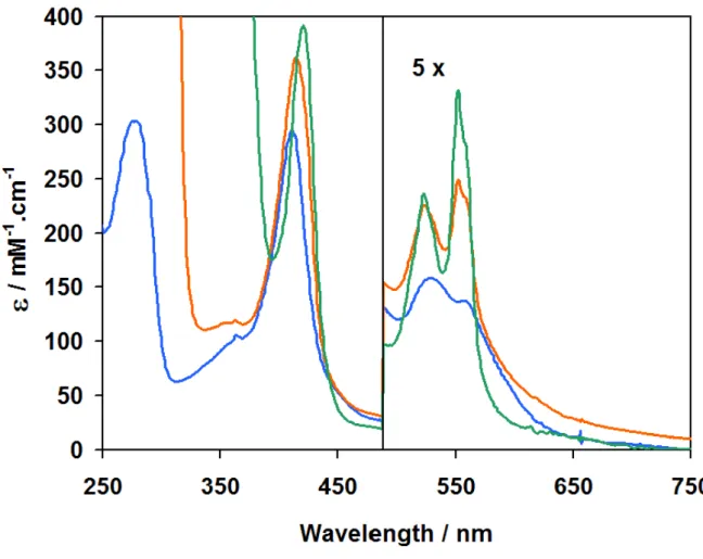
nautica NOR in different redox states. 55
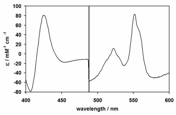
Figure 2.5 UV-visible difference spectrum of the Pseudomonas nautica NOR 56
Figure 2.6 The EPR spectra at 9.653 GHz of the as-isolated (A),
dithionite reduced Ps. nautica cNOR. 60
Figure 2.8 Three-dimensional predicted structure for the Ps. nautica cNOR
subunits.
61
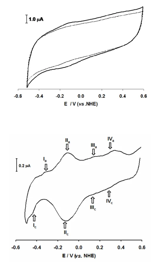
Figure 3.1 Plot of the Pseudomonas nautica NOR cyclic voltammograms at
different scan rates.
73
Figure 3.2 Cyclic voltamogram of the immobilized Pseudomonas nautica NOR. 75
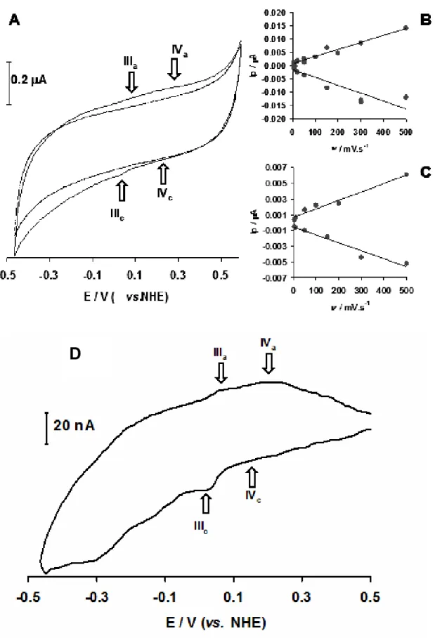
Figure 3.3 Low–spin electron transfer heme redox processes. 79
Figure 3.4 Cyclic voltamogram of the immobilized recombinant Pseudomonas
nautica rNorC subunit. 82
Figure 3.5 The Pseudomonas nautica NOR binuclear active site redox processes. 86
Figure 3.6 pH dependence of the midpoint redox potential for the
Pseudomonas nautica redox co-factors
91
Figure 3.7 Oxygen reduction using the Pseudomonas nautica NOR modified
electrodes. 93
Figure 3.8 Nitric oxide reduction using the Pseudomonas nautica NOR modified
electrodes. 94
Figure 3.9 Determination of the number of electrons involved in the NO and
O2 reduction, by the Pseudomonas nautica NOR 97
Figure 3.10 Oxidoreductase activity assays for the Pseudomonas nautica NOR,
combined with comercial peroxidase. 98
Figure 4.1 Oxygen reduction by Pseudomonas nautica NOR, using different c
-type cytochromes. 111
Figure 4.2 Nitric oxide reduction by Pseudomonas nautica NOR, using reduced
cyt. c552 as the electron donor 113
Figure 4.3 Sequential binding mechanism for NO reduction. 113
Figure 4.4 Oxygen reduction by Pseudomonas nautica NOR, using reduced cyt.
c552 as the electron donor. 115
Figure 4.5 Mechanism for oxygen reduction. 116
Figure 4.6 Cathodic current increase in the presence of NO and O2 obtained
with the Ps. nautica NOR modified graphite RDE 119
Figure 4.7 Nitric oxide reduction by Pseudomonas nautica NOR using the
Oxygen reduction by Pseudomonas nautica NOR using the enzyme
immobilized on a graphite RDE 123
Figure 4.10 pH dependence of the nitric oxide maximum reduction velocity. 126
Figure 4.11 Nitric oxide reduction mechanism assuming two protonable
residues in the Pseudomonas nautica NOR. 126
Figure 4.12 pH dependence of the oxygen maximum reduction velocity. 129
Figure 4.13 Working hypotheses for an oxygen reduction mechanism
assuming two protonable residues in the Pseudomonas nautica NOR. 130
Figure 5.1 Midpoint redox potentials for the NOR metal centers. 140
Figure S.1 Primary sequence alignment from different cNOR catalytic
subunits NorB. 151
Figure S.2 Low-spin heme b simulation spectrum. 152
Figure S.3 CuEDTA EPR spectrum. 153
Figure S.4 Cloning the Pseudomonas nautica NorC subunit. 155
Figure S.5 Nucleotide sequence alignment of the pAD11 vector with the
Pseudomonas nautica NorC coding sequence. 156
Figure S.6 The Pseudomonas nautica NORactivity using ascorbate /PMS as the
electron donor. 159
Figure S.7 The Pseudomonas nautica NOR activity using reduced Pseudomonas
nautica cyt.c552. 159
Figure S.8 The Pseudomonas nautica NOR oxidoreductase specific activity
using TMPD as the electron donor. 160
Figure S.9 The Pseudomonas nautica NOR oxidoreductase activityusing
different concentration of reduced Pseudomonas nautica cyt. c552. 161
Figure S.10 SO-NO sensor setup. 165
Figure S.11 Cyclic voltammogram of the immobilized Pseudomonas nautica
NOR, positive redox potentials. 167
Figure S.12 Cyclic voltammogram of the immobilized Pseudomonas nautica
NOR, negative redox potentials. 168
Figure S.13 Cyclic voltammogram of the immobilized recombinant NorC
performed at different pH values. 171
Figure S.15 Nitric oxide reduction using the Pseudomonas nautica NOR modified
graphite RDE. 172
Figure S.16 Nitric oxide reduction using the Pseudomonas nautica NOR modified
graphite RDE. The figure shows a detail from the previous figure
Table 2.1 Purification table for the Pseudomonas nautica NOR. 51
Table 2.2 Metal quantification results obtained for the isolated NOR. 53
Table 2.3 Ratios determined with the Mössbuer and EPR results. 59
Table 3.1 The Pseudomonas nautica NOR midpoint redox potentials. 87
Table 3.2 pKox and pKred of the Pseudomonas nautica NOR iron centers. 89
Table 4.1 Kinetic parameters obtained for the Pseudomonas nautica NOR with
NO or O2 as substrates. 117
Table 4.2 Kinetic parameters obtained for immobilized Pseudomonas nautica
NOR with NO and O2 as substrates. 124
Table S.1 Spin quantitation for the low-spin heme b and heme c. 153
Table S.2 Solutions for tricine sodium dodecyl sufhate gel electrophoresis. 162
Table S.3 Volumes required for tricine sodium dodecyl sulfate gel
preparation. 163
Table S.4 Reagents and corresponden amounts for Luria broth and S.O.C.
solutions. 166
Table S.5 Values determined for the ΔEp and Epw,1/2 from the anodic and
cathodic peak, achieved from the analysis of several
C
C
H
H
A
A
P
P
T
T
E
E
R
R
1
1
I
Chapter 1 Introduction
1.1. Nitric Oxide: Chemistry and Role in Biology 3
1.2. The Nitrogen Cycle 4
1.2.1. Overview of the Nitrogen Cycle 4
1.2.2. The Denitrification Pathway 6
1.2.2.1. The Nitric Oxide Reductase 15
1.2.2.1.1. The Heme Copper Oxidases Superfamily 15
1.2.2.1.2. The Nitric Oxide Reductase Subclasses 18
1.3. Nitric Oxide Reduction by Nitric Oxide Reductases 25
1.4. NO Related Enzymes: NO-Synthases and Reductases 29
1.4.1 Model Compounds 32
1.4.2. Rational Design in Alternative Proteins 33
1.5. The State of the Art and Aims 34
1.6. References 36
1.
Introduction
1.1.
Nitric Oxide: Chemistry and Role in Biology
Nitric oxide (NO) is a diatomic molecule among the simplest molecules. Its structure
and reaction chemistry has been the subject of study by chemists for many years [1]. NO
was long thought of as a poisonous, pungent-smelling gas, an unpleasant and dangerous
product of the oxidation of ammonia and of the incomplete combustion of gasoline in
motor vehicle exhausts.
From the chemical point of view, the NO molecule is a stable free radical with the molecular orbital diagram showing an unpaired electron residing in a π* molecular orbital.
This electronic configuration explains the high reactivity of the NO. Its oxidation leads to
the nitrosoniumion (NO+) and its reduction to the nitroxide ion (NO−). Its reactivity
reaches beyond the simple ionization and NO is extremely reactive with other simple
molecules such as oxygen (O2) or halogens, and metals, like iron, and it is why it is used to
reveal structural and mechanistic insights in metalloproteins [2].
Several N-oxides were identified by bacteria fermentation of plant material in the
second half of the 19th century, and these microorganisms were designated as denitrifiers
[3]. In the 60’s decade, NO was suggested to play a role as an intermediate of this
pathway, and at the same time, it was identified as a crucial biological intermediate of the
denitrification processes by the marine bacterium Pseudomonas perfectomarinus [4]. Years
after, in the 80’s, NO was discovered to be one of the most important physiological
regulators [2]. Nitric oxide synthase (NOS) was found to synthesize this signalling and
protective molecule that could be extremely helpful in different processes in mammals.
NO was identified as an endothelium relaxing factor, as a key cytotoxic agent of the
immune system, or even as a signalling molecule in the nervous system [4]. In bacteria,
NO also play an important role. Not only is an intermediate of the nitrogen (N) cycle, as
it is a signalling molecule, for example in bacterial biofilm dispear [5].
Nevertheless, above certain concentration levels, NO and its reactive species may
prove to be toxic to cells, and this phenomenon is designated by nitrosative stress. Some
nitrous oxide (N2O) or to ammonia (NH4+), by the denitrification pathway or by other
detoxification mechanisms. [3].
1.2.
The Nitrogen Cycle
1.2.1.
Overview of the Nitrogen Cycle
Nitrogen (N) is the fifth most abundant element in the solar system, and it is also well
represented in essential biomolecules, being an important constituent of nucleic acids and
proteins, the two most important polymers in life. The N-cycle is the process by which
nitrogen is converted between in its different molecular forms (figure 1.1). The chemistry
of this element is almost entirely dependent on reduction-oxidation (redox) reactions [6].
The chemical forms are transformed via specific enzymes, able to oxidize and reduce the
N-compounds. In the cycle, nitrate (NO3−) and ammonia (NH4+) present the higher (+5)
and lower (-3) redox states for the N atom, respectively, and the other molecules of the
cycle present different intermediate redox states (figure 1.1).
Earth’s earliest N-cycle was tightly controlled by a robust natural negative feedback
mechanism between atmospheric reactions, slow geological and microbial processes. In
the previous century, human intervention drastically disrupted the N-cycle by developing
industrial processes to reduce dinitrogen (N2) to NH4+, by implementing new agricultural
practices in order to boost crop yields, and by burning fossile fuels [6]. In particular there
has been an abusive use of nitrogen fertilizers which does not translate to a higher yield in
agricultural products, since only a part of the inorganic material is used to produce
biomass. Fertilizers are mainly composed of NH4+, and aerobic nitrification can not be
avoided, causing an accumulation of NO3− and nitrite (NO2−) at the Earth’s crust [6].
These particular reactions are controlled by three enzymes: the ammonium
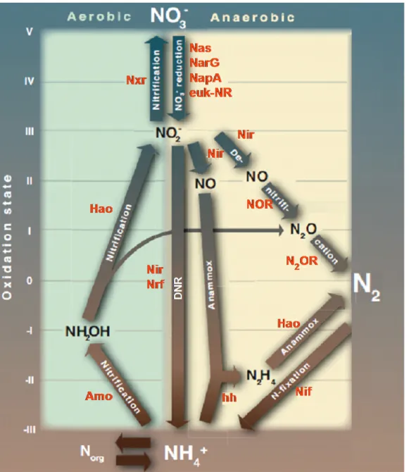
monooxygenase (Amo), the hydroxyalanine oxidoreductase (Hao) and the nitrite
Figure 1.1 – The N-cycle schematic representation. Arrows describe the principle pathways and the enzymes involved in each reaction are in red. Nitrate reductase: Nas - cytoplamatic, prokaryote assimilatory pathway; euk-NR- cytoplamatic, eukaryote – assimilatory pathway; NarG- membrane bound, dissimilatory pathway; NapA – periplasmic, dissimilatory pathway; Nitrite reductase, Nir - various kinds, Nrf - associated with periplasmic NR’s (Nap); nitric oxide reductase, NOR; nitrous oxide reductase N2OR; nitrogenase, Nif; ammonium monooxygenase, Amo; hydroxylamine oxidoreductase, Hao; nitrite
oxidoreductase, Nxr; hydrazine hydrolase, hh. Adapted from [6].
As a compensatory mechanism for the human production of oxidized molecules,
anaerobic microorganisms are able to reduce of the amount of NO3− and NO2− by three
pathways: denitrification, dissimilatory nitrite reduction and anaerobic ammonium
oxidation.
Denitrification is the pathway where NO3− is reduced to N2. It is composed by four
reductases (NRs); NO2− is reduced to NO by the nitrite reductases; NO is reduced to
N2O by nitric oxide reductase (NOR); and in a last step N2O is reduced to N2, by the
nitrous oxide reductase (N2OR). The denitrification pathway will be discussed in the
following section.
Anaerobic ammonium oxidation, currently called anammox, describes the reduction
of the N-oxides as NO3− and NO2− to NO, by nitrate reductases (NRs) and nitrite
reductases (Nir), respectively, and further reduction to hydrazine (N2H4). Alternatively, it
is also possible to oxidize NH4+ to N2H4, under the hydrazine hydrolase (hh) control.
Subsequent N2H4 oxidation is possible, catalyzed by hydroxylamine oxidoreductase
(Hao).
Direct NO2− reduction to NH4+ can be accomplished by microorganisms that harbour
the nitrite reductases (Nir or Nfr) coupled to the nitrite reductase system. This pathway is
designated as dissimilatory nitrite reduction (DNR), or dissimilatory reduction of nitrite in
anoxic conditions. As an example there is the penta-heme cytochromes c nitrate reductase
(ccNir or Nrf), able to reduce NO2−or NO directly to NH4+.
The N2 reduction to NH4+ is performed by nitrogenase (Nif), a multimeric enzyme
that catalyzes this exergonic reaction, since this reduction consumes 16 molecules of ATP
per reduced N2 molecule [6]. These enzymes are characteristic of the symbiotic
connection between bacteria and plant roots.
1.2.2.
The Denitrification Pathway
Denitrification, or dissimilative nitrate reduction, is an anaerobic process used by
some bacteria for energy generation. The study of this metabolic pathway is relevant,
since NO3− and NO2−became one of the worst water pollutants, a major concern being
the removal of NO3− from the water before it can be supplied to costumers [7].
Reduction of NO3− to nitrogen gas (N2) is done in four different steps:
NO3− + 2 e- + 2 H+ NO2− + H2O (eq. 1.1)
NO2− + e- + 2 H+ NO + H2O (eq. 1.2)
2 NO + 2 e- + 2 H+ N
2O + H2O (eq. 1.3)
NO3− is reduced to NO2− in a two electron reaction, followed by one electron reduction
to NO, and by a two electron-two proton reduction to N2O, and a final two electron
reduction to N2. Equations 1.1 to 1.4 show the electrons involved in each reaction
process. These processes are catalyzed by different metalloenzymes as pointed in figure
1.2.
Figure 1.2 – The denitrification pathway. Different resolved structures for the metalloenzymes involved in this metabolic route: A- Cupriavidus necator Nar (PDB:3ML1) [8], B- Escherichia coli Nar (PDB:1Q16) [9], C- Achromobacter cycloclastes CuNir (PDB:2BW4) [10, 11], D- Paracoccus pantotrophus cyt. cd1 (PDB:1H9X)[12],
E- Pseudomonas aeruginosa NOR (PDB: 3O0R) [13] and F- Pseudomonas nautica 617 N2OR (PDB: 1QNI) [14].
The PDB files were edited with Pymol software.
NO
3-NO
2-NO
N
2O
N
2Nar
Nap
CuNir
The Nitrate reductases (NRs) catalyze the reduction of NO3− to NO2−and they are
ubiquitous, from bacteria to eukaryotes. Organisms reduce NO3− for three main reasons:
i) incorporation of N into molecules (assimilatory ammonification), ii) to generate energy
for cellular functions (respiration, denitrification) and iii) to eliminate energy excess
generated by cell metabolism (dissimilatory ammonification) [15]. The NRs are mainly
molybdenum (Mo)-containing enzymes. Additional to the catalytic Mo center, the
enzymes carry redox co-factors such as iron-sulfur clusters or hemes that mediate the
electron transfer. Classification of these proteins have been made according to different
criteria, such as cell localization, protein structure, catalytic center molecular properties,
metabolic routes and others. According to their localization, they are divided in four
groups: the eukaryotic NO3− reductases (euk-nar), the assimilatory NO3− reductases (Nas),
the respiratory NO3−reductases (Nar) (figure 1.2 B) and the periplasmic (Nap) (figure 1.2
A) [16]. All NRs present an active center which is similar to the center of dimethyl
sulfoxide (DMSO) reductase family, with exception for the eukaryotic enzymes which are
part of the sulfite oxidase family [7]. Eukaryiotic NRs and Nas are cytoplasmatic enzymes
involved in the NO3− assimilation, whereas Nars are periplasmic and are involved
exclusively in the denitrification pathway.
Focusing in the prokaryotic NRs, these enzymes are involved in generating a proton
motive force across the membranes. They are constituted by three subunits: NarGHI (αβγ, respectively), narG subunit harbouring the Mo active center, and the remaining
subunits maintaining the electron transfer centers. They can be isolated from different
bacteria such as Escherichia coli [17], Ps. nautica [15] and Cupriavidus necator [18]. The isolated form can be a trimeric or a dimeric form, since the anchored γ-subunit (NarI) can be isolated or not, depending on the purification procedure [7]. NarI is the small subunit,
presenting a molecular weight of 19-26 kDa and comprising two heme b groups (bD and
bP). Moreover, Pseudomonas nautica 6171 revealed the presence of an unexpected c-type heme in this subunit [15]. The β subunit, has a molecular weight of 55-64 kDa, carries
four iron-sulfur clusters: three [4Fe-4S] designated FS1 FS2 and FS3, and one [3Fe-4S]
named FS4. The larger subunit NarG (118-150 kDa), harbours the Mo catalytic center,
coordinated to a molybdopterin guanine dinucleotide (MGD) as observed typically in the
1 Is also known as
DMSO mononuclear Mo enzymes. The other Mo ligands are from the protein peptide
chain, normally sulfur ligands from Cys or selenocysteine (Se-Cys) residues. There is an
additional electron transfer center in this subunit, a [4Fe-4S] cluster named FS0. All the
redox cofactors are located along an electron transfer pathway
(bD→bP→FS4→FS3→FS2→FS1→FS0→Mo) from which the nitrate receives the
electrons provided by the quinol pool (figure 1.3) [7, 15].
Figure 1.3 – Escherichia coli NarGHI complex (PDB:1Q16) [9]. Overall three-dimensional structure is presented. For better visualisation, the diagram of the enzyme electron transfer co-factors was shifted to the right. The PDB file was edited with Pymol software.
The reaction mechanism suggested for nitrate reductases is still a matter of
controversy. Originally a unique reaction mechanism was suggested for all nitrate
reductases regardless of their subclassification. This reaction mechanism, which was
desulfuricans [20], implies the replacement of the sixth coordinating ligand to molybdenum
(originally proposed to be a hydroxyl/water molecule) by nitrate, the transfer of two
electrons from Mo(IV) to nitrate, and the release of nitrite. The presence of an Asp
residue coordinated to molybdenum in a bidentate fashion in NarGHI from E. coli [9]
suggests that this mechanism can be feasible only if the bidentate coordination is opened
and an oxygenic species enters in the sixth coordinating position of the molybdenum
atom, as was determined in the crystal structure of NarGH [21]. This hypothesis is also
supported by the EPR results for both as-isolated and nitrate-reacted Pseudomonas nautica
617 Nar that point to a molybdenum ion coordinated to a hydroxyl/water ligand, which
could act as the labile group in a mechanism involving a direct nitrate–molybdenum
interaction [15]. Most recently, theoretical and computational tools were used to revise
the catalytic mechanism of NRs. The basis was the crystallographic data from the NapA
isolated from Desulfovibrio desulfuricans (PDB: 2v3v). Results show that both Mo species
have an active role on the mechanism but in different phases. The MoVI is required for
the NO3− reduction to NO2− and the MoV is involved in the second part of the
mechanism where one water molecule is formed and enzyme turnover occurs [22].
The Nitrite reductases can be divided in two major groups, according the nature of
their metal co-factor: there are the copper–containing nitrite reductases (Cu-Nir, figure
1.2 C) and the heme or Fe containing nitrite reductases (Fe-Nir, figure 1.2 D), also known
as cytochrome cd1. While the Fe-Nirs are more abundant in nature, Cu-Nirs are found in
a greater variety of ecological systems and therefore demonstrate more physiological
diversity. To date, no biological system has been shown to contain both Fe- and Cu-Nirs.
Nirs catalyze the one electron reduction of NO2− to NO [23].
Cu-Nir enzymes have been isolated from several organisms, such as Achromobacter
cycloclaste [10, 11], Ps. aureofaciens and Alcaligenes xylosodixans [7]. The enzyme presents a
trimeric structure with six copper atoms, divided in two types: the T1Cu, buried inside the
protein core and the T2Cu, the catalytic center, located in the interface of each two
subunits. The enzymes can be divided in two groups, according to the spectroscopic
properties of their T1Cu centers. The green reductases present an axially flattened
tetrahedron with an axial EPR signal and characteristic visible band at 590 nm. The blue
visible absorption spectra with different characteristic features (at 460 and 600 nm) [7,
24].
The electrons are donated by the physiological donor, presumably the cyt. c551 or
pseudozurin, to the T1Cu center and then transferred to the catalytic T2Cu center
through a chemical path involving conserved residues. The model for the catalytic
mechanism of Cu-Nir supposes that NO2− binds to the oxidized form of the T2Cu
center, displacing a solvent molecule. After reduction of the T2Cu center with an electron
from the T1Cu center, a intermediate compound, O=N-O-H is formed, and
consequently the product (NO) is released [7]. Figure 1.4, left side shows the proposed
mechanism for the NO2− reduction by the Cu-Nir.
Figure 1.4 – Nitrite reduction mechanism proposed for the Cu-Nir (left side) and for the cytochrome cd1
(right side), adapted from [7].
Cytochrome cd1 is a periplasmic soluble homodimer, with a molecular weight of 60
kDa and two hemes, one heme c and one heme d1 per monomer. It was isolated from
Pseudomonas strains like aeruginosa [25] and stutzeri, and Pa. denitrificans [7]. The heme c is
located in the N-terminal region; it is six-coordinated with two His ligands and is involved
in the electron transfer between the physiological donor and the catalytic heme d. The
heme d is the site for NO2− reduction, and presents specific spectroscopic features,
namely distinct visible absorption spectra. EPR spectroscopy proves that heme d is also
produces the lost of the Tyr axial ligand. The putative catalytic mechanism for
NO2−reduction points for the binding of a NO2− molecule to the ferrous heme d, in a
high-spin conformation. Dehydration leads to the formation of a nitrosyl intermediate,
and release of NO and concomitant intramolecular electron transfer from heme c to heme
d, completing the mechanism cycle (figure 1.4) [7].
The Nitric oxide reductases (NOR) catalyze the third step in the denitrification
pathway. Protein sequence alignment with members from this classe can be checked in
the supporting information S1. They are divided in three different classes, according to
their physiological electron donor: the cytochromes (cNOR), quinol (qNOR) and the
copper quinol (qCuNOR) [7] (figure 1.5). These enzymes are integral membrane proteins,
and they belong to the heme copper oxidase (HCuO) superfamily, sharing a high
structural homology of the catalytic subunit. In this class of enzymes, there is variation in
the type of electron transfer co-factors and in the number of subunits, but very well
conserved is the unusual binuclear diiron center, composed by a b-type heme (heme b3)
bridged to a non-heme FeB [26].
The cNORs present two subunits and receive electrons from soluble cytochromes or
cupredoxin. The qNORs lack one electron transfer co-factor and are composed by a
unique subunit, they receive electrons directly from the periplasmic quinol pool. The
qCuNORs are composed by two subunits, one comprising a binuclear CuA center similar
to the present in some HCuO members, and the other is the catalytic subunit. These last
can accept electrons directly from menaquinol or from soluble cytochromes via the CuA
center.
Mechanisms for NO reduction are issue in of intensive discussion. However, all the
proposed models consider substrate reduction in the binuclear Fe center with electron
transfer from the other metal co-factors to the binuclear cluster [7, 27]. Since the protein
Figure 1.5 – Schematic representation of the known NOR classes. Grey broken arrows represent the proposed electron transfer pathways from a periplasmic electron donor towards the active site, adapted from [7].
The Nitrous oxide reductase (N2OR) is the last enzyme of the denitrification
pathway, catalyzing the N2O reduction to N2. It has been intensively characterized and
isolated from different denitrifying organisms such as Ps. nautica [14], stutzeri [28, 29], Pa.
denitrificans [30], Achromobacter cycloclastes [31] and Wolinella succinogenes [32, 33]. The isolated
periplasmic enzymes are purified in a homodimeric form with approximately 65 kDa per
monomer. Each subunit contains two Cu centers: a multi-copper catalytic center (CuZ)
and a binuclear Cu site (CuA) similar to the ones observed in some members of the
isolated from Wolinella, which presents an additional extension of approximately 200
aminoacids with c-type heme motif in its C-terminal [7, 34], and ii) the enzyme from
Flexibacter canadensis, which interacts with the cytoplasmatic membrane [7]. The
homodimeric form presents a large dimerization interface, in such a way that the CuA
center from one subunit is very close to the catalytic CuZ center from the other subunit
(figure 1.6). In the CuA center, the two Cu atoms are bridged by two Cys ligands, the CuI
metal presents an addition His and Met residue coordination, and CuII presents a His and
a carbonyl group from a Trp residue. The catalytic CuZ center, is a µ4-sulfide bridged
tetranuclear copper center, coordinated by His residues [7, 14].
Figure 1.6 – The Pseudomonas nautica N2OR (PDB: 1QNI) [14] structure. Monomers are coloured in blue
and grey. Pink and orange spheres are evidence the CuA and CuZ centers, respectively. The PDB files were
edited with Pymol software.
Several mechanisms for the N2O reduction have been proposed [7, 34-36]. The
density functional theory (DFT) calculations suggested the binding of a N2O molecule
between CuI and CuIV atoms from the CuZ center. The cycle seems to involve two
sequential proton-electron transfer steps after N2 release, followed by the restoration of
the enzyme active form by release of one H2O molecule. Recent reports describe a
catalytic mechanism with emphasis in the interconversion between the active and inactive
form of the enzyme, where relevant catalytic intermediate species were detected and
1.2.2.1.
The Nitric Oxide Reductase
The NOR belongs to the Heme copper oxidase superfamily (HCuO), because this
type of enzymes presents a high structural homology, in means of their catalytic subunit,
as well as in the ligands that coordinate the redos centers. The major difference between
them is the catalytic center. In oxidases it is composed by a mixed cluster of Fe and Cu
while in NOR is composed by a binuclear Fe center. Some authors believe that NOR was
present in several ancestral organisms. During evolution, the disappearance of a reductive
environment and appearance of an oxidative atmosphere, produced the insolubility of Fe,
and induce species to replace Fe by Cu [3, 37].
1.2.2.1.1.
Heme Copper Oxidases Superfamily
Terminal oxidases of membrane-bound electron transfer chains catalyze the reduction
of dioxygen (O2) to water, coupling the redox energy to proton translocation through the
cytoplasmatic or mitochondrial/chloroplast membrane. Most of the terminal oxidases
belong to the HCuO. This large family can present different electron donors, subunit
composition, and heme type. Common to all the members is the presence of a catalytic
subunit harbouring a six-coordinated low-spin heme and an unusual binuclear catalytic
center, giving rise to its name, composed by a high-spin heme and a copper ion CuB.
The HCuO catalytic subunit or subunit I is composed, at least, by 12 transmembrane α-helices, carrying the binuclear active center and the immediate low-spin electron
transfer heme. These three metal centers present six well conserved His residues ligands:
two coordinate the low-spin heme, one coordinates the catalytic high-spin heme and the
remaining three ligands support the catalytic CuB. Prokaryotic oxidases may contain quite
diverse heme, such as heme a, b or o, commonly used to designate the enzymes. However,
the type of heme does not correlate with the organisms’ phylogeny, the type of electron
donor, nor with sequence similarities, and therefore it is inappropriate to classify the
oxidase family based on the heme types [38]. A very simple way of illustrating all the
HCuO was described by Garcia-Horsman [39], which separates the superfamily in the
and the quinol oxidases, where the physiological electron donor is the quinol molecule
(figure 1.7).
Figure 1.7 – Schematic illustration showing the similarities and differences between five subclasses of the heme-copper oxidase superfamily. Panels A-C, cytochromes c oxidases (CcO); panels D and E, quinol oxidases [39].
The His ligands coordinating the co-factors in the HCuO catalytic subunit are
extremely well conserved as it can be seen in figure 1.8. Sequence alignment show protein five sequences of HCuO members, including a NOR. The transmembrane α-helixes are
these are inside the proposed pores created by the helixes special arrangement [23, 38]. A
schematic representation of the helices disposition is illustrated in the inset of figure 1.8.
10 20 30 40 50 60 ....|....|....|....|....|....|....|....|....|....|....|....|
CcO aa3 Pa. denitrifans -MSAQISDSIEEKRGFFTRWFMSTNHKDIGVLYLFTAGLAGLISVTLTVYMRMELQHPGV
CcO cbb3 Ps. stutzeri ---MSTAISETAYNYKVVRQFAIMTVVWGIIGMGLGVFIAAQLVWPSL
CcO aa3 Rh. sphaeroides MADAAIHGHEHDRRGFFTRWFMSTNHKDIGVLYLFTGGLVGLISVAFTVYMRMELMAPGV
CcO aa3 B. taurus ---MFINRWLFSTNHKDIGTLYLLFGAWAGMVGTALSLLIRAELGQPGT
cNOR Ps. nautica ---MKYESQRVAMPYFIFALILFAGQIVFGLILGLQYVVGDF 70 80 90 100 110 120 ....|....|....|....|....|....|....|....|....|....|....|....|
CcO aa3 Pa. denitrifans QYMCLEGMR---LVADAAAECTPNAHLWNVVVTYHGILMMFFVVIPALFGGFG
CcO cbb3 Ps. stutzeri NLD---LPWTSFGRLRPLHTNAVIFAFGGCALFATS-
CcO aa3 Rh. sphaeroides QFMCAEHLESGLVKGFFQSLWPSAVENCTPNGHLWNVMITGHGILMMFFVVIPALFGGFG
CcO aa3 B. taurus LLG---DDQIYNVVVTAHAFVMIFFMVMPIMIGGFG
cNOR Ps. nautica LFP---EIPFNVARMVHTNLLIVWLLFGFMGATY- 130 140 150 160 170 180 ....|....|....|....|....|....|....|....|....|....|....|....|
CcO aa3 Pa. denitrifans NYFMPLHIGAPDMAFPRLNNLSYWLYVCGVSLAIASLLSPGGSDQPGAGVGWVLYPPLS-
CcO cbb3 Ps. stutzeri -YYVVQRTCQARLFSDGLAAFTFWGWQAVIVLAVITLP---MGYTSSKEYA-
CcO aa3 Rh. sphaeroides NYFMPLHIGAPDMAFPRMNNLSYWLYVAGTSLAVASLFAPGGNGQLGSGIGWVLYPPLS-
CcO aa3 B. taurus NWLVPLMIGAPDMAFPRMNNMSFWLLPPSFLLLLASSMVEAG---AGTGWTVYPPLAG
cNOR Ps. nautica --YMVPEEAQTELHSPLLAWILFWVFAAAGTLTILGYL---FVDYATLA- 190 200 210 220 230 240 ....|....|....|....|....|....|....|....|....|....|....|....|
CcO aa3 Pa. denitrifans -TTEAGYAMDLAIFAVHVSGATSILGAINIITTFLNMRAPGMTLFKVPLFAWAVFITAWM
CcO cbb3 Ps. stutzeri ---ELEWPIDILITLVWVSYIAVFFGTI---MKRKAKHIYVGNWFFGAFI
CcO aa3 Rh. sphaeroides -TSESGYSTDLAIFAVHLSGASSILGAINMITTFLNMRAPGMTMHKVPLFAWSIFVTAWL
CcO aa3 B. taurus NLAHAGASVDLTIFSLHLAGVSSILGAINFITTIINMKPPAMSQYQTPLFVWSVMITAVL
cNOR Ps. nautica ---EVTMNKLLPTMGREFLEQPTITKIG---IAVVVVAFLYNIAMTALK 250 260 270 280 290 300 ....|....|....|....|....|....|....|....|....|....|....|....|
CcO aa3 Pa. denitrifans ILLSLPVLAGGITMLLMDRNFGTQFFDPAGGGDPVLYQHILWFFGHPEVYMLILPGFGII
CcO cbb3 Ps. stutzeri LVTAMLHIVN---NLEIPVSLFKSYSIYAGATDAMVQWWYGHNAVGFFLTTGFLGM
CcO aa3 Rh. sphaeroides ILLALPVLAGAITMLLTDRNFGTTFFQPSGGGDPVLYQHILWFFGHPEVYIIVLPAFGIV
CcO aa3 B. taurus LLLSLPVLAAGITMLLTDRNLNTTFFDPAGGGDPILYQHLFWFFGHPEVYILILPGFGMI
cNOR Ps. nautica GRKTVVNIVLITGLVGLAVLWLFSFYNPGNLATDKYFWWFVV~~~HLWVEGVWELIMGAI 310 320 330 340 350 360 ....|....|....|....|....|....|....|....|....|....|....|....|
CcO aa3 Pa. denitrifans SHVISTFA-RKPIFGYLPMVLAMAAIAFL~GFIVWAHHMYTAGMSLTQQTYFQMATMTIA
CcO cbb3 Ps. stutzeri MYYFVPKQAERPVYSYRLSIVHFWALITL~YIWAGPHHLHYTALPDWAQSLGMVMSIILL
CcO aa3 Rh. sphaeroides SHVIATFA-KKPIFGYLPMVYAMVAIGVL~GFVVWAHHMYTAGLSLTQQSYFMMATMVIA
CcO aa3 B. taurus SHIVTYYSGKKEPFGYMGMVWAMMSIGFL~GFIVWAHHMFTVGMDVDTRAYFTSATMIIA
cNOR Ps. nautica LAYVLIKLTGVDREVIEKWLYVIIAMALITGIIGTGHHFFWIGPPEYWLWLGSVFSALEP 370 380 390 400 410 420 ....|....|....|....|....|....|....|....|....|....|....|....|
CcO aa3 Pa. denitrifans VPTGIKVFSWIATMWGGSIEFKTP--MLWAL--AFLFTVGG-VTGVVIAQGSLDRVYHDT
CcO cbb3 Ps. stutzeri APSWGGMINGMMTLSGAWHKLRTDPILRFLVVSLAFYGMST-FEGPMMAIKTVNALSHYT
CcO aa3 Rh. sphaeroides VPTGIKIFSWIATMWGGSIELKTP--MLWALGFLFLFTVGG-VTGIVLSQASVDRYYHDT
CcO aa3 B. taurus IPTGVKVFSWLATLHGGNIKWSPA--MMWALGFIFLFTVGG-LTGIVLANSSLDIVLHDT
cNOR Ps. nautica LPFFMMVVFAFNMINRRRRNHPNKAAMLWAMGTTVMAFLGAGVWGFLHTLAPVNWYTHGS 430 440 450 460 470 480 ....|....|....|....|....|....|....|....|....|....|....|....|
CcO aa3 Pa. denitrifans YYIVAHFHYVMSLGALFAIFAGTYYWIGKMSGRQYPEWAGQL--HFWMMFIGSNLIFFPQ
CcO cbb3 Ps. stutzeri DWTIGHVHAGALGWVAMITIGSMYHLIPKVFGREQMHSVGLINAHFWLATIGTVLYIASM
CcO aa3 Rh. sphaeroides YYVVAHFHYVMSLGAVFGIFAGIYFWIGKMSGRQYPEWAGKL--HFWMMFVGANLTFFPQ
CcO aa3 B. taurus YYVVAHFHYVLSMGAVFAIMGGFVHWFPLFSGYTLNDTWAKI--HFAVMFVGVNMTFFPQ
Figure 1.8 – Sequence alignment for the catalytic subunit of five members from the HCuO superfamily.
CcO aa3-type from Paracoccus denitrificans, cbb3 type from Pseudomonas stutzeri, aa3-type from Rhodobacter
sphaeroides, aa3-type from Bos taurus and cNOR from Pseudomonas nautica 617. Bottom inset: schematic top
view of the HCuO superfamily catalytic subunit; roman numerals indicate the helixes number and green circules indicate the helixes with the His ligands (adapted from [23]). 1 stands for the low-spin heme electron transfer center, 2 is the catalytic high-spin heme center and MB is either a Cu or a non-heme Fe.
1.2.2.1.2.
The Nitric Oxide Reductase Subclasses
The nitric oxide reductase (NOR) subclass is characterized by a diiron center. This is
the major difference between NORs and other HCuO members that have a mixed
binuclear center (Fe/Cu). The ligands that coordinate the metal centers are, nevertheless,
conserved. The non-heme FeB prefers an six-coordination environment instead of the
tetrahedic like Cu typical for the HCuO. For many years, assumptions were made for a
possible ligand. Molecular modelling and sequence alignment predicted a Glu residue near
the catalytic center to stabilize the FeB [3, 40]. The first crystal structure for a NOR was
obtained only recently, confirming the existence of an extra Glu ligand to the non-heme
FeB center [13]. Figure 1.9 (red rectangles) shows the sequence alignments with the Glu
residue conserved in several NORs, extremely important for substrate reduction [40, 41].
490 500 510 520 530 540 ....|....|....|....|....|....|....|....|....|....|....|....|
CcO aa3 Pa. denitrifans HFLGR-QGMPRRYIDYPVEFSYWNNISSIGAYISFASFLFFIGIVFYTLFAGKPVNVPNY
CcO cbb3 Ps. stutzeri WVNGITQGLMWRAINEDGTLTYS----FVEALEASHPGFIVRAVGGAFFLAGMLLMAYNT
CcO aa3 Rh. sphaeroides HFLGR-QGMPRRYIDYPEAFATWNFVSSLGAFLSFASFLFFLGVIFYTLTRGARVTANNY
CcO aa3 B. taurus HFLGL-SGMPRRYSDYPDAYTMWNTISSMGSFISLTAVMLMVFIIWEAFASKREVLTVDL
cNOR Ps. nautica LFLTAAGVLQVWLQRIP---ESGEALSFMAGQDQIALFYWMRFVAGAFFMAGLVVYFGSF 550 560 570
....|....|....|....|....|....|....|..
CcO aa3 Pa. denitrifans WNEHADTLEWTLPSPPPEHTFETLPKPEDWDRAQAHR
CcO cbb3 Ps. stutzeri WR---TVRAAKSAQYDTAAQIA---
CcO aa3 Rh. sphaeroides WNEHADTLEWTLTSPPPEHTFEQLPKREDWERAPAH-
CcO aa3 B. taurus T---TTNLEWLNGCPPPYHTFEEP----TYVNLK---
![Figure 1.2 – The denitrification pathway. Different resolved structures for the metalloenzymes involved in this metabolic route: A- Cupriavidus necator Nar (PDB:3ML1) [8], B- Escherichia coli Nar (PDB:1Q16) [9], C- Achromobacter cycloclastes CuNir (PDB:2](https://thumb-eu.123doks.com/thumbv2/123dok_br/16483960.732646/39.892.117.804.310.983/denitrification-different-structures-metalloenzymes-cupriavidus-escherichia-achromobacter-cycloclastes.webp)