Ciências
Evaluation of the role of miR-9 and miR-29 in
amyloid pathway of Alzheimer's disease
Micaela Sofia Ribeiro Riscado
Dissertação para obtenção do Grau de Mestre em
Bioquímica
(2º ciclo de estudos)
Orientador: Profª. Doutora Fani Pereira de Sousa
Co-orientador: Doutora Patrícia Alexandra Nunes Pereira
ii
Acknowledgments
It’s been a long year full of struggle, growth, friendship and most of all things, learning. What makes about this difficult process to be easiest to accomplish is the people involved in my world, the teachers, the coworkers, the friends and above all the family. It is to them that that I must thank for all the patient and support given.
First a I would like to thanks my whole family (parents, brother, grandparents, uncles and cousins) for all the patient to put up with my bad mood after a bad day in the lab or for missing important events like family dinners or birthday parties due to the already integrated in the family, N2a cells, that were like a baby, making me do extra hours and work on the weekends. In particularly, I must thanks my parents, Paulo Riscado and Helena Riscado, for all the financial support that they gave me, all the conversations about the future, all the support in decisions that could have changed everything and must of all for the love that no matter what I absolutely know that is always there. And of course, my little brother, Tiago Riscado, is the best! Always trying to cheer me up, in your special way that only we can understand.
Also, I like to express my sincere gratitude to my recently add family, Ines Vicente and Ines Figueiredo because they chose me as someone to help in their first year of colleges but yet the roles reversed and they have been there in the most comprehensive way that there can be and I know that they are truly happy for watching me deliver this thesis.
I’m deeply grateful to the ones that made possible the execution of this thesis despite all the downs encounter in the way. I want to thank my supervisor Prof. Doctor Fani Sousa for accepting me in her group, supporting me in this process, for all the talks that were important to me and special to seeing the potential in me to pursue this line of work. Also, I must thank my co-supervisor Doctor Patrícia Pereira, that despite the short time spent together had the ability to transfer to me the knowledge that I needed so much to elaborate this thesis and for the real-life talks that just someone with her life and professional experience could have. Last but surely not least I like to thank Bruno Baptista for the availability shown to teach me all the techniques, without him the start of this work it would have been very difficult.
Also want to thanks to all the investigators in the CICS that receive me with an open smile and conversations that made the lab a friendly workplace, in special to Marília Figueira, Tiago Santos, Jorge Ferreira, Tiago Carvalho and Doctor Augusto Pedro, that share tips or experience that showed me that investigations work is not always easy that is normal to encounter obstacles, you just have to keep trying.
iii
Clearly I have to thanks to all my friends, the old ones that always been there both in the lows and the in highs, the ones that I call for a simple conversation or to put out all my melancholy thoughts, in specially Sofia Carneiro, Maria Santos, Vanda Nogueira, Carolina Rosa, Irís de Passos, Daniela Gonçalves, Inês Valado, to the new ones that embrace me like I was part of the group from the beginning, support me and endured with me all the worries that a thesis may bring, Adriana Pinto, Ana Raquel Neves, Diana Pereira, Diana Gomes, Diana Esteves, André Furtado, Pedro Ferreira, Sofia Oliveira and Pedro Vicente. To this last three I have to thanks especially for the new energy and joy that they brought to lab when it was most needed.
Then, are people that you met at the beginning of your life and you thought that they will be nothing but acquaintances, but I was wrong because people like Rita Carapito, appear right back in the moment that you need the most. This is the person that I have the most to thanks for, she was my rock, my source of laughter, my lab partner (we are a pretty good team by the way) and the person who saw me cry the must. Most important she was there even when she was not in the best places herself and she is the only person that knows what the meaning of “The year of my Thesis” really stands for because we were there all the way, side by side. I have to thank her for all of it, without her I can say for sure that I had given up halfway.
Must more could be said because what is described in here is just a little representation of the impact that the mention people made in my life this year, from the bottom of my heart I appreciate all the support and patient given to me.
iv
Resumo Alargado
A doença de Alzheimer (DA) é uma doença neurodegenerativa e a forma mais comum de demência, ocupando o terceiro lugar das patologias que causam incapacidade e morte nos idosos. Apesar do seu impacto, as causas desta doença são, ainda hoje, pouco compreendidas. Entender as vias e mecanismos que conduzem a uma transição entre um estado de saúde até à demência pode ajudar a desenvolver e estabelecer novos tratamentos mais efetivos. A DA caracteriza-se pela expressão desregulada de diversas proteínas envolvidas na formação de péptidos β-amiloide, nomeadamente a proteína precursora amiloide (PPA), a proteína β-secretase (BACE1) e a proteína presenilina 1 (PS1), sendo detetados níveis elevados nesta condição. Como estas proteínas fazem parte da complexa via patológica da DA, têm vindo a ser intensamente estudadas como alvos terapêuticos. No entanto, as terapias que utilizam inibidores dessas proteínas têm mostrado efeitos adversos e são muitas vezes descontinuadas, o que torna difícil, mas urgente, a criação de novas estratégias terapêuticas para controlar a progressão da DA.
Os microRNAs (miRs ou miRNAs) são moléculas de RNA não codificante, de baixo peso molecular, que têm como principal função regular o processo de tradução, com ação sobre o RNA mensageiro, permitindo uma diminuição dos níveis de expressão da proteína codificada. Os trabalhos de investigação utilizando miRNAs como estratégias terapêuticas para a doença de Alzheimer (DA) têm vindo a aumentar ao longo dos últimos anos. A ideia de utilizar os miRNAs como uma nova abordagem terapêutica nesta doença foi motivada essencialmente por dois fatores. Em primeiro lugar, estes miRNAs podem regular os RNA mensageiro (mRNAs) pós-transcricionalmente, e, consequentemente, controlar a expressão das proteínas responsáveis pelo surgimento da DA e, em segundo lugar, porque já foi amplamente descrito que os níveis de determinados miRNAs se encontram também desregulados, ocorrendo tendencialmente uma diminuição da sua expressão, na DA. Deste modo, os estudos que preveem o estabelecimento de uma relação entre os miRNAs e os seus mRNAs alvo, começaram a surgir e mostraram que certos miRNAs podem de facto controlar as principais proteínas relacionadas com os biomarcadores da DA. Os principais biomarcadores são os depósitos extracelulares de péptidos β-amiloide juntamente com a acumulação intracelular da forma hiperfosforilada da proteína Tau associada aos microtúbulos. Apesar de alguns estudos mostrarem a regulação de proteínas pelos miRNAs na DA, esta é uma área vasta onde existem muito alvos ainda por descobrir, devido às múltiplas vias e funções que um só miRNA pode desempenhar.
Assim, o objetivo deste trabalho é estudar a nível celular, o papel de miRNAs cuja expressão se encontra diminuída na DA, dos quais foram selecionados o miR-29 previamente utilizado no grupo de investigação e o miR-9 que foi também relacionado com a regulação de vários mRNAs que codificam proteínas envolvidas em diferentes vias patológicas da DA. O foco
v
foi neste caso, nos miRNAs que apresentam níveis diminuídos na DA, considerando a possibilidade de entrega destes miRNAs às células, reestabelecendo a sua função e consequentemente contrariando assim o ambiente patológico existente na doença. Para isso foram utilizados miRNAs produzidos por métodos diferentes (síntese química ou enzimática), e apresentando a sua forma madura ou a forma precursora (pre-miRNAs), de modo a verificar a existência de um efeito biológico distinto entre eles. Os pre-miRNAs foram produzidos em laboratório através de transcrição in vitro utilizando um DNA plasmídico (pDNA) como template. Adicionalmente, foi também estudada a vantagem ou possível influência na atividade biológica, da introdução de um processo de purificação dos pre-miRNAs produzidos. De forma a avaliar a sua atividade biológica, as células N2A695 (linha celular de neuroblastoma de rato que expressa APP mutada, de maneira a replicar a via patológica da DA) foram transfetadas com os pre-miRNAs produzidos ou os miRNAs sintéticos, recorrendo à utilização de dois agentes de transfeção, como a Lipofectamina e nanopartículas de quitosano, que permitem igualmente a comparação destes sistemas. Após extração do RNA das células foi feita a avaliação dos níveis de expressão dos mRNAs que codificam as proteínas APP, BACE1 e PS1. Foram globalmente verificados efeitos de silenciamento para a APP pelo pre-miR-9 e para a BACE pelo miR-29b e pelo pre-mir-29b. A PS1 foi a proteína para a qual se verificou um efeito mais predominante, em termos de silenciamento, uma vez que todos os tipos de miRNAs/pre-miRNAs utilizados conduziram ao seu silenciamento.
Em suma, um melhor entendimento de como os miRNAs atuam e quais os seus principais alvos, poderá ter um grande impacto na indústria farmacêutica, pois em patologias onde estes são reguladores das vias patológicas, como é o caso da DA, poderão ser utilizados como uma nova abordagem terapêutica, de forma a regular a expressão de proteínas alteradas nesse ambiente patológico.
Palavras-Chaves
vi
Abstract
Alzheimer’s disease (AD) is a neurodegenerative disorder which occupies the 3rd place
of the diseases that cause disability and death in the elderly, but their causes are yet in need of a better understanding. Also, the search of the etiopathological path leading from a healthy state to full-blown dementia could help with the establishment of more effective treatments. MicroRNAs (miRs or miRNAs) are small regulatory non-coding RNAs (sncRNA), which are involved in post-transcriptional gene expression regulation. Diverse studies have shown that the expression levels of several miRNAs are decreased in AD patients and, there is also evidence that certain miRNAs can control the pathologic hallmarks related to the biomarkers of AD. The main biomarkers of AD are extracellular deposits of amyloid-β peptide (Aβ) along with an accumulation of intraneuronal hyperphosphorylated form of the microtubule-associated protein Tau.
The aim of this dissertation is to study the role of two under-expressed miRNAs in AD, miR-29 and miR-9, that are related to the regulation of messenger RNA (mRNAs) that encode proteins involved in various pathological pathways of AD. The selection of miRNAs with decreased levels was based on the possibility of performing its delivery into the cells, in order to contradict the pathological environment of AD. To accomplish this issue, synthetic miRNAs and precursors of miRNAs (pre-miRNAs) produced by two different methods (chemical and enzymatic synthesis, respectively) were used, in order to evaluate if the preparation method had influence in the biological activity. Thus, the pre-miRNAs were produced in the laboratory through in vitro transcription using a plasmid DNA (pDNA) as a template and were further subjected to a purification step. To evaluate the biological activity of the miRNAs, N2a695 cells (mutated APP expressing neuroblastoma mouse cell line that mimics the pathological pathway of AD) were used as in vitro model. Two different methods were also used for cells transfection, Lipofectamine and chitosan-nanoparticles for RNA encapsulation and delivery. After RNA extraction, the mRNA expression levels were evaluated for the APP, BACE1 and PS1 proteins. The findings demonstrated a silencing effect on the expression of APP mRNA by pre-miR-9 and of the BACE mRNA by miR-29b and pre-miR-29b. In turn, PS1 mRNA levels were decreased after cells transfection with all types of miRNAs/pre-miRNAs used in this study, being the most prominent result obtained by RT-qPCR. Western blot assays were also performed, in order to quantify the expression levels of the proteins in study.
In conclusion, a better understanding of how miRNAs act and what are their specific targets may have a major impact on the pharmaceutical industry, because being regulators in the pathologic pathways in certain diseases, such as AD, they can be used as new therapeutic approaches to regulate altered proteins expression in this pathological environments.
vii
Keywords
viii
Table of Contents
Chapter I ... 1 Introduction ... 1 1.1 Ribonucleic Acid ... 2 1.1.1 RNA Interference ... 2 1.1.2 MicroRNA ... 4 1.2 Dementia ... 8 1.2.1 Alzheimer’s Disease ... 8 1.2.2 Amyloid Hypothesis ... 11 1.2.3 Tau Hypothesis ... 131.2.4 Alzheimer’s Disease Treatments ... 13
1.2.5 MicroRNAs and Alzheimer’s Disease ... 15
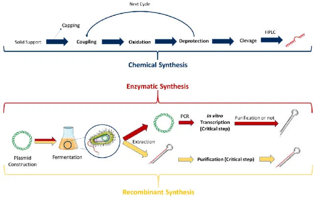
1.3 MicroRNA Manufacturing ... 19
1.4 MicroRNA Delivery ... 21
Chapter II ... 24
Aim of the work ... 24
2. Aim of the work ... 25
Chapter III ... 26
Materials and Methods ... 26
3.1 Materials ... 27
3.2 Methods ... 31
3.2.1 Pre-miRNAs synthesis ... 31
3.2.1.1 Escherichia coli DH5α growth ... 31
3.2.1.2 Plasmid DNA extraction from E. coli DH5α ... 31
3.2.1.3 Conventional polymerase chain reaction ... 32
3.2.1.4 Purification of PCR products ... 33
3.2.1.5 In vitro transcription ... 33
3.2.1.6 Purification and concentration of the RNA transcripts... 34
3.2.2 Cell culture ... 35
3.2.2.1 N2a695 cells lines growth ... 35
3.2.2.2 N2a695 cells transfection assays ... 35
3.2.3 mRNA expression analysis ... 37
3.2.3.1 Total RNA extraction from eukaryotic cells ... 37
3.2.3.2 Complementary DNA synthesis ... 38
3.2.3.3 Real-time quantitative-polymerase chain reaction ... 38
ix
3.2.4.1 Protein extraction from eukaryotic cells ... 39
3.2.4.2 Protein quantification ... 39
3.2.4.3 Western blotting ... 39
3.2.5 Data analysis ... 40
Chapter IV ... 42
Results and Discussion ... 42
4.1 E. coli DH5α growth and plasmid DNA amplification ... 43
4.2 Isolation of plasmid DNA from E. coli DH5α cells ... 44
4.3 Pre-miRNA sequences amplification by PCR ... 46
4.4 Purification of the PCR products ... 51
4.5 In vitro transcription of pre-miRNAs ... 51
4.6 Analysis of the mRNA expression by RT-qPCR ... 53
4.7 Analysis of the protein expression by Western blot ... 57
Chapter V ... 60
Conclusion and Future perspectives ... 60
5. Conclusion and Future perspectives ... 61
Chapter VI ... 63
References ... 63
x
List of Figures
Figure 1. Schematic representation of the miRNA biogenesis ... 5
Figure 2. Overview of the main events leading from healthy state to full-blown dementia ... 10
Figure 3. Schematic representation of the possible processing pathways of APP ... 12
Figure 4. Schematic representation of the miR-29 family and their structural differences .... 17
Figure 5. Schematic representation of the possible targets of miR-9 and miR-29b in AD pathologic pathway. ... 18
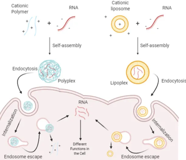
Figure 6. Schematic representation of the three methods currently used to produce microRNAs. ... 21
Figure 7. Schematic representation of the polyplex- and lipoplex-based non-viral strategies for delivering RNA into cell. ... 23
Figure 8. Schematic representation of the methods used to produce the in vitro pre-miRNAs. ... 35
Figure 9. Schematic representation of N2a695 cells transfection. ... 37
Figure 10. Schematic representation of the methods used for the target mRNA expression analysis. ... 39
Figure 11. Schematic representation of the methods used for the target proteins expression analysis. ... 40
Figure 12. Growth curve obtained of E. coli DH5α bacteria modified with plasmid DNA pBHSR1-RM containing the sequence of human pre-miR-9 or -29b. ... 43
Figure 13. Growth curve for E. coli DH5α bacteria modified with plasmid pBHSR1-RM containing the sequence of human pre-miR-29b and production of the pre-miR-29b ... 44
Figure 14. Isolation of plasmid pBHSR1-RM containing the sequence of human pre-miR-9/-29b from E. coli DH5α cellular pellet. ... 45
Figure 15. Optimization of the master mix components used in the PCR technique. ... 47
Figure 16. Effect of the annealing temperature on the PCR reaction. ... 48
Figure 17. Optimization of the DNA concentration used for the PCR technique. ... 49
Figure 18. Final amplification of the pre-miRNAs, after optimization of the PCR conditions. . 50
Figure 19. Electrophoresis after the purification of PCR products. ... 51
Figure 20. Electrophoresis of the pre-miR-9 and pre-miR-29b transcripts obtained by in vitro transcription. ... 52
xi
Figure 22. Electrophoresis to verify the integrity of total RNA extracted from the transfected N2a695 cells.. ... 54 Figure 23. RT-qPCR analysis of the expression levels of APP, BACE1 and PS1 mRNA from N2a695 cells transfected with different types of miRNAs and pre-miRNAs. ... 55 Figure 24. Western blot analysis of APP, BACE1, PS1 and β-actin from N2a695 cells transfected with different types of miRNAs and pre-miRNAs. ... 58
xii
List of Tables
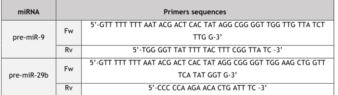
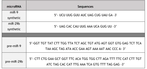
Table 1. Potential therapeutic applications of miRNA drugs currently in clinical trials ... 7 Table 2. Dysregulated miRNAs in Alzheimer’s disease. ... 16 Table 3. Pre-miR-9 and pre-miR-29b primers sequences used in the PCR reaction. ... 27 Table 4. Sequences for miRNAs (miR-9, miR-29b, pre-miR-9 and pre-miR-29b) used in the in
vitro assays. ... 28
Table 5. Primers sequences used in the RT-qPCR experiments. ... 29 Table 6. Characterization of the antibodies used in the Western blot technique. ... 30 Table 7. Experimental condition used in each step of the PCR for pre-miRNA amplification. . 33 Table 8. Summary of the final conditions for PCR after optimization for miR-9 and pre-miR-29b. ... 50
xiii
List of Acronyms
15-LOX Lipoxygenases
ABCA1 ATP-binding cassette transporter
AGO Argonaut protein
AKT Protein kinase B
ApoE4 Apolipoprotein E4
APP Amyloid precursor protein
Aβ β-amyloid peptide
BACE β-secretase
BBB Blood-brain barrier
BSA Bovine serum albumin
CD2AP
CD2-associated protein
CDKN2A Cyclin-dependent kinase Inhibitor 2A
cDNA Complementary DNA
CFH Complement factor H
CNS Central nervous system
CSF Cerebrospinal fluid
DEPC Diethylpyrocarbonate
DMEM Dulbecco’s modified eagles’s medium
DNA Deoxyribonucleic acid
DNAi DNA Interference
dNTPs Deoxyribonucleotide triphosphate
dsRNA Double stranded RNA
E. coli Escherichia coli
EGTA Ethylene glycol tetra acetic acid
EPHA1 Ephrin type-A receptor 1 protein
Fw Forward
GAPDH Glyceraldehyde-3-phosphate
GSK3B Glycogen synthase kinase 3 β
GTP Guanosine triphosphate
IRAK Interleukin-1 receptor-associated kinase
LB Luria broth
Lin-14 Lineage protein 14
Lin-4 Lineage-4
Lipo Lipofectamine 2000
lncRNAs Long non-coding RNAs
LPA Logopenic aphasia
xiv
MAPT Microtubule-associated protein tau
Min Minutes
miR/miRNA microRNA
miRISC microRNA-induced Silencing Complex
MRE microRNA response elements
mRNA Messenger RNA
N2a695 Human mutated APP695 cellular line
ncRNAs Non-coding RNAs
NFH
Neurofilament heavy chain
NFT Neurofibrillary tangles
NMDA N-methyl D-aspartate
NR2E1 Nuclear receptor subfamily 2 group E member 1
Nt Nucleotides
PACT Protein kinase R-activating protein
P-bodies Processing bodies
PCA Posterior cortical atrophy
PCR Polymerase chain reaction
pDNA Plasmid DNA
PEI Poly(ethylenimine)
PET Positron emission tomography
PHFs Paired helical filaments
PI3K Phosphoinositide 3-kinase
piRNAs PIWI-interacting RNA
PLL Poly(L-lysine)
PMSF Phenylmethylsulfonyl fluoride
Pre-miRNA Precursor microRNA
Pri-miRNA Primary microRNA
PS Presenilin
PSA Ammonium persulphate
PSEN1 Presenilin1 gene
PSEN2
Presenilin2 gene
PVDF Polyvinylidene difluoride membrane
qPCR Quantitative polymerase chain reaction
RBPs RNA-binding proteins
RNA Ribonucleic acid
RNAi RNA Interference
RNPs Ribonucleoprotein
Rpm Rotation per minute
rRNA Ribosomal RNA
xv
RT-qPCR Real-time quantitative PCR
Rv Reverse
sAPPα Secreted APPα
SDS-PAGE Sodium dodecyl sulfate – Polyacrylamide gel electrophoresis
Sec Seconds
shRNA Short hairpin RNA
siRNA Small interfering RNA
SIRT1 NAD-dependent deacetylase sirtuin-1
sncRNA Small non-coding RNA
snoRNA
Small nucleolar RNA
snRNA
Small nuclear RNA
SPT Serine palmitoyltransferase
SYN-2 Synapsin II
TB Terrific broth
TEMED Tetramethylethylenediamine
TLR-7 Toll-like receptor 7
TRBP TAR RNA binding protein
tRNA Transfer RNA
xvi
List of scientific communications
Poster Communications related with this Thesis
1. Micaela Riscado, Bruno Baptista, João A. Queiroz, Patrícia Pereira, Fani Sousa. The role of miR-9 and miR-29 biopharmaceuticals in Alzheimer’s disease. III International Congress in Health Sciences Research: Towards Innovation and Entrepreneurship - Trends in Aging and Cancer. 14-16 november 2019, Covilhã, Portugal.
2. Micaela Riscado, Ana Paula Tavares, Patrícia Pereira, Fani Sousa. Studying the interaction between carbon nanotubes and RNA for cellular delivery. XIV Annual CICS-UBI Symposium. 4 july-5 july, 2019, Covilhã, Portugal.
1
Chapter I
2
1.1 Ribonucleic Acid
Ribonucleic acid (RNA) is a polyanionic macromolecule, chemically and structurally similar to deoxyribonucleic acid (DNA), however, its single chain of four different nucleotides (nt), make it a simple source of genetic information, comparing to the double chain of the DNA (Sharp, 2009). Up until 1950, RNA was seen as a simple intermediary in the information flux between DNA and proteins (Alberts et al., 2002). The transcription process depends on a gene (a short sequence of DNA that encodes for a protein), that is recognized and used as a model sequence by the RNA polymerase to produce a complementary RNA strand, called messenger RNA (mRNA). Following the rules of the genetic code, mRNA suffers translation into a protein, where the nucleotide triplets, also known as codons, found in the mRNA correspond to an amino acid in proteins. The genetic code is described as redundant because some amino acids are encoded by more than one triplet. This feature explains how 64 combinations of three nucleotides can be translated into only 20 different amino acids. For this connection between codons in mRNA and amino acids to exist, it is needed an adaptor molecule, the transfer RNA (tRNA) that is composed by 3 loops (Alberts et al., 2002; Nahalka, 2019). One of the loops of the tRNA contains the anticodon - a complementary sequence of three nucleotides to the mRNA’s codon – and, the 3’ end has the complementary amino acid. The protein synthesis takes place in the cytoplasm, in a complex catalytic element (called ribosome), consisting of more than 50 different proteins and several RNA molecules, known as ribosomal RNAs (rRNAs). The ribosomal complex is composed by two subunits: the small subunit providing an accurate match between tRNA and mRNA, while the large subunit catalyzes the peptide bond between the carboxyl group at the end of a growing polypeptide and the amino group of the new amino acid to be incorporated on the sequence (Alberts et
al., 2002; Nahalka, 2019).
In 1990, Andrew Fire and Craig Mello discovered the RNA interference (RNAi) (Fire et
al., 1998), and showed that the RNA is more than a simple intermediate molecule in the
genetic information transfer from DNA to proteins. Thus, RNA is currently recognized as a fundamental molecule for the regulation of gene expression (Sharp, 2009).
1.1.1 RNA Interference
RNAi promotes the post-transcriptional regulation of target genes by inducing translation blockage or degradation of the corresponding mRNA. Several studies have shown that RNAi is a well-conserved, endogenous mechanism in different species. In 1993, Ambros and colleagues identified the first RNAi in the nematode Caenorhabditis elegans, characterizing it as a non-protein-coding transcript named lineage-4 (lin-4). Lin-4 was shown
3
to negatively regulate lineage protein 14 (lin-14) by imperfect complementary binding to its 3’ UTR mRNA (Lee et al., 1993; Wightman et al., 1993). In this species, the downregulation of lin-14 mRNA by the lin-4 helps to progress the embryo from the first to the second larval stage of development (Amakiri et al., 2019; Mockly and Seitz, 2019). In the beginning, it was thought that the double-stranded RNA (dsRNA) had to unwind, so the antisense strand be able to bind to mRNA, however, the complete strand was never detected. In turn, Hamilton and Baulcombe discovered strands of antisense RNA with approximately 25 nt (short forms), which led the researchers to think that these short forms were needed for RNAi final function (Hamilton and Baulcombe, 1999). Posteriorly, several studies demonstrated the same result, suggesting that dsRNA was always cleaved in small intermediates, to which was given the name of small interfering RNAs (siRNAs). siRNAs have a complementary sequence for target mRNA transcripts and they had already been discovered in plants and mammals (Sen and Blau, 2006). Hence, the regulation of the expression of genes by RNAi occurs at a post-transcriptional level, through non-coding RNA molecules (ncRNAs) by blocking the translation or inducing the degradation of their target mRNA via sequence-specific (Ramachandran and Ignacimuthu, 2013).
There is a large group of ncRNAs-regulated gene sequences, some of which playing important roles in a variety of diseases, leading to a line of thinking that the RNAi can be considered for therapeutic proposes. The ongoing research on RNAi-based technology demonstrates that it possesses attractive characteristics such as high specificity, high efficiency, ability to induce a robust silencing of the targeted genes and the possibility to promote long-lasting therapies. In addition, some recent studies suggest that the dosage required for ncRNA therapeutics can be low, which can reduce the occurrence of undesirable adverse effects in the patients, one of the biggest problems encountered in the development of therapeutics (Burnett and Rossi, 2012; Deng et al., 2014).
Although ncRNAs are not protein-coding genes, this does not mean that they are less important. On the contrary, they are important regulatory molecules in several processes, and cellular mechanisms related with development (heterochromatin formation and differentiation), viability (cell cycle regulation, stem cell self-renewal and epigenetic control) and integrity of the cell. For example, in neurodegenerative disorders, ncRNAs can affect the pathology by epigenetic and translational regulation, alternative splicing, mRNA stability change, and molecular decoys (Nahalka, 2019; Svoboda, 2014). ncRNAs can be classified into two main groups according to their length. So, if they have above 200 nt are called long non-coding RNAs (lncRNAs), and if they have bellow 200 nt are named small non-non-coding RNAs (sncRNAs). Both types of ncRNAs are particularly abundant in the central nervous system (Dogini et al., 2014; Nahalka, 2019). LncRNAs appear later than sncRNAs, however, they maintain features common to protein-coding genes including promoter regions, intron-exon and alternative splicing. Compared to the sncRNAs, the lncRNAs are mainly localized in the
4
nucleus, less polyadenylated, more tissue-specific, less conserved and expressed at lower levels. To date, few lncRNAs have been discovered with specific biological functions and biochemical mechanisms, but it is already known that about 40% of lncRNAs are associated with chromatin-modifying complexes, leading transcription factors to activate or repress the gene expression (Nahalka, 2019). In turn, sncRNA has crucial roles in neuronal development and differentiation, synaptic plasticity and memory formation (Nahalka, 2019). For a classification and characterization, sncRNAs can be subdivided via biogenesis and mode of action in infrastructural RNAs [rRNA, tRNA, small nuclear RNA (snRNA), small nucleolar RNA (snoRNA)] and regulatory RNAs [microRNA (miRNA), small interfering RNA (siRNA), short hairpin RNA (shRNA) and PiWI-interacting RNAs (piRNAs)] (Li et al., 2017).
1.1.2 MicroRNA
MicroRNAs are a class of endogenous sncRNAs of approximately 22 nt, with a role on the regulation of gene expression at post-transcriptional level, allowing them to shape the cell’s proteome in a highly regulated manner, sensitive to the outside influence and internal feedback (Long et al., 2019; Mockly, 2019; Nahalka, 2019). Distinct miRNAs are expressed in various cells and tissue types with different expression levels, for example, miR-122 has specificity in the liver, while miR-124 has specificity in the brain (Mott and Mohr, 2016). In turn, the genomic location of miRNA genes also varies, as they can be found in intra- or inter-genetic regions, and, therefore can be divided into two classes: miRNA associated with exons, forming the majority, or with introns (Amakiri et al., 2019).
MiRNAs regulation can occur at several levels, including at the transcriptional level and at each step of its biogenesis. The first step of the biogenesis is the transcription of miRNA genes into a long primary transcript, known as primary-miRNA (pri-miRNA), with 1-4 kilobases by RNA polymerase II. These pri-miRNAs are capped at the 5′ end and polyadenylated at the 3′ end, forming double-stranded stem-loop structures. In the nucleus, the pri-miRNAs are recognized and cleaved by two proteins, namely, enzyme ribonuclease III Drosha and their regulatory subunit, the double-stranded RNA-binding protein called DiGeorge Syndrome Critical Region 8 (DGCR8), which dimerize to form a functional microprocessor complex (Amakiri et al., 2019; Nilsen, 2007; Slota and Booth, 2019).
The cleavage of the pri-miRNAs produces a long stem-loop precursor of miRNA, called precursor miRNA (pre-miRNA), with a hairpin secondary structure of approximately 70 to 100 nucleotides, with two nucleotides overhang at its 3’ end. After nuclear processing, the pre-miRNAs are transported to the cytoplasm by a nuclear transport receptor complex, Exportin-5/Ran-GTP (3' end-recognize). Once in the cytoplasm, pre-miRNAs are digested by a complex that contains the ribonuclease III Dicer, its co-factors TAR RNA binding protein (TRBP) and
5
Protein kinase R-activating protein (PACT) that removes the terminal loop. As a consequence, pre-miRNAs are converted into mature double-stranded miRNAs duplex (miRNA-miRNA* duplexes) with approximately 19-25 nt. After Dicer cleavage, mature miRNAs are associated to Argonaute (Ago) proteins, which are core components of RNA-induced silencing complex (RISC) (Amakiri et al., 2019; Nilsen, 2007; Slota and Booth, 2019). The guide strand is selected based on the thermodynamic stability of the two ends of miRNA duplexes. The strand more stable at the 5’ end (or sense strand, also referred to as miRNA*) is usually degraded by Argonaute-2 protein (Ago2), while the strand less stable at the 5’ end (or antisense strand, also referred to as miRNA) is incorporated into the RISC, forming miRNA-induced Silencing Complex (miRISC), as represented in figure 1. In some cases, both strands can serve as mature functional miRNAs, depending on the cell type (Amakiri et al., 2019; Nilsen, 2007; Slota and Booth, 2019).
Figure 1. Schematic representation of the miRNA biogenesis (Adapted from Saraiva et al., 2017).
For the formation of cytoplasmic ribonucleoprotein (RNPs) complexes, including miRISC, a direct interaction between two families of RNA-binding proteins (RBPs), AGO and GW182, is required. The RBPs are involved in the regulation of gene expression, and GW182 proteins, known for their glycine and tryptophan repeat motifs, are markers of processing bodies (P-bodies), which are dynamic cytoplasmic structures that contain untranslated
6
mRNAs. P-bodies are involved in the cellular stress response and contain proteins involved in diverse post-transcriptional processes (mRNA degradation, nonsense-mediated mRNA decay, translational repression and RNA-mediated gene silencing). In addition of being bound to P-bodies, the GW182 proteins bind to RNA through one or more globular RNA-binding domains and alter their fate or function. Regarding the AGOs proteins, these use single-stranded small nucleic acid molecules for RNA or DNA silencing (Nahalka, 2019; Perconti et al., 2019). For example, prokaryotic AGOs participate in host defense by DNA interference (DNAi), while eukaryotic AGOs control a wide range of processes through the RNAI mechanism. Several studies in eukaryotes have indicated that, among the AGO proteins, AGO2 is catalytically active and involved in the mRNA cleavage process, whereas AGO1, 3 and 4 are catalytically inactive and are mainly involved in the translational repression. If AGOs and GW182 are in the nucleus, they are involved in the transcription and splicing control. However, if in the cytoplasm, they have a role in miRNA-mediated repression (Nahalka, 2019; Perconti et al., 2019).
Thus, the miRISC complex composed by the AGO proteins and miRNA has endonucleolytic activity (PIWI domain), repressing target mRNAs by base-pairing (at least 7 to 8 nt of complementarity) with the “seed sequence” miRNA (corresponding to 2–8 nt localized at the 5’ end of the antisense strand miRNA). However, miRNAs usually bind with partial complementarity to the 3’UTR of the target mRNAs, resulting in their cleavage or inhibition of protein synthesis (Long et al., 2019; Mockly, 2019; Slota and Booth, 2019). Actually, depending on the degree of complementarity between miRNA and the target mRNA different outputs can occur. The mRNA undergoes endonucleolytic cleavage, when the complementarity is perfect, but, if the complementarity is not total, what is the most common in mammalian, the target mRNA is translationally repressed, through interference with the translational machinery (directed to polyribosomes in subcellular compartments, resulting in truncated protein) or by sequestering in cytoplasmic P-bodies (where mRNAs are degraded through exonuclease enzymes or de-adenylated by recruitment of de-adenylating enzymes, poly(A) nucleases). Because of the many ends that the miRNA can give to mRNA in eukaryotes this is a process still in study to better comprehension (Long et al., 2019; Mockly 2019; Nilsen, 2007; Slota and Booth, 2019).
Nowadays, it is known that the main mode of action of mature miRNA is based on the recognition of specific sites [known as microRNA response elements (MRE)], typically present in the 3’UTR of their target mRNAs, however, some miRNAs can bind specific sites in the 5’UTR of the target mRNA with less frequency and efficiency of binding (Amakiri et al., 2019; Long et al., 2019; Mockly, 2019). At present, miRbase lists 1917 precursors and 2654 mature
Homo sapiens miRNAs that recognize the 3’UTR of many genes. These data indicate that over
60% of human protein-coding genes show predicted miRNA target sites, indicating that miRNAs can control the expression of more than half of all proteome. Thus, each miRNA can
7
regulate many, even hundreds of different mRNA molecules, and multiple miRNAs can regulate the same mRNA that is likely to act synergistically (Long et al., 2019; Nahalka, 2019; Selbach et al., 2008; Slota and Booth, 2019). Overall, the miRNAs can regulate a variety of physiological and biological processes including: differentiation, developmental, cell cycle, proliferation, adhesion, apoptosis, stress response, metabolism, stem cell self-renewal, hematopoiesis, embryogenesis, neurogenesis and cellular communication (miRNAs are also released from cells in small extracellular vesicles that can be internalized by other cells). As a result of its versatility in several biologic processes, coupled with its efficacy and potential to inhibit the expression of a specific mRNA, miRNAs can be explored as therapeutic agents in certain pathologies that have genetic origins (Selbach et al., 2008; Slota and Booth, 2019). Indeed, miRNAs-based therapeutics have been proposed in many disorders such as cancer, heart disease, diabetes and neuroinflammation (Nowek et al., 2018; Sethupathy, 2016). Hence, the ability to control the expression of in vivo miRNA will serve as the basis for the development of treatments. Some strategies for controlling these levels have already been developed. For example, in order to raise miRNA levels, it is possible to use miRNAs mimics, small synthetic double-stranded molecules processed into functional miRNA, miRNA expression vectors to induce expression of a miRNA in cells and the delivery of the miRNA itself. In turn, to decrease miRNA levels, antagomirs and miRNA sponges, both synthetic sequences complementary to the miRNAs can be applied, blocking the binding to endogenous targets mRNA (Simonson and Das, 2015; Slota and Booth, 2019). Some of the therapeutic treatments with miRNAs, which are presently used in clinical trials are described in Table 1.
Table 1. Potential therapeutic applications of miRNA drugs currently in clinical trials (Updated as of
19-10-2019 a www.clinicaltrials.gov).
Company/
Institutions miRNAs/Therapy Disease
Status /Development
Chang Gung Memorial Hospital,
Taiwan
miR-29a precursor Shoulder Stiffness Recruiting Clinical Trial
Cancer Prevention Research Institute
of Texas
miR-RX34
Primary Liver Cancer, SCLC, Lymphoma, Melanoma, Multiple Myeloma, Renal
Cell, Carcinoma, NSCLC
Terminated Clinical Trial Phase 1
Mirna Therapeutics,
Inc. miR-34 Melanoma
Withdraw Clinical Trial Phase 1, Phase
8
1.2 Dementia
According to The World Health Organization, dementia is the 5th largest cause of
death in the world (World Health Organization, 2018). Dementia describes a group of symptoms affecting memory, thinking, social abilities, severely enough to interfere with daily life (Lane, 2018). Age is indicated as the biggest risk factor for the development of many forms of dementia. Actually, it is estimated that dementia affects approximately 47 million people worldwide and that by 2050, this number can reach about 131 million people (World Alzheimer Report, 2015). The increased prevalence of these diseases results from the growing aging population due to increased life expectancy, which causes more people living long enough to be affected. This growing number of people affected and the high impact that these diseases have on the quality of life, not only of patients but also of their families, are clearly arguments to make the dementia treatment an attractive opportunity for pharmaceutical companies (Lane, 2018; Reynolds, 2019). The World Health Organization has recognized Alzheimer’s disease (AD) as the most common and devastating form of dementia of our time, where two-thirds (50-75%) of the people affected can face death in approximately 8.5 years after the onset of symptoms (Lane, 2018; Reynolds, 2019).
1.2.1 Alzheimer’s Disease
AD is a multifactorial, progressive, chronic neurodegenerative disorder that occupies the 3rd place in the diseases that causes disability and death for the elderly, after
cardiovascular/cerebrovascular diseases and malignant tumors. The death rate of AD is higher than the rate obtained for breast cancer and prostate cancer. It was estimated that there are about 47 million people with AD worldwide, and due to advances in healthcare, the average life expectancy of the human species has increased, which can contribute to the expected increased number of AD cases in the next few years. As a result, AD is associated with higher healthcare costs, leading to the estimation that the overall cost of health and social care reached 1 trillion dollars in 2018, which is expected to rise to 2 trillion dollars by 2030 (Du et
al., 2018; Martinez and Peplow, 2019; Nahalka, 2019; Wong et al., 2019; World Alzheimer
Report, 2015).
Alzheimer’s disease was first described by Dr. Alois Alzheimer in his patient known only as “Auguste D.”, in the early 20th century. The patient experienced memory loss, paranoia, and psychological changes. Dr. Alzheimer noted in the autopsy that there was shrinkage in and around nerve cells in the patient’s brain (Alzheimer et al., 1991). The pathological hallmarks of AD were first described in 1906 as being extracellular plaques, intercellular tangles and widespread neurodegeneration in the brain. Decades later, the
β-9
amyloid peptide (Aβ) and Tau were identified as the main constituents of these tangles and plaques (Glenner et al., 1984). AD is commonly classified into two types: sporadic AD that can appear at any time in life, but usually appears after 65 and, familial AD that appears early-onset (between 30 and 50). The first form of AD – sporadic - is the most abundant and poorly understood, although it is thought that results from a combination of genetic (70%)
-
ABCA7, APOE and BIN1 -, and environmental (30%) factors (inflammatory, cholesterol metabolism and endosomal vesicle recycling pathways). In this case, when inflammatory pathways occur, microglia activation is initiated in response to amyloid peptide deposition, which is now recognized as a key factor in the pathogenesis of AD. The extremely uncommon form representing 1 to 2% of AD cases is the inherited autosomal, dominant (familial) form with clinical symptoms similar to sporadic AD, namely disease progression, biochemical and neuropathological changes (abnormal overproduction of amyloid-β). This form is caused by mutations in three genes coding for amyloid precursor protein (APP), presenilin1 (PSEN1), presenilin 2 (PSEN2) proteins, which are linked to Aβ processing by γ-secretase complexes (Amakiri et al., 2019; Lane, 2018; Long et al., 2019; Martinez and Peplow, 2019; Reynolds, 2019). Generally, the risk factors for AD can also be divided into two types: modifiable and non-modifiable. The modifiable factors include poorly controlled type 2 diabetes, cardiovascular diseases (like stroke, hypertension), depression, traumatic brain injury, lifestyle and environmental factors (such as stress, alcohol, smoking, high blood pressure, high cholesterol, obesity, lack of exercise). In turn, the non-modifiable factors include genetic mutations, age, sex or genetic polymorphisms. An important example is a genetic mutation in apolipoprotein ε4 allele (APOE4), present in familial and sporadic AD. APOE isoforms have been difficult to determine, but it is known that it can promote Aβ aggregation and impair Aβ clearance in the brain, also participating in the regulation of glucose metabolism, neuronal signaling, and Tau-mediated neurodegeneration. Some examples of polymorphisms can be sortilin-related receptor 1, clusterin, complement component receptor 1, CD2-associated protein (CD2AP), Ephrin Type-A receptor 1 protein (EPHA1) and membrane-spanning 4-domains subfamily A (MS4A6A/MS4A4E) (Amakiri et al., 2019; Reynolds, 2019; Wong et al., 2019).There are seven stages associated with Alzheimer's disease: preclinical (positive biomarkers but no cognitive impairment), prodromal (mild cognitive impairment), mild dementia, moderate dementia, severe dementia and, finally, death. The biological events leading from the first state to dementia are described in figure 2. Nowadays, AD is diagnosed using a combination of several tools, such as clinical examination by magnetic resonance imaging and positron emission tomography (PET) to detect Aβ deposits; Cerebrospinal fluid (CSF) assays to detect biomarkers and neuropsychological tests which include cognitive performance or postmortem gross specimen analysis and histology of brain sections (Martinez and Peplow, 2019; Reynolds, 2019).
10
Figure 2. Overview of the main events leading from healthy state to full-blown dementia (Adaptedfrom Lane, 2018).
Studies of biomarkers and PET scans suggest that signs associated with AD may be found in the patient brain 20 years or less before the first symptoms appear because the initial changes in the brain can be compensated. On the other hand, when the changes are no longer reversible, symptoms can gradually become apparent, thus, the first cognitive decline is visible when disease behavioral changes, impaired mobility, hallucinations and seizures occur. Then, it is visible the memory loss and in more serious cases basic daily functions are affected, leading to the inability of independently living. In the end, other clinical syndromes also emerge like posterior cortical atrophy (PCA), logopenic aphasia (LPA) and the frontal variant of AD, leading to dead (Amakiri et al., 2019; Lane, 2018; Martinez and Peplow, 2019; Reynolds, 2019; Wong et al., 2019). Histopathological and morphological examination of AD postmortem brains in combination with studies on transgenic mouse models with AD show multiple cellular changes, such as: cholinergic neuron damage, mitochondrial fragmentation, mitochondrial DNA damage, oxidative stress, inflammation, dystrophic neurites, astrogliosis, microglial activation, cerebral amyloid angiopathy, hormonal imbalance, altered neurotransmitter levels (mainly acetylcholine), neurofibrillary tangle and senile plaques. Downstream consequences of these processes include neurodegeneration with synaptic and neuronal loss, leading to macroscopic atrophy. These alterations are primarily observed in the learning and memory regions of the brain, including the entorhinal cortex and spread regions of the hippocampus, temporal cortex, frontoparietal cortex and subcortical nuclei (Amakiri et
al., 2019; Lane, 2018; Martinez and Peplow, 2019; Reynolds, 2019). Therefore, the
biomarkers that are commonly used to document the underlying pathologic processes of AD are mainly three: amyloid deposition, pathologic Tau (microtubule-associated protein) and
11
neurodegeneration. Although the clinical characteristics and severity are better correlated with neurofibrillary tangles (NFT), data suggest that Aβ pathology develops many years before clinical symptoms appear and, precedes Tau changes. Once the presence of the pathology of Tau can be related to the normal healthy aging process. It is still unclear how Aβ and Tau are mechanistically linked, but some studies have suggested that this interaction occurs in the immune system, since activated microglia co-localize with amyloid plaques and some AD-risk genes are involved in immune system pathways (Lane, 2018; Nahalka, 2019).
Due to the complexity of this disease, the study of multiple molecular targets, mechanisms and pathways is still necessary, but several hypotheses have been proposed for the better understanding of AD. The most popular hypotheses are amyloid and Tau, where significant evidence supports an Aβ-centered view of AD (Long et al., 2019; Reynolds, 2019; Wong et al., 2019).
1.2.2 Amyloid Hypothesis
The hypothesis of the amyloid cascade was proposed by Hardy in the early 1990s, where Aβ forms, particularly Aβ42, sequentially led to the formation of neurotoxic oligomers,
insoluble amyloid fibrils and, finally, amyloid plaques (Hardy and Allsop, 1991). Previously, in 1984, the scientists George Glenner and Caine Wong identified Aβ as peptides of approximately 40 amino acids, initially described as a “novel cerebrovascular amyloid protein” (Glenner et al., 1984). An amyloid peptide is generated by cleavage of transmembrane APP. This protein has non-pathogenic functions and has a vital physiological role in regulating metals by having metal-associated redox activity and by stabilizing the plasma membrane for iron transport. In the amyloidogenic pathway, APP is cleaved by the β-secretase (BACE), an aspartyl protease with two isoforms BACE1 and BACE2, leading to the formation of sAPPβ, that is then cleaved by γ-secretase complex to produce Aβ. γ-secretase complex is composed by 4 subunits, including presenilins (PS) 1 and 2, nicastrin, anterior pharynx defective 1, and presenilin enhancer 2. Presenilin comprises the catalytic domain of γ-secretase, and the PS1 dysfunction has been directly linked to AD. γ-secretase cleavage is inconsistent, resulting in differences at the C-terminal end. These differences contribute to the existence of a variety of Aβ peptides isoforms: Aβ1–36, Aβ1–37, Aβ1–38, Aβ1–39, Aβ1–40, Aβ1–41,
Aβ1–42, and Aβ1–43. Aβ1–40 and Aβ1–42 are the most common isoforms, corresponding to the
cleavage at the 40 and 42 positions, respectively. Moreover, Aβ1–40 is the most abundant form
and is composed of two protofilaments, while Aβ1–42 is composed of only one protofilament.
Aβ1–42 is slightly longer and less abundant, but is more hydrophobic and fibrillogenic, making it
the main species accumulated in the brain of AD patients (Amakiri et al., 2019; Ham et al., 2018; Nahalka, 2019; Reynolds, 2019). The schematic representation of the possible processing pathways of APP is illustrated in figure 3. Hence, AD is not created by APP protein
12
but by its processing, because the balance between α-secretase/β-secretase is inverted. So far, it is well known that almost all APP cluster mutations occur around the β-secretase and γ-secretase cleavage sites, one example is the autosomal dominant mutation, V717I in the APP-coding genes, as discovered in 1991 (Goate et al., 1991). In the non-amyloidogenic pathway, APP is initially cleaved by α-secretase, generating a soluble APPα ectodomain (sAPPα), which is further processed by γ-secretase in the middle, producing non-amyloidogenic fragments (Long et al., 2019; Reynolds, 2019; Wong et al., 2019). Although the exact pathogenic role of Aβ is unknown, it is well documented that Aβ toxicity depends on size, state of aggregation, diffusion in subcellular compartments and neuronal terminals. The pathogenicity of Aβ is amplified when monomers become oligomers, leading to plaques formation. Several lines of evidence suggest that Aβ accumulates in multiple cellular compartments like small cytoplasmic granules in neurites, perikaryal, trans-Golgi network, Golgi-derived vesicles, early and late endosomes, lysosomes, synaptic vesicles, and mitochondria (Amakiri et al., 2019; Nahalka, 2019; Wong et al., 2019).
Figure 3. Schematic representation of the possible processing pathways of APP (Adapted from Kumar
and Singh, 2015).
The intraneuronal accumulation of Aβ peptides and amyloid plaques lead to a large number of neurotoxic processes such as loss of mitochondrial function, reaction with metal ions in the brain to produce reactive oxygen species (ROS) increasing oxidative stress, disruption of calcium homeostasis by Ca2+ influx to lipid vesicles or triggering of unregulated
calcium flow through the plasma membrane, activation of microglia that release inflammatory mediators (inflammatory cytokines and chemokines) causing neuroinflammation
13
and neuritic alterations/synaptic distortions in cortical regions closer to the Aβ plaques. Furthermore, these neurotoxic issues act as positive feedback, making it impossible to restore the original balance. In turn, several evidence suggest that Aβ plays a role in inducing Tau hyperphosphorylation, leading to protein tangles, the second hallmark feature of AD (Amakiri
et al., 2019; Nahalka, 2019; Wong et al., 2019).
1.2.3 Tau Hypothesis
Tau protein is a highly soluble microtubule-associated protein (MAP) encoded by the MAPT gene. Six Tau isoforms exist in human brain tissue and are mostly found in neurons. One of the Tau main functions is to modulate the stability of axonal microtubules, once the microtubules are naturally unstable, and require the interaction of Tau to maintain their structure. Tau is a phosphoprotein with 79 potential serine and threonine phosphorylation sites on the longest isoform, and its hyperphosphorylation results in disruption of microtubule organization (Guo et al., 2017; Wong et al., 2019). Under pathological conditions, the accumulation of hyperphosphorylated Tau in neurons leads to protein misfold and aggregation in intracellular NFTs, reducing their affinity for microtubules and influencing neuronal plasticity, causing neurodegeneration (Martinez and Peplow, 2019; Nahalka, 2019; Reynolds, 2019; Slota and Booth, 2019; Wong et al., 2019).
1.2.4 Alzheimer’s Disease Treatments
Treatments for AD have two main goals: 1) relieving cognitive symptoms, to improve the quality of life or maintaining cognitive and daily activity skills, and 2) slowing the progress of the disease. The problem is that the last goal is far from reality, because so far there are only six FDA-approved drugs to relieve AD symptoms (Amakiri et al., 2019; Martinez and Peplow, 2019; Wong et al., 2019). In 1970, the susceptibility of the cholinergic system was identified, leading to the emergence of the first effective drug for the treatment of cognitive symptoms of AD (Davies and Maloney, 1976; Francis et al., 1999; Whitehouse et al., 1982). Cholinesterase inhibitors are used in patients with mild to moderate AD, improving neurotransmission by acetylcholinesterase inhibition (hydrolysis of acetylcholine) in the synaptic cleft. Donepezil (Aricept®), Rivastigmine (Exelon®), Galantamine (Razadyne®) are currently approved drugs, with small but valuable clinical benefits. However, they cause adverse effects such as nausea, diarrhea and vomiting (Chu, 2012; Muir et al., 1994; Reynolds, 2019; Sahakian and Coull, 1993; Weller and Budson, 2018). Another drug available to treat the cognitive problems of Alzheimer’s disease is Memantine (Namenda®), an N-methyl-D-aspartate (NMDA) receptor antagonist prescribed for moderate-to-severe AD. The NMDA receptor is abundant in areas involved in cognition, learning, and memory. Memantine,
14
its antagonist, has moderate affinity that allows the physiological action of glutamate (NMDA ligand) without excitotoxicity (Chu, 2012; Lipton, 2006; McShane et al., 2006; Reynolds, 2019; Weller and Budson, 2018).An alternative treatment is the use of antioxidants like selegiline, alpha-tocopherol (vitamin E) and vitamin D, but recent studies do not show consistent benefits. However, nutraceutical huperzine A shows benefits in memory and daily activities (Chu, 2012; Weller and Budson, 2018). In addition, one of the drug classes prescribed for AD patients are antipsychotics due to changes in their behavior, however, haloperidol and other antipsychotics have severe side effects like sedation, leading to physical injuries (Chu, 2012).
Considering all these reasons, limitations and adverse effects, it becomes clear the need to establish new therapeutic strategies, focusing on the pathologic pathway and recognizing the fact that there is an imbalance in the production and clearance of Aβ peptides in the brain of AD patients. Some of the strategies under evaluation involve the use of secretase modulators, immunotherapy, amyloid binders, metal-chelating agents, anti-inflammatory and neuroprotective agents. For example, estrogens, corticosteroids, naproxen, ibuprofen, indomethacin, rofecoxib, tarenflurbil, rosiglitazone, xaliproden, and dimebon are some of the drugs already tested in the treatment of AD which, in the end, had negative results. Nevertheless, immunotherapy, specifically passive immunization with monoclonal antibodies directed to Aβ peptides or Tau protein, have shown good results in the clearance of these proteins. Bapineuzumab has shown cognitive benefit in AD patients, while other antibodies have shown efficacy in animal models and even stimulate positive immune response in patient’s trials (Golde et al., 2013; Cummings et al., 2018). Another therapeutic approach is the inhibition of enzymes involved in Aβ production. Drugs targeting BACE1, like verubecestat, showed acceptable safety at doses that strongly reduce Aβ levels in the plasma and CSF, but showed no cognitive or functional benefit (Kennedy et al., 2016). γ-Secretase was another obvious target for inhibition with semagacestat. Unfortunately, target toxicity is inevitable due to the approximately 40 cellular substrates of the γ-secretase, leading to the closure of the clinical trials (Chávez-Gutiérrez et al., 2012; Doody et al., 2013). Although, γ-secretase modulators (Bursavich et al., 2016; Hall and Patel, 2014) and γ-γ-secretase stabilizers (Szaruga et al., 2017) are still being tested (Chu, 2012; Reynolds, 2019; Weller and Budson, 2018; Wong et al. 2019). On the other hand, there are non-drug treatments that may also be recommended for AD patients, like the Mediterranean diet, regular aerobic exercise, and recreational physical activity (Chu, 2012; Weller and Budson, 2018).
Failures in AD treatment research are inevitable, but all research and information obtained so far can help in the identification of drug targets and the development of therapeutic strategies for this yet incurable disorder. Recent approaches point towards an alternative therapeutic strategy in neurodegenerative diseases, which can pass by the use of miRNAs as the next major therapeutic agents.
15
1.2.5 MicroRNAs and Alzheimer’s Disease
Most reported miRNAs are found in the human brain and appear in complex interactive regulatory networks that govern the normal function of the brain and can be related with sporadic CNS diseases. As previously mentioned, several studies have demonstrated that miRNAs might play important regulatory roles in the molecular control of neuronal development, synaptic functions, neurotransmission, neurite outgrowth and neuroinflammation. Its levels can be detected in blood (mononuclear cells and erythrocytes), CSF, saliva and urine. miRNAs are stable and abundant, but their levels in these fluids are correlated with aging, diets and pathologic states. All of these reasons contribute to their potential as diagnostic biomarkers (Amakiri et al., 2019; Martinez and Peplow, 2019; Nahalka, 2019).
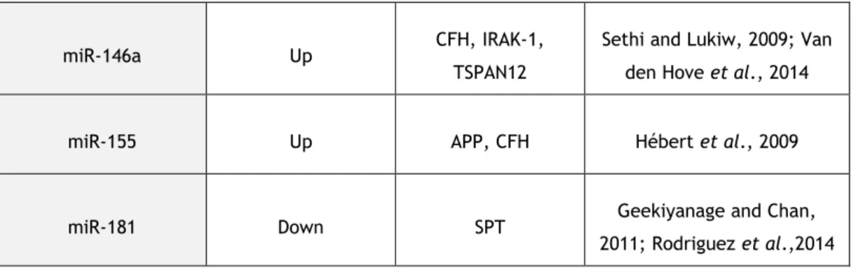
In neurodegenerative disorders, such as AD, brain miRNA profiles are altered,so that their dysfunction may be a cause or a consequence for the onset of the disease, which is not yet well understood. The miRNA pool is controlled by various mechanisms, such as direct modification of miRNA sequences or their precursors that differ in length/sequence, sequestration, miRNA transport modulation and miRNA turnover modulation. Aβ is also a powerful regulator of miRNA levels (Amakiri et al., 2019; Long et al., 2019; Martinez and Peplow, 2019; Nahalka, 2019; Slota and Booth, 2019). It is already clear that miRNAs are involved in many aspects of AD progression, including direct cell senescence, APP expression regulation, APP alternative splicing, BACE and Tau regulation, lipid metabolism and neuroinflammatory processes regulation. However, the identification of specific targets has been difficult due to the several pathways in which miRNAs participate and the interconnections of these paths, making it even more difficult to know if the influence on this target is direct or indirect. Therefore, it is important not to exclude a target just because there are no studies to support it. To try to identify miRNA targets some properties are taken into account, like if the mRNA in study has there expression level repressed in a miRNA-dependent way, if the loss of the mRNA interaction with the miRNA leads to a macroscopic phenotype, if mRNA interact physically with Ago proteins, if exhibits sequence complementarity to the miRNA or if their interaction is phylogenetically conserved due to biologically benefit. These are some common characteristic of the genes that are proven to be regulated by miRNAs. Some of the miRNAs and their targets already involved in or associated to AD are summarized in Table 2 (Long et al., 2019; Mockly, 2019; Nilsen, 2007; Slota and Booth, 2019).
16
Table 2. Dysregulated miRNAs in Alzheimer’s disease.miRNA Expression Levels Target Proteins References
Let-7 Down TLR-7, APP Lehman et al., 2012; Maes
et al., 2009
miR-9 Up/Down
BACE1, PSEN1,
NFH, SIRT1, MAPT, SPT, CFH, NR2E1
Batistela et al., 2017; Delay
et al., 2012; Idda et al.,
2018; Li and Wang, 2018; Maes et al., 2009 miR-15b, -29a, -29b, -29c, -107, -124, -186, -195, -328, -485-5p Down BACE1 Boissonneault et al., 2009; Faghihi et al., 2010; Hébert
et al., 2009; Geekiyanage
and Chan, 2011; Lang et
al., 2016; Li and Wang,
2018; Jiao et al., 2016; Kim
et al., 2016; Yang et al.,
2015; Zhu et al., 2012 miR-16
Up APP Parsi et al., 2015
miR-17, -20a, -27a -3p, -101a-3p, -147, -153, -323-3P, -644, -655 Down APP Barbato et al., 2014; Boissonneault et al.,2009; Delay et al., 2011; Hébert
et al., 2009; Liang et al.,
2012; Lin et al., 2018., 2018; Long et al.,2012; Zhang et al., 2014; miR-21
--- PI3K, AKT, GSK3β Feng et al., 2018
miR-34 Up TAU, SIRT1 Inukai et al., 2012; Mariño
et al., 2010
miR-106b Down ABCA1, APP Noren et al.,2010; Qiu et
al.,2015
miR-125b Up CDKN2A, SYN-2, 15-LOX
Sethi and Lukiw, 2009; Van den Hove et al., 2014
miR-137 Down MAPT, SPT Geekiyanage and Chan, 2011; Smith et al., 2011
17
miR-146a Up CFH, IRAK-1, TSPAN12
Sethi and Lukiw, 2009; Van den Hove et al., 2014
miR-155 Up APP, CFH Hébert et al., 2009
miR-181 Down SPT Geekiyanage and Chan, 2011; Rodriguez et al.,2014
This study is focused on miR-29b previously used in the research group and miR-9 which is related to the regulation of various mRNAs encoding proteins involved in pathological pathways of AD. MiR-29 is a highly conserved family in humans, mice and rats. The elements of this family are miR-29a and miR-29b-1 transcribed from chromosome 7 (7q32.3), miR-29b-2 with mature sequences identical to those of miR-29b-1 (both called miR-29b) and miR-29c transcribed from chromosome 1 (1q32.2), both clusters are transcribed together. All of these miRNAs share identical sequences at the 2-7 nt position at the 5’ end and have the same “seed region”. Although they have similar sequences, they are located in different cell compartments, miR-29a is located in the cytoplasm, while miR-29b and miR-29c are mainly in the nucleus, because of these differences, their targets may also vary, leading to different biologic effects (Kwon et al., 2019; Slusarz and Pulakat, 2015). One of the causes of this distribution is the hexanucleotide segmented sequence unique to miR-29b, that makes this miRNA to be mainly present in the nucleus, but its nuclear function is still not well understood. Another difference is the presence of a Tri-uracil sequence that makes miR-29a more stable and with a longer half-life, while miR-29c has a very low expression, perhaps due to the high turnover rate (Kwon et al., 2019; Slusarz and Pulakat, 2015; Zong et al., 2015). The differences in miR-29 structures are illustrated in figure 4.
Figure 4. Schematic representation of the miR-29 family and their structural differences (Adapted
from Kwon et al., 2019).
This family has been shown an important role in several pathological disorders, like cardiovascular, AD and cancer
(
Roshan et al., 2009). In turn, the dysregulation of this family has implications for neuronal differentiation, proliferation, communication and death. MiR-29b is downregulated in brains of AD patients and several studies have shown that it may be18
involved in the regulation of APP and BACE1 expressions (Pereira et al., 2016; Zong et al., 2015). As mentioned above, the BACE1 protein levels are elevated in AD brains, suggesting that BACE1 dysregulation is directly implicated in AD pathogenesis through the formation of toxic Aβ peptides. Particularly, the work of Herbert and co-workers, in 2008, showed that the decreased levels of the miR-29a/29b-1 cluster were correlated with increased levels of BACE1 expression in patients with sporadic form of AD and that the introduction of synthetic miR-29 reduced the secretion of Aβ peptides (Kwon et al., 2019; Zong et al., 2015).
MiR-9 has different origins from the 5’ end of the precursor, named miR-9-5p (miR-9) or from the 3’ end called miR-9-3p (miR-9*). However, the precursor and mature sequences are the same for both types. In mammals, the genes encoding miR-9 are located on chromosomes 1 (1q22) 5 (5q14.3) and 15 (15q26.1), highly conserved during evolution (Nowek
et al., 2018; Saraiva et al., 2017). MiR-9 is highly expressed in the brain and its expression is
often related to neurodegenerative disorders. Several studies have shown that miR-9 is downregulated in AD, more specifically in the hippocampus and cortex, but also in the blood serum and CSF (Nowek et al., 2018; Saraiva et al., 2017). In 2017, Riancho and collaborators demonstrated by microRNAome analysis that 14 miRNAs are differentially expressed in CSF fluid of AD patients, with miR-9 being one of them. When compared to the control group, miR-9 was detected in 50% of the control group, while in the AD group nothing was detected (Riancho et al., 2017). Schonrock and co-workers also showed that the increase Aβ levels leads to a considerable miRNA dysregulation, with miR-9 being one of the downregulated miRNAs (Schonrock et al., 2012). In addition, some studies have suggested that miR-9 could regulate BACE1 (Hebert et al., 2008) and PS1 (Krichevsky et al., 2003; Reddy et al., 2017). So, if this miRNA is able to negatively regulate targets that play a key role in AD, such as PS1 and BACE1, the same logical thinking given for miR-29 can be applied to miR-9, making this miRNA potential therapeutics for AD. The possible targets of miR-9 and miR-29b in AD pathologic pathway studied in this work are represented in figure 5.
Figure 5. Schematic representation of the possible targets of miR-9 and miR-29b in AD pathologic pathway.