Systems/Circuits
Activation of Dopaminergic D2/D3 Receptors Modulates
Dorsoventral Connectivity in the Hippocampus and Reverses
the Impairment of Working Memory after Nerve Injury
Helder Cardoso-Cruz,
1,2* Margarida Dourado,
1,2,3* Clara Monteiro,
1,2Mariana R. Matos,
1,2and Vasco Galhardo
1,2 1Departamento de Biologia Experimental, Faculdade de Medicina, Universidade do Porto, 4200-319 Porto, Portugal,2Instituto de Biologia Molecular eCelular, Grupo de Morfofisiologia do Sistema Somatosensitivo, Universidade do Porto, 4150-180 Porto, Portugal, and3Programa Doutoral em
Neurocieˆncias, Faculdade de Medicina, Universidade do Porto. 4200-319 Porto, Portugal
Dopamine plays an important role in several forms of synaptic plasticity in the hippocampus, a crucial brain structure for working
memory (WM) functioning. In this study, we evaluated whether the working-memory impairment characteristic of animal models of
chronic pain is dependent on hippocampal dopaminergic signaling. To address this issue, we implanted multichannel arrays of
elec-trodes in the dorsal and ventral hippocampal CA1 region of rats and recorded the neuronal activity during a food-reinforced spatial WM
task of trajectory alternation. Within-subject behavioral performance and patterns of dorsoventral neuronal activity were assessed
before and after the onset of persistent neuropathic pain using the Spared Nerve Injury (SNI) model of neuropathic pain. Our results show
that the peripheral nerve lesion caused a disruption in WM and in hippocampus spike activity and that this disruption was reversed by the
systemic administration of the dopamine D2/D3 receptor agonist quinpirole (0.05 mg/kg). In SNI animals, the administration of
quin-pirole restored both the performance-related and the task-related spike activity to the normal range characteristic of naive animals,
whereas quinpirole in sham animals caused the opposite effect. Quinpirole also reversed the abnormally low levels of hippocampus
dorsoventral connectivity and phase coherence. Together with our finding of changes in gene expression of dopamine receptors and
modulators after the onset of the nerve injury model, these results suggest that disruption of the dopaminergic balance in the
hippocam-pus may be crucial for the clinical neurological and cognitive deficits observed in patients with painful syndromes.
Key words: awake animal physiology; dopamine; hippocampus; neuropathy; pain; working memory
Introduction
The differential processing of working memory (WM) between
hippocampal regions is a topic of renewed interest because it is
still not understood how cognitive salient information is
distrib-uted along the dorsoventral axis of the hippocampus (
Fanselow
and Dong, 2010
). Although neuroanatomical (
Swanson and
Cowan, 1977
;
Witter and Amaral, 2004
), functional (
Bannerman
et al., 1999
;
Kjelstrup et al., 2002
), and gene expression (
Thomp-son et al., 2008
;
Dong et al., 2009
) studies support the notion of a
functional separation between the entorhinal-dorsal
hippocam-pus circuit and the amygdala-ventral hippocamhippocam-pus circuit
(
Moser and Moser, 1998
), few studies have simultaneously
re-corded both areas during spatial WM tasks (
Schmidt et al., 2013
).
We have shown recently that chronic pain disrupts place cell
activity in the hippocampus (
Cardoso-Cruz et al., 2011a
) and
leads to a decrease in functional connectivity between the medial
prefrontal and the hippocampus during performance in spatial
WM tasks (
Cardoso-Cruz et al., 2013a
); however, these results
relate only to the dorsal hippocampus and it is still not known
whether the neural activity in ventral hippocampus is also
af-fected in chronic pain models.
Dopaminergic transmission has a vital influence on WM
(
Arnsten et al., 1995
;
Cai and Arnsten, 1997
;
Castner et al., 2000
)
and dopamine within the dorsal hippocampus is known to be
particularly important for the establishment of the longer forms
of LTP (
Lisman et al., 2011
). Interestingly, it has been shown in
both animal and human chronic pain syndromes that there is a
decrease in the tonic levels of dopamine in brain areas such as the
prefrontal cortex, striatum, and hippocampus (
Wood et al.,
2007a
;
Wood et al., 2007b
;
Pais-Vieira et al., 2009
), whereas there
is some evidence of a correlation between a decrease in reduced
dopamine activity and a decrease in hippocampal gray matter in
chronic pain patients (
Wood et al., 2009
).
Pharmacological studies have shown that specific D2-family
agonists can improve memory retention and WM in
delay-Received Jan. 2, 2014; revised Feb. 25, 2014; accepted Feb. 28, 2014.Author contributions: H.C.-C., M.D., C.M., and V.G. designed research; H.C.-C., M.D., C.M., and M.R.M. performed research; H.C.-C., M.D., C.M., M.R.M., and V.G. analyzed data; H.C.-C., M.D., C.M., and V.G. wrote the paper.
This work was supported by FEDER funds through the Operational Competitiveness Programme–COMPETE, National Funds through Fundac¸a˜o para a Cieˆncia e a Tecnologia (Project FCOMP-01– 0124-FEDER-011202, Grant PTDC/SAU-NEU/100773/2008; Project FCOMP-01– 0124-FEDER-029686, Grant PTDC/NEU-SCC/1516/2012; and Doctoral Grant SFRH/42500/2007), and the BIAL Foundation (Project 126/08).
The authors declare no competing financial interests. *H.C.-C. and M.D. contributed equally to this work.
Correspondence should be addressed to Vasco Galhardo, PhD, Departamento de Biologia Experimental, Fac-uldade de Medicina da Universidade do Porto, Alameda Prof Hernani Monteiro, 4200 –319 Porto, Portugal. E-mail: galhardo@med.up.pt.
DOI:10.1523/JNEUROSCI.0021-14.2014
remains unclear whether chronic pain affects the hippocampal
dopaminergic transmission and if that translates to a disruption
of the information processing that is crucial needed for spatial
mnemonic processing. Therefore, in this study, we
investi-gated whether activation of dopamine D2/D3 receptors during
performance in a spatial alternation WM task affects
hip-pocampus dorsoventral functional connectivity and
individ-ual neuron encoding.
Materials and Methods
Animals and ethical statement
Experiments were performed in 31 Sprague Dawley adult male rats (weight 275–325 g; Charles River Laboratories). Before surgery, animals were housed in collective cages (three per box) and kept on a 12 h light/ dark cycle. Training and recording sessions were performed at approxi-mately the same time each day during the light portion of the cycle. During the course of the experiments, all animals were food deprived to 90 –95% of their ad libitum feeding body weights. All electrophysiological and behavioral studies were performed in accordance with the guidelines of the European Union (2010/63/CE) and with the Research and Ethical Issues of the International Association for the Study of Pain ( Zimmer-mann, 1983). The experimental protocols were also approved by the local Ethical Committee for Animal Use and national Direcc¸a˜o Geral de Ali-mentac¸a˜o e Veterina´ria board (Lisbon, Portugal). At least one category C Federation of European Laboratory Animal Science Associations (FELASA)-certified experimenter was present when animals were manipulated.
Implantation of electrodes and nerve lesion model
The procedure for the surgical implantation of intracranial multielec-trode arrays has been described previously in detail (Cardoso-Cruz et al., 2011b). Briefly, animals were anesthetized with a ketamine/medetomi-dine mixture (75 and 0.5 mg/kg in saline, respectively, i.p.). Anesthesia was maintained with small additional injections of ketamine (one-third of the initial dosage). Depth of anesthesia and paralysis of the muscula-ture were assessed by regular testing of corneal blink, hind-paw with-drawal, and tail-pinch reflexes. Core body temperature was measured with rectal thermometer and maintained at 37°C using a homeothermic blanket system (Harvard Apparatus). Animals were secured in a stereo-taxic frame using ear bars and the skull was exposed and cleaned using hydrogen peroxide. Holes were bored in the skull for entry points of the two microelectrode arrays and to secure four to five screws necessary to hold the arrays and for grounding purposes. Each microelectrode array contained eight filaments of isonel-coated tungsten wire (35m in di-ameter; California Fine Wire Company) with impedances varying be-tween 0.5 and 0.7 M⍀ at 1 kHz. The multielectrode arrays were assembled in a 4⫻ 2 architecture, with spacing of 250m between filaments and 450m between each row (Cardoso-Cruz et al., 2011a). The arrays were oriented rostrocaudally and mounted in the holder of a hydraulic micropositioner (FHC) and subsequently slowly driven (50 m/min) to the dorsal hippocampus (dCA1) and ventral hippocampus (vCA1). The following coordinates in millimeters relative to bregma (Paxinos and Watson, 1998) were used to place the electrode arrays: dCA1,⫺3.2 rostrocaudal, ⫾ 2.2 mediolateral, ⫺2.7 dorsoventral; vCA1, ⫺6.0 rostrocaudal, ⫾ 5.5 mediolateral, ⫺7.5 dorsoventral. The multi-electrode arrays were implanted unilaterally and counterbalanced be-tween animals.
(Chaplan et al., 1994;Cardoso-Cruz et al., 2011a).
After surgery, rats were allowed to recover for 7 d before behavioral and recording sessions began. During this recovery period, each animal was placed in the testing room daily and connected to a W16 wireless head-stage transmitter (Triangle Biosystems) to adapt to the recording conditions in the laboratory environment.
Behavioral procedures
Figure-8 maze. In this study, we evaluated spatial WM performance using
a food-reinforced spatial alternation task on a figure-8-shaped maze (Cardoso-Cruz et al., 2013a;Fig. 1B). The total dimension of the arena
was 90⫻ 60 cm, with Plexiglas corridors 15 cm wide and opaque walls 30 cm high. Starting from the center of the maze (C), the rats were trained to alternately visit two reward sites (R) to obtain one chocolate-flavored pellet (45 mg) that was automatically delivered by a pellet dispenser (Coulbourn Instruments). After visiting one of the reward locations, the animal had to continue forward and cross again the central corridor before visiting the opposite reward site; food rewards were not dispensed if the animal failed to cross the central corridor immediately before ar-riving at the reward sites or if the animal made two consecutive visits to the same reward site. Control of pellet dispensers was fully automated using the OpenControl software adapted to this particular task (Aguiar et al., 2007). In all of the analyses in this study, we considered three different zones in the behavioral arena: “reward zones” were the 15⫻ 15 cm corner areas where the animal received a pellet upon a correct alterna-tion; the “delay zones” were the area between the reward zones and the central corridor; and the “choice zone” was the area preceding the reward zones and immediately after the central corridor (Cardoso-Cruz et al., 2013a).
Experimental timeline. After the 7 d recovery period following surgery,
each rat was habituated to the maze in 5 daily sessions of 15 min in which the animal was allowed to explore and consume chocolate-flavored pel-lets that were distributed throughout the maze. In the last habituation session, the chocolate pellets were placed only in the vicinity of the re-ward dispensers. The experimental protocol timeline had two phases per animal of both sham and SNI groups (Fig. 1A). The first phase consisted
in a training period of 10 sessions during 10 consecutive days in which only the behavioral performance was evaluated. After this period, each animal performed two recording sessions of hippocampus activity, the first one after intraperitoneal saline injection (control recording session) and the second after intraperitoneal administration of quinpirole, a do-pamine D2/D3 receptor agonist (0.05 mg/kg, dissolved in saline; Sigma). The second physiology recording session was performed 10 d after the control session and was preceded by 2 d of retraining in the figure-8 maze to keep and stabilize the performance level. In all sessions, we alterna-tively tested animals from control and experimental groups to prevent unpredictable bias effects. Vehicle and quinpirole were administered in-traperitoneally 20 min in advance of the behavioral testing session. Given the extensive amount of electrophysiology sessions that are required to complete our experimental protocol, we opted to test only one drug and a single dosage. Quinpirole at 0.05 mg/kg was elected based on performance-only results from preliminary experiments in this maze, from other ongoing studies in the laboratory using a decision-making task, and from a comparative analysis of other similar studies (Horvitz et al., 2001;Huang et al., 2013). In our preliminary sessions, we found that higher doses of quinpirole (0.1 and 0.5 mg/kg) caused severe decreases in locomotion in this task.
Electrophysiological recordings. Single-cell and local field potential
(LFP) recordings were obtained from the implanted multielectrode ar-rays during behavioral performance in the figure-8 maze. The 16-channel multielectrode array was connected to a wireless head-stage transmitter (W16; Triangle Biosystems) that sent continuous analog sig-nals to a Multineuron Acquisition Processor system (16-MAP’ Plexon). Neural signals were preamplified (10,000 –25,000⫻) and digitized at 40 kHz. Voltage-time threshold windows were used to identify single-unit waveforms and up to two neuronal action potentials per recording chan-nel were sorted online (SortClient 2.6; Plexon) and validated by offline automatic and manual sorting techniques (Offline Sorter 2.8; Plexon) according to the cumulative criteria described in detail previously (Cardoso-Cruz et al., 2011a;Pais-Vieira et al., 2012;Cardoso-Cruz et al., 2013a). Each electrode was manually checked for artifacts (e.g., 50 Hz noise). Extracellular LFPs were simultaneously recorded from the same implanted microwires by low-frequency (0.5–200 Hz) filtering of the raw signals. LFPs were preamplified and digitized at 500 Hz. An overhead video-tracking system (CinePlex 2; Plexon) was used to provide infor-mation about the animal position on the maze and to synchronize the video recordings with the acquired neuronal data. After the end of last recording session, the rats were deeply anesthetized with a ketamine/ xylazine mixture and the recording site was marked by injecting DC current (10 –20A for 10–20 s) through one microwire per array. After this step, the animals were transcardially perfused with 0.01Mphosphate buffer, pH 7.2, in a 0.9% saline solution, followed by 4% paraformalde-hyde. Brains were removed and postfixed in 4% paraformaldehyde for 4 h and stored in 30% sucrose before they were frozen, sectioned into 40 m slices, and counterstained to identify the recording sites.
mRNA levels of dopamine receptors in hippocampus. Rats were deeply
anesthetized by intraperitoneal injection of 200 mg/kg sodium pentobar-bital solution. Brains were immediately removed after decapitation, bathed in RNALater (Ambion) for RNA preservation, and kept refriger-ated during further processing. Thick coronal slabs were cut and each individual brain area was then carefully dissected under a surgical micro-scope, resulting in bilateral brain samples from dorsal and ventral hip-pocampus from 20 animals (sham, n⫽ 10, and SNI, n ⫽ 10; 21 d after
lesion). All tissue samples were flash frozen in liquid nitrogen and stored at⫺80°C until further processing. After mechanical tissue disruption, total RNA was isolated using the TRIzol Plus RNA Purification System (Life Technologies) according to the manufacturer’s protocol; 1g of RNA was immediately used for cDNA synthesis with the RevertAid First Strand cDNA Synthesis Kit (Thermo Fisher Scientific) according to the manufacturer’s instructions. PCR amplification was performed in a StepOnePlus Real-Time PCR System (Applied Biosystems) using SYBR Select Master Mix (Applied Biosystems) and the results were analyzed using StepOne analysis software (Applied Biosystems). Real-time PCRs were performed in triplicate and the primer sequences were as follows:
Drd1 (accession number NM_012546.2), 5
⬘-TGCTGCTGGCTC-CCTTTC-3⬘ and 5⬘-GTTAATGCTCACCGTCTCTATGG-3⬘; Drd2 (ac-cession number NM_012547.1), 5⬘-TGGAGGTGGTGGGTGAG-3⬘ and
5⬘-CAGCAGAGTGACGATGAAGG-3⬘; Drd3 (accession number
NM_017140.1), 5⬘-GTCCGCACGCCTACTACG-3⬘ and 5⬘-CTCCAC-CTGTCACCTCCAAG-3⬘; Th (accession number NM_012740.3)
5⬘-CGTCCCCAAGGTTCATC-3⬘ and 5⬘-GGCTTCAAATGTCTCAAA
TACTT-3⬘; Maoa (accession number NM_033653.1),
5⬘-CAGTATG-GAAGGGTGATTCGCC-3⬘ and 5⬘-CAGACCAGGCACGGAAGG-3⬘;
Gapdh (accession number NM_017008.4), 5
⬘-ATGATTCTACCCACG-GCAAG-3⬘ and 5⬘-TTCACACCCATCACAAACAT-3⬘. Specificity of PCRs was confirmed using the melting curves of each studied gene. Am-plifications were performed starting with a 2 min step for enzyme acti-vation at 95°C, followed by 2 min at 50°C, and 40 cycles at 95°C for 30 s, 60°C for 45 s, and 72°C for 45 s. For each animal, semiquantitative expression of each gene of interest was performed according to the del-ta-Ct method, using GAPDH as housekeeping gene. Significant differ-ences in gene expression between experimental groups were statistically assessed using a nonparametric Mann–Whitney test.
Data analyses
Behavioral data. Custom MATLAB and Python scripts were used to
clas-sify behavioral intervals based on previous or next trial outcome (correct vs error), maze navigation zone (choice and delay zones), and experi-mental session (sham vs SNI, vehicle vs D2/D3r-agonist treatment).
Figure 1. Experimental timeline, arena, and behavioral performance. A, Timeline of experimental protocol. Briefly, each animal was implanted with multielectrode arrays in the dorsal and ventral hippocampus and subjected to a sham or SNI lesion surgery. After recovery, the animals were trained for 10 d and then had electrophysiological recording sessions before and after injection of quinpirole. B, Diagram of the figure-8 maze of spatial alternation used in the study. Starting from the center of the maze, the animal had to visit two reward sites (R) in alternation to obtain chocolate-flavored pellets. C, Gain in performance during the 10 training sessions. The nerve-lesioned animals (SNI: n⫽7)showedasignificantlylowerperformancecomparedwithcontrolanimals (sham: n⫽ 4). D, Level of mechanical sensitivity measured by withdrawal response to stimulation with Von Frey filaments. As expected, a large decrease was observed in the threshold required to induce a paw response in SNI group and the sensitivity level was stable throughout the training period. Values are presented as mean⫾SEM.Comparisonsbetweenexperimentalgroupsarebased on two-way ANOVA (group⫻ time), followed by post hoc Bonferroni. *p ⬍ 0.05; **p ⬍ 0.01; ***p ⬍ 0.001.
These classifications were calculated based on tracking navigation vec-tors obtained from behavior video recordings. Several parameters con-cerning the behavioral performance and navigation maps were examined across experimental sessions: percentage of correct alternations, total number of performed trials, and average running speed (Cardoso-Cruz et al., 2013a;Cardoso-Cruz et al., 2013b).
Spiking activity. Neural data were processed offline using
NeuroEx-plorer 4 (NEX; Plexon) and exported to our own MATLAB R14 routines for additional analysis (MathWorks). Populational average of dCA1 and vCA1 neurons firing activity was examined across choice zone and delay zone navigation, comparing sham and SNI groups after vehicle or quin-pirole administration. To characterize the temporal structure of spiking activity for correct versus error trials, population perievent time histo-grams were computed for each recorded region and plotted in a 2 s range centered at choice boundary (resolution of 50 ms per bin). The choice boundary was defined as the transition between central corridor and reward locations, indicated in maze diagram by the dashed black line (Fig. 1B). A two-side Kolmogorov–Smirnov (KS2) test ( p⬍ 0.05) was
used to identify differences in the firing distributions between experi-mental groups. All recording sessions selected for neuronal activity com-parisons had at least 5% of error trials.
LFP analysis. For statistical comparison, five bands of the LFP
oscilla-tions were considered: 1– 4 Hz (␦), 4–9 Hz (), 9–15 Hz (␣), 15–30 Hz (), and 30–50 Hz (␥). The Kolmogorov–Smirnov (KS) test (with Dallal-Wilkinson-Lilliefor-corrected p-value) was used to determine whether datasets were normally distributed (Prism 6.0; GraphPad). For single comparisons, we used the nonparametric Mann–Whitney test for unpaired samples; for multiple comparisons of normally distributed samples, we used two-factor ANOVA (with post hoc Bonferroni test); and for non-normal samples, we used the non-parametric Kruskal–Wallis analysis of ranks (KW) with post hoc Dunn’s test. The level of significance was set as 5%. Results are expressed as mean⫾ SEM.
Spectral analysis. Global spectral characteristics of dCA1 (Pxx) and
vCA1 (Pyy) LFP signals were calculated in respect to choice zone and
delay zone. The power spectral densities (PSD) of the LFPs signals were computed using fast Fourier transform analysis (512-point), using Welch’s method as a spectral estimator (MATLAB native function) over the 0 –50 Hz range of frequencies (0.19 Hz of resolution). Histograms were smoothed using a three-point Gaussian process and are presented as percentage of total PSD in respect to frequency bands. To examine the relationship between power activity and behavioral performance, we plotted the averaged dCA1-vCA1 ratio (as dB relative to 1 V) during choice zone navigation for all correct and error trials. To maintain a balanced number of trials per condition, only a maximum of eight cor-rect trials per recording session were randomly selected and included for this analysis.
To measure phase coherence (⌽xy), hippocampus LFPs were filtered
in the (4–9 Hz) and ␥ (30–50 Hz) range using a zero-phase forward and
reverse digital four-pole Butterworth band-pass filter to yield LFP/LFP␥. Phase coherence was then evaluated by calculating the Hilbert transform of each LFP segment. The phase angles of each signal segment were extracted and wrapped between 0° and 360° and are displayed as a polar plot histogram in bins of 6° per segment. The Rayleigh test of uniformity ( p⬍ 0.01) was used to assess the resulting phase distributions for devi-ations from circular uniform distribution. The degree of phase coherence was determined calculating the concentration around the preferred phase in the circular distribution, with its value lying between 0 and 1, where⌽xy⫽ 0 indicates that phase values at a particular frequency range
are randomly distributed across the time interval and⌽xy⫽ 1 indicates
that phase values are the same across the time interval. Phase coherence values are inversely related to the Rayleigh p-value, with⌽xy⫽ 0 standing
for uniform distribution. The CircStat MATLAB toolbox (Berens, 2009) was used for circular plotting and circular statistics were calculated ac-cording to common methods (Fisher, 1993).
To determine the spectral coupling among LFP signals from recorded areas, we calculated the quadratic coherence [Cxy( f )] applying the
equa-tion equivalent to Cxy( f )⫽ 兩Pxy( f )兩2/(Pxx( f ).Pyy( f )), where the
coher-ence is equal to the averaged cross-power spectrum [Pxy( f )} normalized
matrix of cross-power spectral density of the two signals. A higher coher-ence value (near 1) indicates that phases of signals are identical and both are totally phase locked at this frequency; otherwise, lower coherence values (near 0) indicate that phases are dispersed.
The statistical method of partial directed coherence (PDC) was used to quantify the frequency-domain connectivity between dorsal and ventral hippocampus. The PDC method has been described in detail previously (Sameshima and Baccala´, 1999;Baccala´ and Sameshima, 2001; Cardoso-Cruz et al., 2011b). Briefly, PDC is an alternative representation of Granger causality involving multivariate processes to uncover direct in-fluences in the frequency domain. Higher PDC values indicate a strong connectivity between recorded structures, which can be interpreted as the existence of information flow between them.
Results
Behavioral performance
Pain-related performance behavior
To evaluate the effect of chronic pain on spatial WM
perfor-mance, we compared the controls and animals with a
neuro-pathic pain model in a spatial alternation maze (sham control
group, n
⫽ 4; SNI group, n ⫽ 7;
Fig. 1
). All animals of the
SNI-treated group developed mechanical allodynia after nerve lesion,
as indicated by the von Frey filament stimulation test (
Fig. 1
C).
Significant statistical differences were observed between groups
(F
(1,90)⫽ 161.20, p ⬍ 0.0001) and training sessions (F
(9,90)⫽
3.30, p
⫽ 0.0015). Post hoc analysis revealed that the threshold of
response to stimulation in the SNI group was lower than the
Figure 2. Effect of quinpirole injection on neuropathic nociception and behavioral performance. A, Quinpirole did not affect the already significant difference in mechanical threshold between sham (n⫽ 4) and SNI (n ⫽ 7) animals. B, Injection of quinpirole reversed the behavioral performance between sham and SNI animals. The percentage of completed correct alternations between reward sites was larger in the sham animals after vehicle administration, but it was larger in the SNI animals after quinpirole. The major effect of quinpirole was a decrease in the performance of the sham animals. C, Quinpirole decreased the running speed of the sham animals to the same range of the SNI animals, but did not affect the running speed of the SNI group. D, Quinpirole reduced the total number of trials (visits to the reward site after completing a full turn in the lateral corridors) executed by the sham animals. Note that despite the low number of completed trials by the SNI animals, their percentage of correct trials was larger (B). Values are presented as mean⫾ SEM. Comparisons between experimental groups are based on two-way ANOVA (group ⫻ treatment), followed by post hoc Bonferroni. *p⬍ 0.05; **p ⬍ 0.01; ***p ⬍ 0.001.
threshold of the sham group at all training sessions ( p
⬍ 0.001,
Bonferroni test).
Testing sessions in the figure-8 maze were initiated 7 d after
the sham or SNI intervention. ANOVA indicated significant
dif-ferences between groups (F
(1,90)⫽ 14.48, p ⫽ 0.0052) and across
training sessions (F
(9,90)⫽ 63.84, p ⬍ 0.0001). In addition, post
hoc analysis revealed a lower performance behavior of the SNI
group compared with control group at day 3 (Bonferroni test,
p
⬍ 0.05), day 4 (p ⬍ 0.05), day 5 (p ⬍ 0.01), and day 6 (p ⬍ 0.05;
Fig. 1
D). These results indicate that SNI animals had lower
per-formance levels, particularly at early testing sessions.
Effect of quinpirole on behavioral performance
Systemic administration of quinpirole did not change pain
per-ception thresholds significantly. ANOVA revealed differences
be-tween sham and SNI animals (F
(1,18)⫽ 10.92, p ⫽ 0.0039) and no
significant effect for drug treatment (F
(1,18)⫽ 1.56, p ⫽ 0.2281;
Fig. 2
A).
Behavioral results in the figure-8 maze showed an important
recovery of spatial WM performance of SNI-treated animals after
quinpirole administration (
Fig. 2
B). ANOVA revealed significant
differences between experimental groups (F
(1,18)⫽ 6.24, p ⫽
0.0224) and drug treatment (F
(1,18)⫽ 12.25, p ⫽ 0.0026). Post hoc
analysis revealed that the SNI group exhibited an increase of
per-formance after quinpirole administration ( p
⬍ 0.05, Bonferroni
test), whereas the sham group had an opposite effect ( p
⬍ 0.001).
An essential issue of spatial alternation tasks success is the
motor performance. We found significant differences between
experimental groups in terms of running speed parameter during
maze navigation (F
(1,18)⫽ 10.02, p ⫽ 0.0054) and drug treatment
(F
(1,18)⫽ 8.49, p ⫽ 0.0093). Moreover, post hoc analysis revealed
that running speed of SNI-treated group were lower compared
with sham for vehicle administration, as expected ( p
⬍ 0.001;
Fig.
2
C). It is important to note that quinpirole injection induces a
decline of motor activity; however, that reduction was not
corre-lated with performance impairment in the SNI group. In fact, SNI
animals after quinpirole injection illustrated better performance
even with lower navigation running speed. This premise were
also supported by the total number of trials performed per
ses-sion in the figure-8 maze after quinpirole injection, which for SNI
animals were reduced but had a better performance ratio (
Fig.
2
D). Data from the total number of performed trials showed clear
differences between experimental groups (F
(1,18)⫽ 13.06, p ⫽
0.0020) and a significant effect for treatment factor (F
(1,18)⫽
11.56, p
⫽ 0.0032). Post hoc analysis revealed a decrease in the
total number of trials performed per session in SNI-treated
ani-mals compared with sham aniani-mals for vehicle administration
( p
⬍ 0.001, Bonferroni test) and also indicateda similar response
after quinpirole treatment shared by both experimental groups
(
Fig. 2
D).
Neuronal activity
Hippocampal firing patterns during behavioral performance
A total of 197 neurons were recorded from dorsal (dCA1, n
⫽ 100
cells) and ventral hippocampus (vCA1, n
⫽ 97 cells).
Table 1
Figure 3. Peridecision neuronal activity during correct and error trials. A, Color-coded raster plots of neuronalactivity(binresolutionof50ms)foralldorsalhippocampusrecordedneuronsduringthe4speriod centeredinthemomentofcrossingfromthedelaytothechoiceareainthefigure-8mazeduringcorrecttrials. dCA1neuronsofshamanimalspresentapeakofneuronalfiringrateimmediatelyprecedingtheapproachof thedecisionpointthatisfollowedbyasuddendecreaseinfiringrate.Incontrast,dCA1neuronsofSNIanimals showamorestablefiringrateduringtheperidecisionperiod.Injectionofquinpirolereversesthiselectrophys-iologicalpattern,withtheSNIanimalsnowpresentingapeakofactivityandshamanimalsshowingastable firing rate. B, C, Detailed peridecision histograms of population neuronal activity for both dorsal (B) and ventral(C)hippocampusduringeithercorrect(top)orerror(bottom)trials.Valuesarepresentedasmean⫾ SEM.ComparisonsoffiringdistributionsofexperimentalgroupsarebasedontheKS2test;p⬍0.05.
Table 1. Neuronal average firing rates of dorsal and ventral CA1 hippocampus recorded units during execution of the figure-8 maze WM task
dCA1 vCA1 Sham (43 neurons) SNI (57 neurons) Sham (41 neurons) SNI (56neurons)
Mean SEM Mean SEM Mean SEM Mean SEM
Delay zone Vehicle 4.34 0.29 4.11 0.14 5.13 0.31 5.20 0.21 Quinpirole 3.81 0.31 5.03* 0.37 5.02 0.24 6.03 0.37 Choice zone Vehicle 5.24 0.34 4.54 0.51 5.71 0.28 6.17 0.11 Quinpirole 5.31 0.28 6.41* 0.27 5.77 0.44 5.76 0.24 Values represent averaged spikes per second for all of the periods that the animal spent in either the delay zone or the choice zone during the entire recording session. Comparisons between experimental groups are based on two-way ANOVA (group⫻ treatment), followed by post hoc Bonferroni (*p ⬍ 0.05).
presents the averaged populational firing rate activity for the two
recorded hippocampal areas. In the case of dCA1, significant
differences were observed between experimental groups (delay
zone: F
(1,196)⫽ 6.12, p ⫽ 0.0142; choice zone: F
(1,196)⫽ 3.73, p ⫽
0.0495) and pharmacological treatment (delay zone: F
(1,196)⫽
3.85, p
⫽ 0.0428; choice zone: F
(1,196)⫽ 7.21, p ⫽ 0.007).
Quin-pirole administration caused an increase of firing rate in dCA1
neurons in SNI-treated animals (both navigation zones p
⬍ 0.05,
Bonferroni test). For vCA1 neurons, no significant differences
were observed in either behavioral task between experimental
groups (delay zone: F
(1,190)⫽ 3.24, p ⫽ 0.0735; choice zone:
F
(1,190)⫽ 0.69, p ⫽ 0.4075) or after pharmacological treatments
(delay zone: F
(1,190)⫽ 1.44, p ⫽ 0.2316; choice zone: F
(1,190)⫽
0.42, p
⫽ 0.5193).
Firing activity in correct and error trials
To analyze whether the firing activity was affected when the
ani-mal approached the point of decision between the two alternative
reward locations, we calculated the population activity by
re-corded area for either correct or error trials for each animal (
Fig.
3
). The spiking activity for both dCA1 and vCA1 neurons
re-vealed different temporal activity patterns centered at the choice
line of the maze in which the animals decided between left or
right turn. Our data showed that sham animals present an
in-crease of population firing rate of dCA1 neurons immediately
preceding the arrival at the point of left/right decision, and that
this decision-related increase in activity was abolished by the
quinpirole administration (
Fig. 3
A, top rasters). Interestingly,
dCA1 neurons of SNI group of animals presented the inverse
results, with an increase of firing rate occurring only after the
administration of quinpirole (
Fig. 3
A, bottom raster). Statistical
analysis of the normality of the temporal profiles of neuronal
activity showed that, in the dCA1, there were differences in the
firing rate between experimental groups during the correct trials
(vehicle: KS2
⫽ 0.487, p ⬍ 0.001; quinpirole: KS2 ⫽ 0.480, p ⬍
Figure 4. Spectral analysis of dorsal and ventral CA1 hippocampus LFP signals. A, Example trace of raw recording representing 5 s of ongoing LFP activity in a sham animal recorded during delay zone navigation. Blue trace represent the dorsal hippocampus filtered signal (4–9 Hz), whereas the red trace represent the ventral hippocampus filtered signal (4–9 Hz); the black traces undern each colored trace are the unfiltered LFP signal of each area. B, Full spectrograms for the same example of 5 s recording used in A show the predominance ofoscillationsinboth areas of the hippocampus during active navigation. C, D, Power spectra density plots of dorsal and ventral LFP activity during navigation in the delay zone (C) and choice zone (D) for both sham (n⫽ 4) and SNI (n ⫽ 7) animals. Data were calculated for each entire recording session independently of correct or error trials. PSD traces show that quinpirole does not affect the overall pattern of predominance.
Figure 5. Dissociation between dorsal and ventral CA1 field power activity during correct and error trials. Scatter plots of the distribution of dorsoventral ratios per trials of sham (blue dots) and SNI animals (red dots). A, During correct trials, sham animals presented higher power in dorsal hippocampus, whereas SNI animals presented higher power in ventral hip-pocampus; this pattern of dorsoventral ratio was reversed by quinpirole (shaded scatter plot in
A). B, In contrast, the during error trials, there was no preferential dorsoventralratioineither
0.001;
Fig. 3
B, top row), and also during the error trials, but only
after quinpirole treatment (KS2
⫽ 0.462, p ⬍ 0.001;
Fig. 3
B,
bottom row). In the case of the vCA1 neurons, our data showed
differences between experimental groups during the correct trials
(vehicle: KS2
⫽ 0.436, p ⫽ 0.007; quinpirole: KS2 ⫽ 0.564, p ⬍
0.001;
Fig. 3
C, top row), whereas during the error trials, there
were differences in the firing rate only after vehicle injection
(KS2
⫽ 0.359, p ⫽ 0.009;
Fig. 3
C, bottom row).
Spectral analysis
Changes in PSDs
The qualitative comparison of PSD values for sham and SNI
groups in function of drug treatment and maze navigation zones
revealed the expected profile of power oscillations with the major
power peak occurring in the
frequency band (4–9 Hz) in both
recorded areas. The administration of quinpirole also did not
cause major differences in the PSD profile (
Fig. 4
).
To analyze whether the
rhythm was affected with the
per-formance of the animal, we calculated the
power ratio and the
absolute value of the
power difference between the signals of the
dCA1 and vCA1 from correct versus error trials (
Fig. 5
).
Interest-ingly, the plots of the dCA1/vCA1
ratio show that sham and SNI
groups do not have overlapping power ratios during the correct
trials (
Fig. 5
A), but they overlap during error trials (
Fig. 5
B).
Moreover, the administration of quinpirole did not affect the
separation of
ratios during the correct and the overlap during
the error trials, although it reversed the magnitude of the power
ratios, with the SNI animals presenting larger
power ratios in
both areas after quinpirole during correct trials (
Fig. 5
A, B).
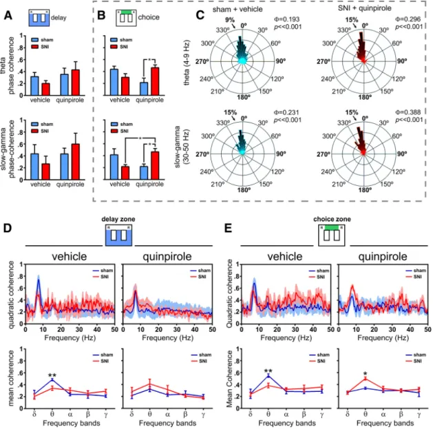
Figure 6. Spectral analysis of and ␥ LFP activity in dorsal and ventral hippocampus during maze navigation. A, B, dCA1-vCA1 phase coherence (⌽) activity for (4–9 Hz) and ␥ (30–50 Hz) frequency bands during delay and choice zones navigation. No significant differences were observed in delay zone between experimental groups or quinpirole administration (A). In contrast, during choice zone navigation (B), quinpirole reversed the phase coherence between sham and SNI animals in both theand␥bandsoffrequency.C,ExamplesofroseplotsofLFPphasedistributionsafter vehicle (in a sham animal) and quinpirole (in an SNI animal). The number in the upper right corner of each rose plot represents the value of⌽.Allcircularconcentrationdistributionsaresignificantly nonuniform (Rayleigh test, p⬍ 0.01). D, E, Spectral quadratic coherence between CA1 and vCA1 LFP signals show in all cases a strong coherence in the band of frequency (top). Detailed analysis of the quadratic coherence per band of frequency (bottom) shows that sham animals in both areas of maze have the largest quadratic coherence, whereas after quinpirole, the maximal coherence occurs in the SNI animals. Frequency bands:␦, 1–4 Hz; , 4–9 Hz; ␣, 9–15 Hz; , 15–30 Hz; ␥, 30–50 Hz. Values are presented as mean ⫾ SEM. Comparisons between experimental groups are based on two-way ANOVA (group⫻ treatment) or (group ⫻ frequency band), followed by post hoc Bonferroni. *p ⬍ 0.05; **p ⬍ 0.01.
LFP phase coherence in dorsoventral hippocampus
To determine whether
and ␥ oscillations were modulated by the
behavioral flexibility required for strategic navigation, we
per-formed phase coherence analyses of LFP signals from correct
trials. Our results showed no statistical differences during
navi-gation in the delay zone (
Fig. 6
A); however, during navigation in
the choice area, we found significant differences between groups
(: F
(1,18)⫽ 6.28, p ⫽ 0.0220;
␥: F
(1,18)⫽ 6.47, p ⫽ 0.0204) and
between pharmacological treatments (
: F
(1,18)⫽ 9.40, p ⫽
0.0066;
␥: F
(1,18)⫽ 13.89, p ⫽ 0.0015;
Fig. 6
B). Post hoc analyses
indicated an enhancement of
and ␥ phase coherence (both p ⬍
0.05, Bonferroni test) after quinpirole injection in SNI-treated
animals compared with the control group (
Fig. 6
B). Illustrative
examples of phase distributions are given in
Figure 6
C. The rose
plot histograms cover a narrow range of phase coherence for
and
␥ frequencies (all phase distributions are significantly
non-uniform, p
⬍ 0.01 Rayleigh test), suggesting a broader
coordina-tion of hippocampal activities during decision-making processes.
LFPs coherence in dorsoventral hippocampus
The quadratic coherence between dorsal and ventral FLP signals
was separately quantified for periods of navigation in delay or
choice zones. In all cases, the peak of dCA1::vCA1 coherence
occurred at the
frequency band (
Fig. 6
D, E).
In the analysis per frequency bands, our data showed no
sta-tistical differences between experimental groups (delay zone:
F
(1,45)⫽ 0.03, p ⫽ 0.8626; choice zone: F
(1,45)⫽ 0.06, p ⫽ 0.8132),
but a significant effect between frequency bands (delay zone:
F
(4,45)⫽ 35.98, p ⬍ 0.0001; choice zone: F
(4,45)⫽ 50.66, p ⬍
0.0001); post hoc analyses revealed that, after saline injection, the
sham group presented higher
coherence in both navigation
areas (delay zone: p
⬍ 0.01; and choice zone: p ⬍ 0.01;
Fig. 6
D, E,
Figure 7. Dorsoventral functional connectivity during maze navigation. A, B, Bidirectional traces of PDC across the spectral range of frequencies for both the sham and SNI groups before and after quinpirole administration. In all cases, there is a peak of PDC values in therange,reflectingthathippocampusdorsoventralfunctionalconnectivityisparticularlyimportantinthisbandoffrequency.
C, D, Bidirectional analysis of PDC per band of frequencies in both sham and SNI groups before and after quinpirole administration. The functional connectivity is only different between experimental
groups during navigation in the delay zone, not in the choice zone. During navigation in the delay zone, quinpirole reversed the functional connectivity between experimental groups: before quinpirole, the sham animals presented the largest dorsoventral connectivity, whereas after quinpirole, the largest connectivity occurs in the SNI animals. Frequency bands:␦, 1–4 Hz; , 4–9 Hz; ␣, 9–15 Hz; , 15–30 Hz; ␥, 30–50 Hz. Values are presented as mean ⫾ SEM. Comparisons between experimental groups are based on two-way ANOVA (group ⫻ frequency band), followed by
bottom row). In contrast, quinpirole injection reversed the levels
of dorsoventral hippocampus coherence and the SNI animals
presented higher levels of coherence than sham animals; we
found a difference between groups during navigation in the
choice zone (F
(1,45)⫽ 4.72, p ⫽ 0.0351) and differences across
frequency bands for both navigation zones (delay zone: F
(4,45)⫽
9.87, p
⬍ 0.0001; and choice zone: F
(4,45)⫽ 5.63, p ⫽ 0.0009). In
contrast to vehicle administration, post hoc analysis revealed an
increase of
oscillations coherence activity for SNI-treated
ani-mals during choice zone navigation ( p
⬍ 0.05;
Fig. 6
D, E, bottom
row).
Modulation of hippocampus dorsoventral functional connectivity
The occurrence of high coherence between two LFP signals
sug-gests synchronization, but does not disambiguate whether the
synchrony is due to synchronous fluctuations in power or to
consistent maintenance of phase relationships. To further study
the time-frequency-dependent relationship between the dorsal
and ventral regions of hippocampus, we estimated the level of
functional connectivity by PDC analysis (
Baccala´ and
Sameshima, 2001
). The quantification of bidirectional PDC
lev-els between recorded regions during task navigation is presented
in
Figure 7
and clearly shows that changes in circuit connectivity
occurred mainly at the
frequency band.
In the case of vehicle administration, significant differences
were observed between groups (dCA1 to vCA1 direction: delay
zone: F
(1,45)⫽ 36.46, p ⬍ 0.0001; choice zone: F
(1,45)⫽ 25.20, p ⬍
0.0001; vCA1 to dCA1 direction: delay
zone: F
(1,45)⫽ 51.21, p ⬍ 0.0001; choice
zone: F
(1,45)⫽4.40, p
⫽ 0.0417), as well as
across frequency bands (dCA1 to vCA1
direction: delay zone: F
(4,45)⫽ 23.75, p ⬍
0.0001; choice zone: F
(4,45)⫽ 25.84, p ⬍
0.0001; vCA1 to dCA1 direction: delay
zone: F
(4,45)⫽ 28.57, p ⬍ 0.0001; choice
zone: F
(4,45)⫽ 5.89, p ⫽ 0.0007;
Fig.
7
C,D). Moreover, post hoc analysis
re-vealed a decrease of PDC activity of
SNI-treated animals compared with control
animals in the delay zone (dCA1 to vCA1
direction:
, p ⬍ 0.001; vCA1 to dCA1
di-rection:
, p ⬍ 0.01, ␥, p ⬍ 0.05;
Fig. 7
C).
Importantly, quinpirole injection
re-versed the levels of intrahippocampus
functional connectivity by increasing the
abnormally reduced connectivity in SNI
animals to levels close to that observed in
normal conditions (cf.
Fig. 7
C,D, left vs
right, respectively). ANOVA showed
sta-tistical differences between groups (dCA1
to vCA1 direction: delay zone: F
(1,45)⫽
31.49, p
⬍ 0.0001; vCA1 to dCA1
direc-tion: delay zone: F
(1,45)⫽ 6.16, p ⫽
0.0159; choice zone: F
(1,45)⫽ 8.31, p ⫽
0.0060), and between frequency bands
(dCA1 to vCA1 direction: delay zone:
F
(4,45)⫽ 49.07, p ⬍ 0.0001; choice zone:
F
(4,45)⫽ 12.69, p ⬍ 0.0001; vCA1 to dCA1
direction: delay zone: F
(4,45)⫽ 19.62, p ⬍
0.0001; and choice zone: F
(4,45)⫽ 14.47,
p
⬍ 0.0001).
Hippocampus dorsoventral connectivity in
correct and error trials
We expanded the analysis of decision-related neuronal activity by
separating correct from error trials and repeating the PDC
anal-ysis, now focusing only on the
and ␥ frequency bands. The
results presented in
Figure 8
show that the PDC levels of
func-tional connectivity were in all cases larger in the correct versus
error trials; similarly, all of the PDC differences were observed in
the signals from the correct trials, with the majority of differences
occurring in the sham versus SNI comparison rather than on the
vehicle versus quinpirole administration. In the dCA1 to vCA1
direction, statistical significances were obtained for experimental
groups in the
frequency band (KW ⫽ 19.11, p ⫽ 0.0003). Post
hoc analysis revealed a decrease in SNI versus sham animals ( p
⬍
0.01; Dunn’s test) and an increase in SNI after quinpirole
treat-ment ( p
⬍ 0.01;
Fig. 8
A). In the vCA1 to dCA1 direction, data
showed differences in the
(KW ⫽ 19.29, p ⫽ 0.0002) and ␥
frequencies during correct trials (KW
⫽ 15.89, p ⫽ 0.0012;
Fig.
8
B). Post hoc analysis revealed that SNI animals increased their
connectivity level in respect to sham animals after quinpirole
administration ( p
⬍ 0.01; Dunn’s test), whereas in the
␥ band,
there was a difference in the sham versus SNI comparison
(vehi-cle: p
⬍ 0.05; quinpirole: p ⬍ 0.05) and a reduction of
connectiv-ity in the sham animals in the vehicle versus quinpirole
comparison ( p
⬍ 0.05).
mRNA levels of DA receptors in dorsal and ventral hippocampus
Finally, we evaluated whether the neuropathic pain condition
could produce changes in the expression of DA receptors mRNA
Figure 8. Differences in dorsoventral functional connectivity between correct and error trials. A, B, Values of bidirectional PDC separated by group, treatment, and correct versus error trials. The separation of correct from error trials reveal distinct patterns of connectivity: whereas during correct trials, there are high connectivity PDC values in and ␥ bands and a quinpirole-induced reversal of connectivity between sham and SNI animals (as described previously inFig. 7), the error trials are characterized by low dorsoventral connectivity, no difference between sham or SNI animals, and no change after quinpirole administration. Frequency bands:, 4–9 Hz; ␥, 30–50 Hz. Values are presented as mean ⫾ SEM. Comparisons between experimental groups are based on nonparametric Kruskal–Wallis test, followed by Dunn’s post hoc test for multiple comparisons. *p⬍ 0.05; **p ⬍ 0.01.
levels in the hippocampus CA1 field. This quantification was
done in a separate group of animals (sham, n
⫽ 10; SNI, n ⫽ 10).
We observed an increase in the mRNA levels of the dopamine D1
receptor in the ventral hippocampus (Mann–Whitney U test
⫽ 6,
p
⫽ 0.0047), an increase in the D2 receptor in both areas (dCA1:
U
⫽ 72, p ⫽ 0.0350; vCA1: U ⫽ 43, p ⫽ 0.0009), a decrease in the
D3 receptor in both areas (dCA1: U
⫽ 64, p ⫽ 0.0151; vCA1: U ⫽
2, p
⫽ 0.0097), and finally an increase in the tyrosine hydroxylase
in the ventral hippocampus (U
⫽ 55, p ⫽ 0.0051;
Fig. 9
).
Discussion
In this study, we report how the induction of chronic neuropathic
pain affects neuronal activity and LFP reward-related oscillations
across the dorsoventral axis of the hippocampus while rats
per-formed a spatial alternation WM task. We report that the
periph-eral nerve lesion caused a disruption in WM performance and
that the activation of dopamine D2/D3 receptors by systemic
quinpirole restored the behavioral performance and the
neuro-physiological patterns of hippocampus activity to the range
ob-served in sham animals. We also report that peripheral nerve
injury changes the gene expression of dopamine receptors and
modulators in dorsal and ventral hippocampus, suggesting that
disruption of the hippocampus dopaminergic system is an
important side effect that may be crucial for the clinical
neuro-logical deficits observed in patients with painful neuropathic
syndromes.
Mesencephalic dopaminergic innervation of the
hippocam-pus terminates in both the dorsal and ventral hippocamhippocam-pus (
Gas-barri et al., 1997
), with a larger predominance of ventral
tegmental area and substantia nigra afferents in the ventral CA1
(
Verney et al., 1985
;
Gasbarri et al., 1994
). Previous studies have
also demonstrated that dopamine D2 receptor (D2r) is
abun-dant in the ventral hippocampus region and that D2r agonist
injection in this region can restore memory impairment via
D2r–acetylcholine interactions (
Fujishiro et al., 2005
).
More-over, 6-hydroxydopamine lesions of hippocampal dopaminergic
innervations cause selective deficits in spatial WM (
Gasbarri et
al., 1996
), whereas the present and previous studies have shown
that the administration of the dopamine D2/D3 receptor agonist
quinpirole improves both memory performance (
Gasbarri et al.,
1993
;
Wilkerson and Levin, 1999
), and memory retention (
Pack-ard and White, 1991
). Future studies will be needed to fully
ascertain whether direct intrahippocampus application of
quin-pirole administered systemically has the same effect that we
describe here.
Electrophysiological studies have shown that dopamine
re-ceptors have differential effects over the expression of LTP
de-pending on the brain region of interest: activation of D2r
enhances LTP in the amygdala and dentate gyrus but not in CA1
(
Frey et al., 1989
;
Manahan-Vaughan and Kulla, 2003
;
Amano et
al., 2008
), whereas LTP in striatum is enhanced by the blockade of
D2 receptors (
Calabresi et al., 1997
). Conversely, activation of
D1r promotes LTP across all tested brain areas and is particularly
well studied in hippocampus CA1 (
Frey et al., 1990
;
Frey et al.,
1991
). This distinct effect of each dopamine receptor certainly
results from different regional distributions (
Meador-Woodruff
et al., 1991
;
Weiner et al., 1991
;
Bruinink and Bischoff, 1993
),
especially from the opposite effect that D1-like and D2-like
re-ceptors have on intracellular cAMP production and subsequent
PKA and DARPP-32 signaling (
Greengard et al., 1999
;
Neve et al.,
2004
).
Our present results are in agreement with the hypothesis that
chronic pain causes a wide-brain decrease in dopamine levels and
changes in dopaminergic signaling (
Wood, 2006
;
Pais-Vieira et
al., 2009
;
Wood et al., 2009
), because the administration of a D2
agonist reversed the behavioral and neurophysiological
altera-tions induced by the onset of the SNI neuropathic pain model.
This effect was particularly visible in the restoring of peridecision
peak of neuronal firing rate during correct alternations that was
abolished in the SNI animals (
Fig. 3
). Therefore, our results
sup-port the notion that the dopaminergic system is crucial for
reward-related learning in the hippocampus network (
Pennartz
et al., 2011
;
Baudonnat et al., 2013
). It is well known that
dopa-minergic modulation of hippocampus activity affects
perfor-mance in spatial learning tasks (
O’Carroll et al., 2006
;
Granado et
al., 2008
) and disrupts the stability of hippocampus place cells
(
Martig and Mizumori, 2011
) and that VTA inactivation
sup-Figure 9. Nerve injury associated changes in dopamine-related gene expression in dorsal and ventral hippocampus. Gene expression was assessed by real-time PCR using GAPDH as a housekeeping gene. Each bar represents the average of individual tissue samples run separately (10 animals per group). Comparisons between experimental groups are based on nonparametric Mann–Whitney test for unpaired samples. TH, tyrosine hydroxylase; MAO, monoamine oxidase A. *p⬍ 0.05; **p ⬍ 0.01; ***p ⬍ 0.001.
presses CA1 LTP (
Ghanbarian and Motamedi, 2013
). It has also
been reported recently that the dorsal hippocampus conveys
rel-evant information to the VTA concerning goal-directed
behav-iors to promote the rapid activation of dopaminergic neurons
and improve the context response (
Luo et al., 2011
).
Only a few studies have addressed the issue of information
spread along the dorsoventral axis of the hippocampus by
simul-taneously recording both areas in awake unrestrained animals
(
Sabolek et al., 2009
;
Royer et al., 2010
;
Patel et al., 2012
;
Penley et
al., 2013
;
Schmidt et al., 2013
). Overall, these previous studies
have shown that, during decision making, the
power presents
an increase in dorsal hippocampus and a decrease in ventral
hip-pocampus, whereas the coherence between both areas increases
only when new spatial learning occurs; both observations are also
confirmed by the present results. Moreover, our present study
expands previous results by showing the role that dopaminergic
modulation plays in restoring the correct balance of dorsoventral
connectivity in chronic pain animals. In addition, we found
dif-ferences in dorsoventral
power ratios between trials with
cor-rect and incorcor-rect performance (
Fig. 5
); curiously, quinpirole
administration reversed the
ratios in correct trials but did not
affect the ratios in the incorrect trials, suggesting that episodic
performance errors may result from instantaneous changes in
dorsoventral balance of activity. This effect was also extended to
dorsoventral CA1 LFP spectral coherence activity, particularly in
frequency. A similar intensification of coherence across
hip-pocampal and frontal regions has been described during the
ex-ecution of WM tasks using human EEG recording (
Tesche and
Karhu, 2000
;
Onton et al., 2005
) and these patterns were greatest
just before a correct decision (
Jones and Wilson, 2005
). In
addi-tion,
oscillations can interact with other frequencies—namely ␥
oscillations—and this interplay has been associated to a possible
hippocampal control mechanism for temporal segregation of
in-formation to other brain regions during memory processing
(
Colgin et al., 2009
). To further study the relationship of activity
between the dorsal and ventral hippocampal CA1 fields, we
cal-culated the information flow dynamics using PDC analysis,
which revealed that the main changes in dorsoventral functional
connectivity occurred in
oscillations.
A final note should be given on the recent insights that have
demonstrated a central role for dopaminergic neurotransmission
modulating pain perception and analgesia within supraspinal
re-gions. There is ample evidence from animal models that DA plays
a critical role in antinociception. This occurs primarily via
acti-vation of dopamine D2 receptors in ventral striatum/nucleus
ac-cumbens (
Altier and Stewart, 1999
), and insular cortex (
Coffeen
et al., 2010
). Microinjection of quinpirole within nucleus
accum-bens and dorsolateral striatum inhibits the persistent phase of
formalin-induced nociception in a dose-dependent fashion and
this effect is blocked by preadministration of the selective D2r
receptor antagonist raclopride (
Altier and Stewart, 1999
). Lesion
of striatal dopaminergic terminals significantly decreases
laten-cies of several nociceptive reflexes and accelerates the time of
onset of chronic deafferentation pain after peripheral
neurec-tomy (
Saade´ et al., 1997
). Bilateral VTA lesion also enhances
pain-related behaviors (
Sotres-Bayo´n et al., 2001
), whereas
stim-ulation of the VTA–substantia nigra produces analgesia (
Sarkis et
al., 1984
;
Sotres-Bayo´n et al., 2001
). Together, these results
sug-gest that dopamine depletion results in hypersensitive
mechani-cal pain thresholds and that modulation of dopamine receptors is
an important component of pain perception.
In summary, we show here that changes in dorsoventral
hip-pocampus CA1 neuronal activity are associated with the
impair-ment in spatial WM performance that occurs after a peripheral
nerve injury and that this behavioral disruption is reversed by
systemic administration of the D2/D3 receptor agonist
quin-pirole. Further characterization of the role that dopaminergic
modulation plays in hippocampus activity of chronic pain
syn-dromes may be clinically relevant for the understanding of the
cognitive impairment that accompanies nociceptive stressful
conditions (
Moriarty et al., 2011
).
References
Aguiar P, Mendonc¸a L, Galhardo V (2007) OpenControl: a free opensource software for video tracking and automated control of behavioral mazes. J Neurosci Methods 166:66 –72.CrossRef Medline
Altier N, Stewart J (1999) The role of dopamine in the nucleus accumbens in analgesia. Life Sci 65:2269 –2287.CrossRef Medline
Amano T, Wada E, Yamada D, Zushida K, Maeno H, Noda M, Wada K, Sekiguchi M (2008) Heightened amygdala long-term potentiation in neurotensin receptor type-1 knockout mice. Neuropsychopharmacology 33:3135–3145.CrossRef Medline
Arnsten AF, Cai JX, Steere JC, Goldman-Rakic PS (1995) Dopamine D2 receptor mechanisms contribute to age-related cognitive decline: the ef-fects of quinpirole on memory and motor performance in monkeys. J Neurosci 15:3429 –3439.Medline
Baccala´ LA, Sameshima K (2001) Partial directed coherence: a new concept in neural structure determination. Biol Cybern 84:463– 474.CrossRef Medline
Bannerman DM, Yee BK, Good MA, Heupel MJ, Iversen SD, Rawlins JN (1999) Double dissociation of function within the hippocampus: a com-parison of dorsal, ventral, and complete hippocampal cytotoxic lesions. Behav Neurosci 113:1170 –1188.CrossRef Medline
Baudonnat M, Huber A, David V, Walton ME (2013) Heads for learning, tails for memory: reward, reinforcement and a role of dopamine in deter-mining behavioral relevance across multiple timescales. Front Neurosci 7:175.CrossRef Medline
Berens P (2009) CircStat: a MATLAB toolbox for circular statistics. Journal of Statistical Software 31:1.
Bruinink A, Bischoff S (1993) Dopamine D2 receptors are unevenly distrib-uted in the rat hippocampus and are modulated differently than in stria-tum. Eur J Pharmacol 245:157–164.CrossRef Medline
Cai JX, Arnsten AF (1997) Dose-dependent effects of the dopamine D1 re-ceptor agonists A77636 or SKF81297 on spatial working memory in aged monkeys. J Pharmacol Exp Ther 283:183–189.Medline
Calabresi P, Saiardi A, Pisani A, Baik JH, Centonze D, Mercuri NB, Bernardi G, Borrelli E (1997) Abnormal synaptic plasticity in the striatum of mice lacking dopamine D2 receptors. J Neurosci 17:4536 – 4544.Medline
Cardoso-Cruz H, Lima D, Galhardo V (2011a) Instability of spatial encod-ing by CA1 hippocampal place cells after peripheral nerve injury. Eur J Neurosci 33:2255–2264.CrossRef Medline
Cardoso-Cruz H, Sameshima K, Lima D, Galhardo V (2011b) Dynamics of circadian thalamocortical flow of information during a peripheral neuro-pathic pain condition. Front Integr Neurosci 5:43.CrossRef Medline
Cardoso-Cruz H, Lima D, Galhardo V (2013a) Impaired spatial memory performance in a rat model of neuropathic pain is associated with reduced hippocampus-prefrontal cortex connectivity. J Neurosci 33:2465–2480.
CrossRef Medline
Cardoso-Cruz H, Sousa M, Vieira JB, Lima D, Galhardo V (2013b) Prefron-tal cortex and mediodorsal thalamus reduced connectivity is associated with spatial working memory impairment in rats with inflammatory pain. Pain 154:2397–2406.CrossRef Medline
Castner SA, Williams GV, Goldman-Rakic PS (2000) Reversal of antipsychotic-induced working memory deficits by short-term dopamine D1 receptor stimulation. Science 287:2020 –2022.CrossRef Medline
Chaplan SR, Bach FW, Pogrel JW, Chung JM, Yaksh TL (1994) Quantitative assessment of tactile allodynia in the rat paw. J Neurosci Methods 53:55– 63.CrossRef Medline
Coffeen U, Ortega-Legaspi JM, de Gortari P, Simo´n-Arceo K, Jaimes O, Amaya MI, Pellicer F (2010) Inflammatory nociception diminishes do-pamine release and increases dodo-pamine D2 receptor mRNA in the rat’s insular cortex. Mol Pain 6:75.CrossRef Medline
D2-receptor, blocks a late phase of an electrically induced long-term po-tentiation in the CA1-region in rats. Biomed Biochim Acta 48:473– 476.
Medline
Frey U, Schroeder H, Matthies H (1990) Dopaminergic antagonists prevent long-term maintenance of posttetanic LTP in the CA1 region of rat hip-pocampal slices. Brain Res 522:69 –75.CrossRef Medline
Frey U, Matthies H, Reymann KG, Matthies H (1991) The effect of dopami-nergic D1 receptor blockade during tetanization on the expression of long-term potentiation in the rat CA1 region in vitro. Neurosci Lett 129: 111–114.CrossRef Medline
Fujishiro H, Umegaki H, Suzuki Y, Oohara-Kurotani S, Yamaguchi Y, Iguchi A (2005) Dopamine D2 receptor plays a role in memory function: im-plications of dopamine-acetylcholine interaction in the ventral hip-pocampus. Psychopharmacology (Berl) 182:253–261.CrossRef
Gasbarri A, Introini-Collison IB, Packard MG, Pacitti C, McGaugh JL (1993) Interaction of cholinergic-dopaminergic systems in the regulation of memory storage in aversively motivated learning tasks. Brain Res 627:72– 78.CrossRef Medline
Gasbarri A, Verney C, Innocenzi R, Campana E, Pacitti C (1994) Mesolim-bic dopaminergic neurons innervating the hippocampal formation in the rat: a combined retrograde tracing and immunohistochemical study. Brain Res 668:71–79.CrossRef Medline
Gasbarri A, Sulli A, Innocenzi R, Pacitti C, Brioni JD (1996) Spatial memory impairment induced by lesion of the mesohippocampal dopaminergic system in the rat. Neuroscience 74:1037–1044.CrossRef Medline
Gasbarri A, Sulli A, Packard MG (1997) The dopaminergic mesencephalic projections to the hippocampal formation in the rat. Prog Neuropsycho-pharmacol Biol Psychiatry 21:1–22.CrossRef Medline
Ghanbarian E, Motamedi F (2013) Ventral tegmental area inactivation sup-presses the expression of CA1 long term potentiation in anesthetized rat. PLoS ONE 8:e58844.CrossRef Medline
Granado N, Ortiz O, Sua´rez LM, Martin ED, Cen˜a V, Solís JM, Moratalla R (2008) D1 but not D5 dopamine receptors are critical for LTP, spatial learning, and LTP-Induced arc and zif268 expression in the hippocam-pus. Cereb Cortex 18:1–12.CrossRef Medline
Greengard P, Allen PB, Nairn AC (1999) Beyond the dopamine receptor: the DARPP-32/protein phosphatase-1 cascade. Neuron 23:435– 447.
CrossRef Medline
Horvitz JC, Williams G, Joy R (2001) Time-dependent actions of D2 family agonist quinpirole on spontaneous behavior in the rat: dissociation be-tween sniffing and locomotion. Psychopharmacology (Berl) 154:350 – 355.CrossRef Medline
Huang JJ, Yen CT, Liu TL, Tsao HW, Hsu JW, Tsai ML (2013) Effects of dopamine D2 agonist quinpirole on neuronal activity of anterior cingu-late cortex and striatum in rats. Psychopharmacology (Berl) 227:459 – 466.CrossRef Medline
Jones MW, Wilson MA (2005) Phase precession of medial prefrontal corti-cal activity relative to the hippocampal theta rhythm. Hippocampus 15: 867– 873.CrossRef Medline
Kjelstrup KG, Tuvnes FA, Steffenach HA, Murison R, Moser EI, Moser MB (2002) Reduced fear expression after lesions of the ventral hippocampus. Proc Natl Acad Sci U S A 99:10825–10830.CrossRef Medline
Lisman J, Grace AA, Duzel E (2011) A neoHebbian framework for episodic memory; role of dopamine-dependent late LTP. Trends Neurosci 34: 536 –547.CrossRef Medline
Luo AH, Tahsili-Fahadan P, Wise RA, Lupica CR, Aston-Jones G (2011) Linking context with reward: a functional circuit from hippocampal CA3 to ventral tegmental area. Science 333:353–357.CrossRef Medline
Manahan-Vaughan D, Kulla A (2003) Regulation of depotentiation and
93:385– 404.CrossRef Medline
Moser MB, Moser EI (1998) Functional differentiation in the hippocampus. Hippocampus 8:608 – 619.CrossRef Medline
Neve KA, Seamans JK, Trantham-Davidson H (2004) Dopamine receptor signaling. J Recept Signal Transduct Res 24:165–205.CrossRef Medline
O’Carroll CM, Martin SJ, Sandin J, Frenguelli B, Morris RG (2006) Dopa-minergic modulation of the persistence of one-trial hippocampus-dependent memory. Learn Mem 13:760 –769.CrossRef Medline
Onton J, Delorme A, Makeig S (2005) Frontal midline EEG dynamics dur-ing workdur-ing memory. Neuroimage 27:341–356.CrossRef Medline
Packard MG, White NM (1991) Dissociation of hippocampus and caudate nucleus memory systems by posttraining intracerebral injection of dopa-mine agonists. Behav Neurosci 105:295–306.CrossRef Medline
Pais-Vieira M, Mendes-Pinto MM, Lima D, Galhardo V (2009) Cognitive impairment of prefrontal-dependent decision-making in rats after the onset of chronic pain. Neuroscience 161:671– 679.CrossRef Medline
Pais-Vieira M, Aguiar P, Lima D, Galhardo V (2012) Inflammatory pain disrupts the orbitofrontal neuronal activity and risk-assessment perfor-mance in a rodent decision-making task. Pain 153:1625–1635.CrossRef Medline
Patel J, Fujisawa S, Bere´nyi A, Royer S, Buzsa´ki G (2012) Traveling theta waves along the entire septotemporal axis of the hippocampus. Neuron 75:410 – 417.CrossRef Medline
Paxinos G, Watson C (1998) The rat brain in stereotaxic coordinates. San Diego: Academic.
Penley SC, Hinman JR, Long LL, Markus EJ, Escabí MA, Chrobak JJ (2013) Novel space alters theta and gamma synchrony across the longitudinal axis of the hippocampus. Front Syst Neurosci 7:20.CrossRef Medline
Pennartz CM, Ito R, Verschure PF, Battaglia FP, Robbins TW (2011) The hippocampal-striatal axis in learning, prediction and goal-directed be-havior. Trends Neurosci 34:548 –559.CrossRef Medline
Rainer G, Miller EK (2000) Effects of visual experience on the representa-tion of objects in the prefrontal cortex. Neuron 27:179 –189.CrossRef Medline
Royer S, Sirota A, Patel J, Buzsa´ki G (2010) Distinct representations and theta dynamics in dorsal and ventral hippocampus. J Neurosci 30:1777– 1787.CrossRef Medline
Saade´ NE, Atweh SF, Bahuth NB, Jabbur SJ (1997) Augmentation of noci-ceptive reflexes and chronic deafferentation pain by chemical lesions of either dopaminergic terminals or midbrain dopaminergic neurons. Brain Res 751:1–12.CrossRef Medline
Sabolek HR, Penley SC, Hinman JR, Bunce JG, Markus EJ, Escabi M, Chrobak JJ (2009) Theta and gamma coherence along the septotemporal axis of the hippocampus. J Neurophysiol 101:1192–1200.CrossRef Medline
Sameshima K, Baccala´ LA (1999) Using partial directed coherence to de-scribe neuronal ensemble interactions. J Neurosci Methods 94:93–103.
CrossRef Medline
Sarkis D, Souteyrand JP, Albe-Fessard D (1984) Self-stimulation in the ven-tral tegmental area suppresses self-mutilation in rats with forelimb deaf-ferentiation. Neurosci Lett 44:199 –204.CrossRef Medline
Schmidt B, Hinman JR, Jacobson TK, Szkudlarek E, Argraves M, Escabí MA, Markus EJ (2013) Dissociation between dorsal and ventral hippocampal theta oscillations during decision-making. J Neurosci 33:6212– 6224.
CrossRef Medline
Sotres-Bayo´n F, Torres-Lo´pez E, Lo´pez-Avila A, del Angel R, Pellicer F (2001) Lesion and electrical stimulation of the ventral tegmental area modify persistent nociceptive behavior in the rat. Brain Res 898:342–349.
CrossRef Medline