217
Brazilian Archives of Biology and Technology Vol.50, n. 2 : pp.217-223, March 2007
ISSN 1516-8913 Printed in Brazil BRAZILIAN ARCHIVES OF
BIOLOGY AND TECHNOLOGY
A N I N T E R N A T I O N A L J O U R N A L
Production of Extracellular Protease by a Brazilian Strain of
Beauveria bassiana
Reactivated on Coffee Berry Borer,
Hypothenemus hampei
Eliana Tiemi Ito, Geni Varéa-Pereira1*, Dalva Tomoe Miyagui1, Maria Helena Pimenta
Pinotti1andPedro Manoel Oliveira Janeiro Neves2
1
Departamento de Bioquímica e Biotecnologia; 2Departamento de Agronomia; Universidade Estadual de Londrina;
Campus Universitário; C. P. 6001; 86051-990; Londrina - PR - Brasil
ABSTRACT
Studies were carried out on extracellular protease production by Beauveria bassiana CG432 in liquid medium containing glucose and yeast extract. B. Bassiana presented active growth after lag period of 24 h., produced 80%
of the total of the extracellular protease activity in 48 h which was maximum on the 5th culture day. The
extracellular protease presented optimum activity at 60oC, was stable up to 1M Cl-, maintained the stability during
15 day at 4oC and -18oC, but was not stable if frozen repeatedly.
Key words: Protease activity, Beauveria bassiana, enzyme stability
*
Author for correspondence INTRODUCTION
The entomopathogenic fungus Beauveria bassiana
is commonly isolated from dead insects in the environment (Boucias and Pendland, 1998; Cottrell and Shapiro-Ilan, 2003) and its spores are frequently used as biological control agent against pest infestation in agriculture (MacLeod, 1954; Furlong and Pell, 2001). During multiplication on the insect, the conidia produce different virulence factors, identified as extracellular enzymes, especially proteases characterized as chymotrypsin-like serine proteinases with activity on casein, hide protein azure (HPA), azocoll, elastin Congo Red and collagen substrates (Chrzanowska et al., 2001), chitinases (Samsinakova et al., 1971; St-Leger et al., 1986) and lipases (Giraldo-Cardozo et al., 2001).
presented better conidia production and more than 70% of mortality, with 108 conidia/mL concentration among 61 Brazilian strains when reactivated on the same insect. Previous studies showed that extracelullar proteases produced by this strain were active at pH 4.5, 7.0 and 9.5, suggesting the production at least three different type of proteases (Stürmer et al., 2003/2004). There are many papers about protease production and their characteristics, but more information is necessary about B. bassiana strains adapted at Brazilian conditions. Thus, the objective of this study was to determine the culture time for the maximum production of extracellular proteases by
B. bassiana CG432 reactivated on coffee berry borer and the effect of some reaction and storage conditions on the activity and stability of these enzymes.
MATERIALS AND METHODS
Fungal strain
B. bassiana CG432 was isolated from adult insects of the family Membracidae (Homoptera), Rio Grande do Norte State, northeast, Brazil, supplied to the Entomopathogen Bank in the Department of Agronomy at Londrina State University by the Brazilian Corporation for Agricultural Research – EMBRAPA/Cenargen.
Conidium reactivation
The fungus conidiawas maintained in Petri dishes containing the solid culture medium for B. bassiana: 20g agar, 10g anhydrous D-glucose, 5g yeast extract, 1.58g NaNO3, 1.05g
Na2HPO4.7H2O, 1g KCl; 0.6g MgSO4.7H2O and
0.36g KH2PO4, distilled water to one liter (Alves,
1998), at 25°C, 12h photophase, for ten days. Media produced conidia were sprayed on H. hampei alive adults previously treated with 0.5% sodium hypochlorite solution placed on Petri dishes and incubated at 25oC until insect death. Newly emerged conidia from the insect were subcultured not more than four times, at ten days intervals in solid culture medium and used to prepare the reactivated inoculum suspension containing 108conidia/mL (Neves and Hirose, 2005).
Fungal growth conditions
The fungus was cultivated at 28oC, 150 rpm during for nine days in 250 mL erlenmeyers flacks, with 100 mL of liquid culture medium (as above culture medium) without agar with 1% of the suspension of 108conidia/mL as the inoculum, in duplicate. One un-inoculated culture medium was used as control. At 24 h, a group of the three flacks (duplicate and control) was filtrated through previously weighed Whatman № 1 filter papers and the filtrate, called crude extract (CE), was dialysed against 5 mM phosphate buffer pH 7 at 4oC, served as the enzyme source for determination of the extracellular protease activity and the soluble protein content.
Biomass determination
Biomass was determined from the residue material on the filter paper. The biomass obtained from the 1st to the 9th day of cultivation was washed with two aliquots (50 mL each) of distilled water and dried at 70ºC until constant weight.
Extracellular protease assay
Extracellular protease was determinated (Ito, 2003), using 200µl aliquots of CEs obtained from the first to the ninth day of culture were incubated at 37oC for 30 minutes with 100µl of the 50mM phosphate buffer pH 7 and 250µL bovine serum albumin 5mg/mL dissolved in the same buffer. The reaction was interrupted with 250µL 10% TCA and centrifuged at 1100g for 15 minutes The protease activity was considered as the absorbance lecture at 650 nm corresponding to the released soluble peptide content of the supernatant (Hartree, 1972). Each 0.1 increased in absorbance was considered as one unity of protease activity.
Effect of temperature and íons Cl- on protease activity
Temperature and Cl- effect on protease activity was determined using the CE obtained after 5 days of cultivation in 50 mM phosphate buffer pH 7 at temperatures from 20 to 70°C. The effect of 0.1 to 1.0 M NaCl concentration was determinated at 37ºC in 50 mM acetate, phosphate and Glycine-NaOH buffer at pH 4.5, 7 and 9.5, respectively (Stürmer et al., 2003/2004).
Protease stability on storage conditions
Production of Extracellular Protease by a Brazilian Strain of Beauveria bassiana
Brazilian Archives of Biology and Technology
219
cultivation day after stay storing at 4oC or frozen at -18oC for 30 days. The effect of successive freezing and thawing of the sample was also evaluated 6 times (each 5 days) during the same time period.
RESULTS AND DISCUSSION
Protease production and fungal growth
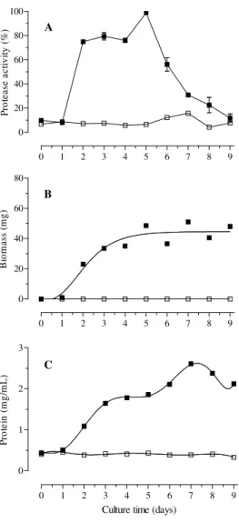
The highest extracellular protease activity by B bassiana CG432 (Fig. 1A) was observed on the 5th day (Abs 650 nm 0.489) but around 80% of this activity was obtained after 2nd day of the cultivation. After the 5th day, there was an abrupt
reduction of the activity until the 9th day. Variable time courses of extracellular protease production have been related in literature, reflecting the variability in protease production on different media. The stimulatory effects of a series of 24 nitrogen sources including inorganic, organic nonprotein, proteins and complex natural media on the production of proteases in submerged cultures demonstrated that the maximum amount of protease into the maize meal, yeast extract, ground maize and wheat bran broth was released 3 days after inoculation (Kucera, 1971).
0 1 2 3 4 5 6 7 8 9 0
1 2 3
Culture time (days)
P
ro
te
in
(
m
g
/m
L
)
0 1 2 3 4 5 6 7 8 9 0
20 40 60 80 100
P
ro
te
a
se
a
c
ti
v
it
y
(
%
)
0 1 2 3 4 5 6 7 8 9 0
20 40 60 80
B
io
m
a
ss
(
m
g
)
B A
C
Others studies that used inducer substrates to produce proteases from B. bassiana, stopped the cultures in between 3rd to 6th day of the cultivation as 0.2% lyophilized porcine blood plasma and 0.5% ground larval of Apis mellifera with higher release at 3rd day by B. Bassiana 278 (Chrzanowska et al., 2001), 1% gelatin at 3rd and 4th day by B bassiana GK2016 (Bidochka and Khachatourians, 1987 and 1988), 1% gelatin and casein, at 4th day by B. bassiana 11892A (Urtz and Rice, 2000), ground migratory grasshopper cuticle, at 5th day by B bassiana GK2016 and GK2018 (Bidochka and Khachatourians, 1990, 1991, 1993), colloidal chitin, at 6th day (Havukkala et al. 1993), and liquid medium with cuticle ground from the own coffee berry borer, Hypothenemus hampei at 5th to 7th days after the inoculation (Giraldo-Cardozo et al., 2001).
The biomass production (Fig. 1B) and the concentration of soluble protein (Fig. 1C) of the culture medium increased after 2nd day and coincided with 80% of the total of the extracellular protease production, after a small lag period of 24 h shorter than obtained by two isolates of B. Bassiana (Arcas et al., 1999). These results could be related with the production of complex proteolityc enzymes during the reactivation of the inoculum on coffee berry borer alive, using liquid medium without inducer substrates such as the cuticle (Havukkala et al. 1993; Urtz and Rice,
2000) or extract from insects (Bidochka and Khachatourians, 1990, 1991, 1993; Giraldo-Cardozo et al., 2001).
The active growth continued until the 5th day when the enzyme was released in the culture medium reaching the maximum extracellular protease activity. After that, the biomass production was stable when the protease release was reduced (Fig. 1A and 1B). These results confirmed the importance of the extracellular protease activity on the substrate utilization during the multiplication celular phase of this fungus as related to other microorganisms (Fu-Chu et al., 2004). The concentration of soluble protein of the culture medium was highest on the 7th day probably related with biomass (Fig. 1C).
The previous reactivation of the fungus on insects proposed in this work presented the advantage of less laborious laboratory work to produce the inoculum than to prepare different substrates and less cultivation time, considering that 80% of the total activity was obtained on 2nd day.
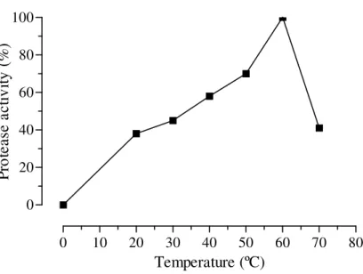
Temperature effect
The extracellular proteases were more active between 40 and 60oC (Fig. 2). This result was better than those by Bidochka and Khachatourians (1987), with optimum temperature between 37 to 42oC.
0 10 20 30 40 50 60 70 80 0
20 40 60 80 100
Temperature (ºC)
P
ro
te
a
se
a
c
ti
v
it
y
(
%
)
Figure 2 – Effect of the temperature on the extracellular protease activity produced by Beauveria
Production of Extracellular Protease by a Brazilian Strain of Beauveria bassiana
Brazilian Archives of Biology and Technology
221
0.0 0.2 0.4 0.6 0.8 1.0 0
25 50 75 100
Cl
-concentration (M)
P
ro
te
as
e
ac
ti
v
it
y
(
%
)
Figure 3 – Effect of chloride ion concentation at pH 4.5 (□), 7.0 (■) and 9.5 (▲) pH on the extracellular protease activity produced by Beauveria bassiana CG432.
0 5 10 15 20 25 30 0
20 40 60 80 100
Time (days)
P
ro
te
a
se
a
c
ti
v
it
y
(
%
)
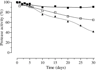
Figure 4 – Stability of extracellular protease produced by Beauveria bassiana CG432 stored at 4ºC refrigeration (□), -18ºC freezing (■) and submitted to repetitive freezing/thawing(▲).
Cl- ion concentration effect
The CE proteases remained active when analyzed in increasing concentrations of chloride ions of 0.1 to 1.0 M at pH 4.5, 7.0 and 9.5 (Fig. 3). This could be important information, because chloride ions
Protease stability in storage
It was observed that the proteases maintained above 90% of their activity up to the 30ty day under freezing and above 80% until the 15th day under refrigeration. The activity fell down to 50% at the 5th times of freezing/thawing (Fig. 4). Thus, repetitive freezing of the extracts should be avoided.
At least, the extracellular proteases produced in these conditions presented optimum temperature activity at 60°C, without interferences from Cl- ion and was stable for many days on refrigeration or freezing conditions. This fact could be very important in purification procedures. Also, proteases produced under same conditions were stable during 10 days at 25ºC (Stürmer et al., 2003/2004) being too important on insect procedures for bioassay when used as bioinseticide.
ACKNOWLEDGMENTS
Ito, E. T. whishes to thank Fundação Araucária/Pr and Fundação Coordenação de Aperfeiçoamento de Pessoal de Nível Superior (CAPES) for the fellowship for the Master Course in Biotechnology. The authors acknowledge Pro-Reitoria de Pesquisa e
Pós-Graduação (PROPPG-UEL-number
24698/2003), Fundação Araucária (number-213/2003) and Conselho Nacional de Desenvolvimento Científico e Tecnológico (CNPq-number 473595/2003-6) for the financial supports and to Profª Drª Maria Helena Pimenta Pinotti for the English revision.
RESUMO
A cepa brasileira do fungo entomopatogênico
Beauveria bassiana CG432 foi cultivada em meio de cultivo líquido contendo glucose e extrato de levedura a 28°C, 150 rpm, durante 9 dias utilizando 1% de inóculo constituído por 108conídios/mL pré-ativados em insetos vivos de broca-do-café. Avaliou-se diariamente o crescimento do fungo através da quantificação da biomassa e a atividade de proteases extracelulares no filtrado do cultivo pela determinação de
peptídeos solúveis liberados pela hidrólise de soro albumina bovina a 37°C durante 30minutos. O fungo cresceu ativamente após reduzido período lag de 24 horas, produziu 80% da atividade total de proteases em 48 horas e atingiu produção máxima em 5 dias de cultivo, sem adição de substratos indutores no meio de cultivo. As proteases apresentaram atividade ótima a 60ºC, estabilidade até 1M de íons cloreto, mantiveram mais de 80% de atividade durante 15 dias a 4ºC e –18ºC, porém foram sensíveis a congelamentos repetitivos.
REFERENCES
Alves, S. B. (1998), Patologia e Controle Microbiano: vantagens e desvantagens/Técnicas de Laboratório.
In: Controle Microbiano de Insetos, 2 ed. FEALQ,
Piracicaba, pp. 665.
Arcas, J. A.; Días, B. M. and Lecuona, R. E. (1999), Bioinsecticidal activity of conidia and dry mycelium preparation of two isolates of Beauveria bassiana against the sugarcane borer Diatraea saccharalis.
Journal of Biotechnology, 67, 151-158.
Bidochka, M. J. and Khachatourians, G. G. (1992), Growth of the entomopathogenic fungus Beauveria
bassiana on cuticular components from the migratory
grasshopper Melanoplus sanguinipes. Journal of
Invertebrate Pathology, 59, 165-173.
Bidochka, M. J. and Khachatourians, G. G. (1990), Identification of Beauveria bassiana extracellular protease as a virulence factor in pathogenicity toward the migratory grasshopper, Melanoplus sanguinipes.
Journal of Invertebrate Pathology, 56, 362-370.
Bidochka, M. J. and Khachatourians, G. G. (1993), Oxalic acid hyperproduction in Beauveria bassiana mutants is related to a utilizable carbon source but not to virulence. Journal Invertebrate Pathology, 62, 53-57.
Bidochka, M. J. and Khachatourians, G. G. (1987), Purification and properties of an extracellular protease produced by the entomopathogenic fungus
Beauveria bassiana. Applied and Environmental
Microbiology, 53: (7), 1679-1684.
Bidochka, M. J. and Khachatourians, G. G. (1988), Regulation of extracellular protease in the entomopathogenic fungus Beauveria bassiana.
Experimental Mycology, 12, 161-168.
Bidochka, M. J. and Khachatourians, G. G. (1991), The implication of metabolic acids produced by
Beauveria bassiana in pathogenesis of the migratory
grasshopper, Melanoplus sanguinipes. Journal of
Invertebrate Pathology, 58, 106-117.
Boucias, D. G. and Pendland, J. C. (1998), Principles of
Production of Extracellular Protease by a Brazilian Strain of Beauveria bassiana
Brazilian Archives of Biology and Technology
223
Chen, Fu-Chu; Shen, Li-Fen and Chak, Kin-Fu. (2004), A facile analytical method for the identification of protease gene profiles from Bacillus thuringiensis strains. Journal of Microbiological Methods, 56, 125-132.
Cottrell, T. E. and Shapiro-Ilan, D. I. (2003), Susceptibility of a native and an exotic lady beetle (Coleoptera: Coccinellidae) to Beauveria bassiana.
Journal of Invertebrate Pathology, 84, 137-144.
Chrzanowska, J.; Banas, J.; and Kolaczkowska, M. (2001), Purification and characterization of
Beauveria bassiana proteinases. Acta Biotechnology,
21: (1), 73-81.
FURLONG, M. J. and PELL, J. K. (2001), Horizontal transmission of entomopathogenic fungi by the diamondback moth. Biological Control, 22, 288-299. Giraldo-Cardozo, E. M.; López-F, Y.; Delgado-B., F.
and Vélez-A, P. E. (2001), Actividad lipolítica y proteolítica de Beauveria bassiana y Metarhizium
anisopliae y su relación con la patogenicidad sobre
Hypothenemus hampei (Coleoptera: Scolytidae).
Revista Colombiana de Entomología, 27: (1-2),
61-65.
Hartree, E. F. (1972), Determination of Protein: A modification of the Lowry method that gives a linear photometric response. Analytical Biochemistry, 48, 422-427.
Havukkala, I.; Mitamura, C.; Hara, S.; Hirayae, K.; Nishizawa, Y. and Hibi, T. (1993), Induction and purification of Beauveria bassiana chitinolytic enzymes, Journal of Invertebrate Pathology, 61, 97-102.
Hepburn, H. R. (1985), Structure of the Integument. In: Comprehensive Insect Physiology, Biochemistry and
Pharmacology, G. A. Kerkut and L. I.Gilbert, Eds., 3,
Pergamon, Oxford, pp. 1-58.
Ito, E.T. (2003), Estabilidade e perfil de produção de proteases pelo fungo entomopatogênico Beauveria
bassiana (BALS.) VUILL. Monografia
(Especialização em Bioquímica Aplicada), Universidade Estadual de Londrina, Londrina, Paraná, Brasil.
Kucera, M. (1971), Toxins of the entomophagous fungus Beauveria bassiana. II. Effect of nitrogen sources on formation of the toxic protease in submerged culture. Journal of Invertebrate
Pathology, 17, 211-215.
Leopold, J. and Samsinakova, A. (1970), Quantitative estimation of chitinase and several other enzymes in the fungus Beauveria bassiana. Journal of
Invertebrate Pathology, 15, 34-42.
MacLeod, D. M. (1954), Investigations on the genera
Beauveria Vuill. and Tritirachium Limber. Canadian
Journal of Botany,32, 818-890.
Moino Jr., A.; Alves, S. B. And Pereira, R. M. (1998), Efficacy of Beauveria bassiana (Balsamo) Vuillemin isolates for control of stored-grain pests. Journal of
Applied Entomology, 122, 301-305.
Neves, P. M. O. J and Hirose, E. (2005), Seleção de isolados de Beauveria bassiana para o controle biológico da broca-do-café, Hypothenemus hampei (Ferrari) (Coleoptera: Scolytidae). Neotropical
Entomology, 34: (1), 77-82.
Samsinakova, A.; Misikova, S. and Leopold, J. (1971), Action of enzymatic systems of Beauveria bassiana on the cuticle of the greater wax moth larvae
(Galleria mellonella). Journal of Invertebrate
Pathology, 18, 322-330.
Shimizu, S.; Tsuchitani, Y. and Matsumoto, T. (1993), Production of an extracellular protease by Beauveria
bassiana in the haemolymph of the silkworm,
Bombyx mori, Letters in Applied Microbiology, 16,
291-294.
St-Leger, R. J.; Charnley, A. K. And Cooper, R. M. (1986), Cuticle-degrading enzymes of entomopathogenic fungi: Synthesis in culture on cuticle. Journal of Invertebrate Pathology, 48, 85-95. Stürmer, A. T.; Ito, E. T. and Varéa-Pereira, G.
(2003/2004), Estabilidade de proteases produzidas pelo fungo entomopatogênico Beauveria bassiana. Revista UNOPAR Científica - Ciências Biológicas e
da Saúde, 5/6, 13-19.
Urtz, B. E. and Rice, W. C. (2000), Purification and characterization of a novel extracellular protease from Beauveria bassiana. Mycological Research., 104: (2), 180-186.