Gastroprotective effect of Byrsonima sericea DC leaf extract against ethanol-induced
gastric injury and its possible mechanisms of action
PATRÍCIA A. RODRIGUES1, SELENE M. MORAIS1,2, CAROLINA M. SOUZA3, DAVI V. MAGALHÃES1,
ÍCARO G.P. VIEIRA1,4, GEANNE M. ANDRADE3, VIETLA S. RAO3 and FLÁVIA A. SANTOS3 1Programa de Pós-Graduação em Ciências Veterinárias, Faculdade de Veterinária, Universidade Estadual do Ceará,
Av. Paranjana, 1700, Campus do Itaperi, 60740-000 Fortaleza CE, Brasil
2Departamento de Química, Laboratório de Química de Produtos Naturais, Universidade Estadual do Ceará,
Av. Paranjana, 1700, Campus do Itaperi, 60740-000 Fortaleza, CE, Brasil
3Departamento de Fisiologia e Farmacologia, Universidade Federal do Ceará, Faculdade de Medicina,
Rua Coronel Nunes de Melo, 127, Rodolfo Teófilo, 60430-270 Fortaleza, CE, Brasil
4 PADETEC-Parque de Desenvolvimento Tecnológico, Universidade Federal do Ceará,
Av. Humberto Monte, 2977, Bairro Parquelândia, Bloco 310, 60.440-593 Fortaleza, CE, Brasil.
Manuscript received on October 7, 2010; accepted for publication on March 1, 2011
ABSTRACT
Byrsonima sericea leaves are extensively used in folk medicine in Brazil against gastric disorders. This study investigated the chemical constituents of B. sericea leaf ethanolic extract (BSLE) and its potential gastroprotective activity, with its possible mechanism of the action using ethanol to induce gastric mucosal damage in mice. The phytochemical analysis was carried out to identify the active constituents
present in the extract, and the HPLC analysis was performed for the identification of flavonoids. BSLE
at oral doses of 125, 250 and 500 mg/kg markedly attenuated the ethanol-evoked gastric lesions by 53.2, 84.9 and 87.6 %, respectively. The BSLE (250 mg/kg) prevented the depletion of gastric mucus and
gastric mucosal nonproteic-sulfhydryl groups, SOD and CAT, as well as the increase in the MDA content
promoted by absolute ethanol. Moreover, the effect of BSLE against ethanol damage was found to be
significantly reduced in mice pretreated with Capsazepine (i.p.), L-NAME (i.p.) or glibenclamide (i.p.), the respective blockers/inhibitors of TRPV1, NO synthase and K+ATP channel. The phytochemical investigation on BSLE revealed the presence of flavonoids rutin, isoquercitrin, kaempferol 3-O-rutinoside
and quercetin, which are compounds well known for their antioxidant and gastroprotective properties. These results suggest that BSLE affords gastroprotection through multiple mechanisms, which may be helpful in the treatment of pathologies associated with gastric dysfunctions.
Key words: antioxidant, Byrsonima sericea, flavonoids, gastroprotection, mechanisms of action.
Correspondence to: Selene Maia de Morais E-mail: [email protected]
INTRODUCTION
Plants of the genus Byrsonima (Malpighiaceae) are widely distributed in various parts of Brazil where local people call them murici. Leaves and trunk barks from various species the Byrsonima are
popularly employed in folk medicine to treat fever, gastrointestinal dysfunction (diarrhea and gastric ulcer), asthma, skin infections, and snakebites (Lira et al. 2008, Mendes et al. 1999). While chemical investigations on various Byrsonima species have shown the presence of several bioactive compounds
like flavonoids, triterpenes and tannins (Mendes et
al. 1999, Martínez-Vázquez et al. 1999), bioactivity studies have demonstrated the gastroprotective, healing and antidiarrheal activities of B. fagifolia
(Lima et al. 2008), the antimutagenic activity of
B. basiloba (Lira et al. 2008), and the mutagenic and gastroprotective effects of Byrsonima crassa (Cardoso et al. 2006, Sannomiya et al. 2007).
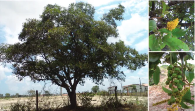
The presence of phenolic compounds possibly explains the gastroprotective effect of the extracts of Byrsonima species. Byrsonima sericea DC (Fig. 1) grows abundantly in the Northeastern states of Brazil, and it is traditionally used to treat gastrointestinal dysfunction. An antioxidant activity of the methanol extract from B. sericea
has recently been described, using the DPPH assay (Boscolo et al. 2007).
MATERIALS AND METHODS
CHEMICALS
Capsaicin, capsazepine, glibenclamide, diazoxide, L-arginine and L-NAME were purchased from
Sigma Chemical Co. (St Louis, MO, USA). Absolute
ethanol was obtained from Synth (Brazil). All other chemicals used were of analytical grade. Quercetin, quercetin 3-O-glycoside (isoquercetin), quercetin 3-O-rutinoside (rutin) and kaempferol 3-O-rutinoside
isolated from the flowers and leaves of Dimophandra
gardneriana, were used as standards.
PLANT MATERIAL AND EXTRACT PREPARATION
B. sericea leaves were collected from the State University of Ceará campus, Ceará State, Brazil, in
April 2008, after their identification by Dr. Afrânio
G. Fernandes, botanist of the Federal University of Ceará. A voucher specimen (#39.451) has been deposited at Prisco Bezerra Herbarium. Fresh leaves (1.24 kg) were macerated with 70% ethanol at room temperature for 7 days. The resultant
ethanolic solution was filtered with filter paper using
a buchner funnel under vacuum and evaporated to dryness at 50°C in a rotary evaporator to yield 153 g of crude ethanolic extract. This extract was then submitted to a chromatographic treatment process using silica gel column being eluted with solvents hexane, ethyl acetate and ethanol. With the evaporation of these solvents, the three correspondent extracts weighed 1.1, 5.6 and 126.2 g
respectively. Only the ethanolic extract (BSLE)
was utilized in the present work.
PHYTOCHEMICALANALYSIS
BSLE was analyzed for the presence of phenols,
tannins, leucoantocianidins, flavonoids, steroids,
triterpenes and alkaloids by the qualitative chemical analysis (Matos 1997).
Fig. 1 - View of fruits and flowers and an adult individual of Byrsonima sericea (by Pablito Augusto Travassos Ferreira) .
To our knowledge, there were no scientific studies
HPLCANALYSIS
The HPLC system (Waters 2690 Alliance) was connected to a Waters 486 tunable absorbance, column detector (Waters C18, 3.9 X 150mm X 4um). The analysis was run under isocratic conditions using the solvent mixture: 80:20 (ACN: H2O, pH 2.8 phosphoric acid), flow rate: 1 mL/min, 20 µL; and
temperature: 21oC. The concentrations of extract and standards were 1 mg/mL, the injection volume was 30 µL and the eluted was monitored at 350 nm.
ANIMALS
Female Swiss mice (20-25g) obtained from the Central Animal House of this university were used. Experimental groups consisted of 8 animals per group. They were housed at 24 ± 2°C under a 12-h light/12-h dark cycle and had free access to standard pellet diet (Purina chow) and tap water. The animals were deprived of food for 15 h before the experimentation, but had free access to drinking water. The Institutional Ethics Committee on the Care and Use of Animals for experimentation approved the experimental protocols (Nº 07520831-8), and all experiments were performed in accordance with the guidelines of the National Institute of Health, Bethesda, USA.
GASTRIC DAMAGE INDUCED BY ETHANOL
Groups of mice (n=8), were pre-treated with the
vehicle (0.5% of DMSO in saline, 10mL/kg),
N-acetylcysteine (NAC, 150 mg/kg, i.p.) or BSLE (125, 250 and 500 mg/kg, v.o.) one hour before the induction of gastric damage by oral administration of absolute ethanol (96%, 0.2 mL/animal) (Robert et al. 1979). After 30 min the animals were
sacrificed and their stomachs excised, opened along
the greater curvature and rinsed with saline (0.9%). The mucosal lesion area (mm2) was measured by planimetry using a transparent grid (area: 1mm2)
placed on the glandular mucosal surface and expressed in percentage (%) in relation to the total area of the corpus. After the evaluation of the lesions induced by ethanol, the glandular parts of
the stomachs were frozen and stored at −70 oC for
further biochemical analyses.
DETERMINATION OF GASTRIC MUCUS
Gastric mucus content was measured according to the methodology described by Corne et al. (1974). The gastric tissues were transferred to 0.1% Alcian Blue solution prepared in 16 mM sucrose and 50 mM sodium acetate (pH 5.8) and kept for 2 h at room temperature. The segments were then rinsed twice with 25 mM sucrose solution for 15 and 45 min, and the dye solution, together with the gastric mucus with 5 mM of magnesium chloride solution, was extracted for 2 h. The extract was then mixed with an equal volume of diethyl ether and centrifuged (1000g) for 10 min. The absorbance was determined at 598 nm. The amount of mucus was calculated using a standard curve of Alcian Blue.
ESTIMATION OF ANTI-OXIDANT PARAMETERS
Gastric strips were cut into small pieces and then homogenized in ice-cold 50 mM Phosphate buffer pH 7.4 to obtain a 10% homogenate. The homogenate was then made into aliquots and used for the assessment of anti-oxidant parameters.
DETERMINATION OF NONPROTEIC
SULFHYDRYLGROUPS(NP-SH)
(2 mL) were then mixed with 4mL of tris buffer
(40 mM, pH 8.9), and 5,5'-ditiobis (2-nitrobenzoic
acid) (DTNB, 10 mM) was added. The absorbance was measured within 5 min after the addition of DTNB at 412 nm against a reagent blank with no homogenate. The absorbance values were extrapolated from a glutathione standard curve and expressed as µg/g of stomach tissue.
SUPEROXIDE DISMUTASE (SOD)ACTIVITY
The SOD activity was assessed through the
measurement of the enzyme capacity for the photochemical inhibition of nitroblue-tetrazolium (NBT) (Beauchamp and Fridovich 1971). The
reduction of NBT by O2− was utilized as the basis
of assays for superoxide dismutase, which shows its presence by inhibiting the reduction of NBT and producing formazan that is absorbed at 560 nm. Aliquots of tissue homogenates were centrifuged
12,000 g for 20 min. In a dark room 40 μl of phosphate
buffer or supernatants were added to glass test tubes containing 1mL of reaction mixture (Phosphate buffer 50 mM, EDTA 100 nM and L-methionine
19.5 μM pH 7.8). Then 150 μL of NBT 750 μM and 300 μL riboflavin 10 μM were added. After
shacking, the tubes were exposed to light (20 Watt) for 15min. The absorbance was measured at 560 nm. The results were expressed in enzyme units, which
represent the amount of SOD necessary to inhibit the
NBT reduction by 50%. The enzymatic activity was expressed as U/µg of protein.
DETERMINATION OF CATALASE (CAT)ACTIVITY
The CAT activity was measured by the method that
employs hydrogen peroxide to generate H2O and O2 (Maehly and Chance 1954). The activity was
measured by the degree of this reaction. In short,
the supernatant (20 μL) was added to a quartz
cuvette containing 980 μL of H2O2 800 μM, EDTA
25 μM and tris buffer HCl (50 mM, pH 8.0). The
change in absorbance was monitored at 240 nm over a 6-min period using a spectrophotometer. The CAT activity was expressed as U/µg of protein.
DETERMINATION PROTEIN CONTENT
The protein concentration was determined by the method of Bradford 1976, using bovine serum albumin as a standard.
DETERMINATION OF TOTAL THIOBARBITURIC ACID-REACTIVE
SUBSTANCES (TBARS)
Total thiobarbituric acid-reactive substances (TBARS) was determined according to the method
of Ohkawa et al. (1979). Gastric strips were cut
into small pieces and then homogenized in ice-cold phosphate buffer (50 mM pH 7.4) to give a 10% homogenate. The homogenates were transferred to test tubes and incubated in a water bath at 37ºC for 60 min. After this period, 35% of perchloric acid was added. The mixture was centrifuged at 15000g for 10 min. Thiobarbituric acid at 0.6% was then added to the upper layer. The mixture was submitted to a water bath at 100ºC for 30 min, after which the absorbance was measured at 532 nm. The standard curve was obtained using several concentrations of MDA solutions, expressed in nmol/g of wet tissue.
ROLE OF TRPV1CHANNEL INTHE
GASTROPROTECTIVE EFFECT OF BSLE
Groups of mice (n=8) were pretreated with vehicle
(0.9% saline in a 0.5% of DMSO, 10 mL/kg), BSLE
ROLE OF NITRIC OXIDE IN THE GASTROPROTECTIVE
EFFECT OFBSLE
Mice (n=8/per group) were pretreated with vehicle
(0.9% saline in a 0.5% of DMSO, 10mL/kg),
BSLE (250 mg/kg, v.o.) and L-arginine (600 mg/ kg, i.p.), alone or in their combinations with L-NAME (20 mg/kg, i.p.) prior to the induction of gastric damage with ethanol (0.2 ml of ethanol, 96%). While BSLE was administered 1h before, L-NAME and L-arginine were given 30 min prior to ethanol (Arrieta et al. 2003).
ROLE OF KATP-CHANNELS IN THE GASTROPROTECTIVE
EFFECT OF BSLE
Mice (n=8/per group) were pretreated with vehicle
(0.9% saline in a 0.5% of DMSO, 10mL/kg), BSLE
(250 mg/kg, v.o.) diazoxide (3 mg/kg, i.p.), alone or in their combinations with glibenclamide (5 mg/ kg, i.p.) prior to the oral administration of 0.2 ml of ethanol (96%). BSLE was given 1h before, whereas diazoxide was administered 30 min prior to ethanol or glibenclamide. Glibenclamide was administered 30 min before BSLE (Peskar et al. 2002).
STATISTICAL ANALYSIS
The results are presented as the mean ± S.E.M. of 8 animals per group. Statistical analysis was carried out using the one way analysis of variance
(ANOVA) followed by Student Newman Keul´s
post hoc test for multiple comparisons. P-values less than 0.05 (P<0.05) were considered as indicative of
statistical significance.
RESULTS
PHYTOCHEMICAL ANALYSIS
The phytochemical analysis of BSLE revealed
the presence of flavones, flavonols, flavanones,
xanthones and hydrolizable tannins. The amounts of
phenolic compounds present in the extract were: total
phenols 0.37 mg/g and flavonoids 0.17 mg/g. For the
HPLC analysis of ethanol extract of B. sericea (Fig.
2), rutin, isoquercitrin, kaempferol 3-O-rutinoside
and quercetin were used as standards. The structural
characteristics of each identified peak in Fig. 2
was determined by comparing its correspondent UV spectrum with those UV spectra available at the HPLC computer library. For dried leaves, the
percentage concentration for each flavonoid was
evaluated in relation to a quercetin standard curve as: isoquercitrin 0.13%, quercetin 0.003%, rutin 0.007% and kaempferol 3-O-ritinoside 0.07%.
EFFECT OF BSLE ON GASTRIC DAMAGE
INDUCED BY ETHANOL
The oral administration of BSLE (125, 250 and 500 mg/kg) exhibited a protective effect against ethanol-induced gastric lesions in comparison to the vehicle group. The inhibition percentages for the respective doses employed were 53.2, 84.9 and 87.6 %. NAC, which was the positive control
included for the study, also offered a significant
protection (Table I).
Fig. 2- HPLC Chromatogram of the ethanolic extract of B. sericea. Column: Waters C18 (150 x 3.9 mm, 4 μm; eluent: water /ACN / fosforic acid / 80:20), Flow-rate: 1ml/min, 20 μl, Temperature 21oC. Detection: 350 nm. 1. unidentified, 2. Rutin, 3. Isoquercitrin,
EFFECT OF BSLE ON ANTI-OXIDANTPARAMETERS
The animals that received only ethanol showed
a significant decrease in gastric catalase (CAT), superoxide dismutase (SOD) and NP-SH levels, and a significant TBARS level increase (Table
II). Animals pretreated with BSLE (250 mg/kg) effectively decreased TBARS levels almost to a similar extent to NAC (150 mg/kg). Animals
treated with BSLE showed a significant enhanced activity of the antioxidant enzyme SOD. The
NP-SH level in gastric mucosa of normal control mice
(280.1 ± 12.7 µg/g) was significantly lowered by
the ethanol treatment (102.3 ± 10.4 µg/g) (Table II). Both BSLE and NAC inhibited the NPSH depletion caused by ethanol (229.1 ± 9.9 and 239.5 ± 6.5 µg/g, respectively regarding the doses employed).
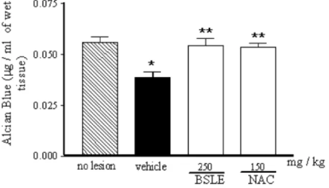
EFFECT OF BSLE ON GASTRIC MUCUS
The amount of gastric mucus was significantly
(p < 0.01) less in the gastric tissues collected from ethanol-treated animals, as compared to non-damaged tissues from the control. The pretreatment of animals with BSLE greatly enhanced the gastric mucus when compared to animals with ethanol injury (Fig. 3).
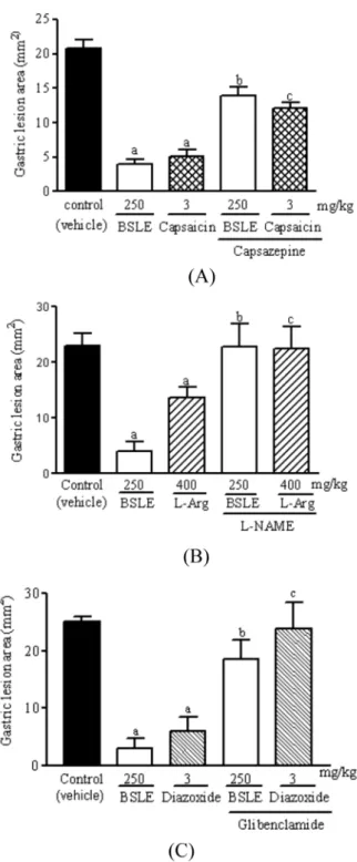
EFFECT OF TRPV1 AND K+ATP-CHANNELS AND THE ROLE OF
NITRIC OXIDE ON THE GASTROPROTECTIVE EFFECT OF BSLE
In mice pretreated with vanilloid antagonist capsazepine, the gastroprotective effect of BSLE (250 mg/kg) and capsaicin (0.3 mg/kg, v.o.) was
significantly reduced (Fig. 4A). These data indicate
that the gastroprotective effect of BSLE is mediated by the activation of capsaicin-sensitive primary afferents. L-NAME (20 mg/kg) pretreatment
significantly blocked the gastroprotection produced
by BSLE and L-arginine (400 mg/kg, i.p.),
suggesting the participation of NO (Fig. 4B) on
the BSLE gastroprotection. The pretreatment with K+ATP channel blocker, glibenclamide (5 mg/kg,
i.p.), also significantly reduced the gastroprotection
produced by BSLE and diazoxide (3 mg/kg, i.p.) (Fig. 4C), which indicates a role for K+ATP channels
in the gastroprotection.
Effect of B. sericea extract (BSLE) on gastric damage induced by absolute ethanol in mice.
Effects of B. sericea leaf extract on the contents of CAT, SOD and
MDA on gastric damage induced by absolute ethanol in mice. Treatment
Treatment
Data are mean ± S.E.M of 8 animals in each group.*** p < 0.001 vs. Vehicle (ANOVA followed by Student Newman Keul’s test).
Data are mean ± S.E.M of 8 animals in each group. a p < 0.01 vs. No lesion; b p < 0.01 vs. Vehicle (ANOVA followed by Student Newman Keul’s test).
Fig. 3 - Effect of the B. sericea extract (BSLE) treatment on the amount of mucus after induction of gastric lesions by ethanol in mice. The results are expressed as mean ± S.E.M. * p < 0.01 vs. no lesion; ** p < 0.01 vs. vehicle (ANOVA followed by Student Newman Keul’s test).
Vehicle
Vehicle No lesion
BSLE
BSLE
(250 mg/kg)
NAC
NAC
(150 mg/kg)
-125 mg/kg
250 mg/kg 500 mg/kg
150 mg/kg
21.8 ± 0.4
10.4 ± 1.0***
4.96 ± 1.01 1.04 ± 0.28 54.68 ± 6.9 280.1 ± 12.7 3.3 ± 0.6***
2.66 ± 0.24a 0.69 ± 0.26a 86.63 ± 17.3a 102.3 ± 10.4a
2.7 ± 0.5***
3.50 ± 0.27 1.08 ± 0.15b 67.76 ± 7.5b 229.1 ± 9.9b
3.0 ± 0.5***
2.78 ± 0.41 1.08 ± 0.12b 62.1 ± 6.96b 239.5 ± 6.5b
Dose
CAT
(U/µg protein)
SOD
(U/µg protein)
TBARS
(U/µg protein)
NP-SH
(U/µg protein)
Ethanol lesion area (mm2)
Table I
DISCUSSION
The phytochemical analysis of BSLE indicated
the presence of flavonoids and tannins, and the HPLC analysis confirmed the presence of rutin
(quercetin 3-O-rutinoside), isoquercitrin (quercetin 3-O-glucoside), kaempferol 3-O-rutinoside and quercetin as the main secondary metabolites.
Several flavonoids and their structurally related
compounds have been shown to inhibit lipid peroxidation (Katsube et al. 2006).
The results of this study show that BSLE at doses of 125, 250 and 500 mg/kg affords a pronounced gastroprotection against ethanol that induced lesions, which is probably due to the presence of
strong antioxidant flavonoids in the extract. The
gastroprotective and antioxidant effects of quercetin and rutin are well known (La Casa et al. 2000, De la Lastra et al. 1994), and there are many studies
showing the antiulcerogenic properties of flavonoids
(Gonzalez and Di Stasi 2002). The role of reactive oxygen species in the pathogenesis of acute ethanol that induced gastric mucosal lesions and the effects of quercetin have been evaluated in a few studies.
It has been confirmed that the quercetin treatment significantly inhibits the gastric erosions induced by
ethanol (Gracioso et al. 2002). Galati et al. (2003) have also stated that there is a correlation between the
antioxidant and the antiulcer activities of flavonoids.
The oxidative stress and impaired prostaglandin synthesis contribute to gastric mucosal damage in experimental models of gastric lesions induced by ethanol (Chattopadhyay et al. 2006). The administration of BSLE (250 mg/kg) to animals increased the levels of gastric NP-SH (GSH) and the amount of mucus. Baggio et al. (2007) demonstrated that the oral administration of the Maytenus ilicifoli
leaf extract that is rich in flavonoids can increase the
mucus production in the ethanol model of gastric damage in mice, which is in agreement with our data. Both gastric mucus and glutathione serve as protective agents against mucosal injury (Chen et al. 2005, Cnubben et al. 2001).
Fig. 4 - (A) The involvement of TRPV1 channel in the gastroprotective effect of BSLE against ethanol that induced gastric damage in mice - (B) The involvement of nitric oxide in the gastroprotective effect of BSLE against ethanol that induced gastric damage in mice - (C) The role of K+ATP channels in the gastroprotective effect of BSLE against ethanol-induced gastric damage in mice. The results are expressed as mean ± S.E.M. ap < 0.001 vs vehicle; bp < 0.001 vs BSLE alone; cp < 0.001 vs L-arginine (L-arg) or diazoxide alone or capsaicin alone
The plant-derived natural antioxidants are extremely useful to combat the oxidative stress, and antioxidant enzymes such as superoxide dismutase
(SOD) and catalase (CAT), in a preventive way, act as the first line of defense against the reactive
oxygen species (Repetto and LLesuy 2002). Acute treatment with ethanol promotes oxidative stress, enhancing consequently the lipid peroxidation and malonaldehyde formation (Peskar et al. 2002). The present study clearly shows that BSLE inhibits lipid peroxidation and lowers TBARS formation, which indicates a cytoprotective function.
Several studies have reported that NO is
involved in the preservation of mucous membrane integrity in experimental models of gastric
ulceration (Ancha et al. 2003, Cho 2001, Kwiecień
et al. 2002). In this study, L-arginine (400 mg/kg) and BSLE (250 mg/kg) induced gastroprotection, but it was reversed by L-NAME, a non-selective
NOS inhibitor, suggesting that the gastroprotective effect of BSLE is mediated, in part, by NO.
It is known that potassium channels are involved in a variety of stomach physiological functions and that gastroprotection can be inhibited by several agents such as indomethacin, K+ATP blockers and glibenclamide (Peskar et al. 2002). Glibenclamide, a K+
ATP channels blocker, significantly antagonized
the protective effect of BSLE and diazoxide. These results support the hypothesis that the K+
ATP
channel opening is involved in the gastroprotective activity of BSLE. Since this protection is also sensitive to indomethacin, it is suggested that endogen prostaglandins activate the K+
ATP channels
and that this mechanism is responsible, in part, for the BSLE gastroprotective action (Campos et al. 2008). The results of this study indicate that BSLE has a cytoprotective role in the gastroprotection against gastric damage induced by ethanol, which is presumably mediated, in part, by the nitric oxide release and K+ATP channel opening.
The extract does not seem to be toxic as consecutive daily oral administrations of BSLE (500
or 1000 mg/kg) over a period of 7 days to mice did not produce any signs of overt toxicity. In conclusion,
these findings suggest that the ethanolic extract of B.
sericea is a safe gastroprotectant to obliterate gastric dysfunction associated with pathologies.
ACKNOWLEDGMENTS
The authors thank Fundação Cearence de Apoio
ao Desenvolvimento Científico e Tecnológico
(FUNCAP) and Conselho Nacional de
Desenvolvimento Científico e Tecnológico (CNPq, Brazil) for financial support.
RESUMO
Folhas de Byrsonima sericea são amplamente utilizadas na medicina popular no Brasil no tratamento de distúrbios gástricos. Este estudo investigou os constituintes químicos do extrato etanólico das folhas de B. sericea (BSLE) e sua atividade gastroprotetora com seus possíveis mecanismos de ação utilizando o modelo de lesão gástrica induzida por etanol em
camundongos. A análise fitoquímica foi realizada para identificar os componentes ativos presentes no extrato e análise por HPLC foi realizada para a identificação de flavonóides. A administração de BSLE (v.o.) nas doses de 125, 250, 500 mg/kg, v.o. atenuou significativamente
as lesões gástricas induzidas por etanol em 53,2, 84,9 e 87,6% respectivamente. BSLE (250 mg/kg) preveniu a
depleção do muco gástrico, de grupamentos sulfidrílicos não-protéicos (GSH), das atividades da SOD e da CAT
assim como o aumento de malonaldeído promovido pelo etanol. Além disso, o efeito gastroprotetor do BSLE foi
significantemente reduzido pelos pré-tratamentos com
capsazepina (i.p.), L-NAME (i.p.) ou glibenclamida (i.p.), respectivamente bloqueadores/inibidores de receptores
TRPV1, NO sintase e canais de K+ATP. A investigação fitoquímica revelou a presença de flavonoides como rutina, isoquercitrina, 3-O-rutinosideo-canferol e
quercetina que são conhecidas por suas propriedades
que BSLE proporciona ação gastroprotetora através de vários mecanismos que podem ser úteis no tratamento de patologias associadas a disfunções gástricas.
Palavras-chave: Antioxidante, Byrsonima sericea, Flavonoides, Gastroproteção, Mecanismos de ação.
REFERENCES
ANCHA H,OJEAS H, TEDESCO D,WARD A AND HARTY RF. 2003. Somatostatin-induced gastric protection against ethanol: involvement of nitric oxide and effects on gastric mucosal blood flow. Regul Peptides 110: 107-113. ARRIETA J, BENITEZ J, FLORES E, CASTILLO C AND
NAVARRETE A. 2003. Purification of gastroprotective triterpenoids from the stem bark of Amphipterygium adstringens; role of prostaglandins, sulfhydryls, nitric oxide and capsaicin-sensitive neurons. Planta Med 69: 905-909.
BAGGIO CH, FREITAS CS, OTOFUJI GM, CIPRIANI T,
SOUZA LM,SASSAKI GL,IACOMINI M,MARQUES MC
AND MESIA-VELA S. 2007. Flavonoid-rich fraction of Maytenus ilicifolia Mart. ex. Reiss protects the gastric mucosa of rodents through inhibition of both H+, K+-ATPase activity and formation of nitric oxide. J Ethnopharmacol 113: 433-440.
BEAUCHAMP C AND FRIDOVICH I. 1971. Superoxide dismutase: improved assays and an assay applicable to acrylamide gels. Anal Biochem 44: 276-287.
BOSCOLO OH, MENDONÇA-FILHO RFW, MENEZES FS AND
SENNA-VALLE L.2007. Potencial antioxidante de algumas plantas de restingas citadas como medicinais. Rev Bras Plant Med 9: 8-12.
BRADFORD MM. 1976. A rapid and sensitive method
for the quantitation of microgram quantities of protein utilizing the principle of protein-dye binding. Anal Biochem 72: 248-254.
CAMPOS DA, DE LIMA AF, RIBEIRO RS, SILVEIRA
ER, PESSOA OD, RAO VS AND SANTOS FA. 2008.
Gastroprotective effect of a flavone from Lonchocarpus araripensis Benth. (Leguminosae) and the possible mechanism. J Pharm Pharmacol 60: 391-397.
CARDOSO CR, DE SYLLOS-CÓLUS IM, BERNARDI CC,
SANNOMIYA M, VILEGAS W AND VARANDA EA. 2006.
Mutagenic activity promoted by amentoflavone and methanolic extract of Byrsonima crassa Niedenzu. Toxicology 225: 55-63.
CHATTOPADHYAY I, BANDYOPADHYAY U, BISWAS K,
MAITY P AND BANERJEE PK. 2006. Indomethacin
inactivates gastric peroxidase to induce reactive-oxygen-mediated gastric mucosal injury and curcumin protects it by preventing peroxidase inactivation and scavenging reactive oxygen. Free Radic Biol Med 40: 1397-1408.
CHEN SH, LIANG YC, CHAO JC, TSAI TH, CHANG CC, WANG CC AND PAN S. 2005. Protective effects of Ginkgo biloba extract on the ethanol-induced gastric ulcer in rats. World J Gastroenterol 11: 3746-3750.
CHO CH. 2001. Current roles of oxide nitric in gastrointestinal disorders. J Physiol Paris 95: 253-256.
CNUBBEN NHP, RIETJENS IMCM, WORTELBOER H, VAN
ZANDEN JPJ AND VAN BLADEREN PJ. 2001. The interplay of glutathione-related processes in antioxidant defense. Environment Toxicol Pharmacol 10: 141-152.
CORNE SJ, MORRISEY SM AND WOODS RJ. 1974. A
method for the quantitative estimation of gastric barrier mucus. J Physiol 224: 116-117.
DE LA LASTRA CA, MARTIN MJ AND MOTILVA V. 1994. Antiulcer and Gastroprotective effects of Quercetin: A Gross and Histologic Study. Pharmacology 48: 56-62.
GALATI EM, MONDELLO MR, GIUFFRIDA D, DUGO
G, MICELI N, PERGOLIZZI S AND TAVIANO MF. 2003. Chemical characterization and biological effects of Sicilian Opuntia ficus indica (L.) mill. Fruit juice: antioxidant and antiulcerogenic activity. J Agric and Food Chem 51: 4903-4908.
GONZALEZ FG AND DI STASI LC. 2002. Anti-ulcerogenic
and analgesic activities of the leaves of Wilbrandia ebracteata in mice. Phytomedicine 9: 125-134.
GRACIOSO JS, VILEGAS W, HIRUMA-LIMA CA AND SOUZA
BRITO AR. 2002. Effects of tea from Turnera ulmifolia
L. on mouse gastric mucosa support the Turneraceae as a new source of antiulcerogenic drugs. Biol Pharm Bull 25: 487-491.
KATSUBE T, IMAWAKA N, KAWANO Y, YAMAZAKI Y,
SHIWAKU K AND YAMANEY. 2006. Antioxidant flavonol
glycosides in mulberry (Morus alba) leaves isolated based on LDL antioxidant activity. Food Chem 97: 25-31.
KWIECIEŃ S, BRZOZOWSKI T, KONTUREK PCH AND
KONTUREK SJ.2002. The role of reactive oxygen species
in action of nitric oxide-donors on stress-induced gastric mucosal lesions. J Physiol Pharmacol 53: 761-73. LA CASA C, VILLEGAS I, DE LA LASTRA CA, MOTILVA V AND
MARTÍN CALERO MJ. 2000. Evidence for protective and antioxidant properties of rutin, a natural flavone, against ethanol induced gastric lesions. J Ethnopharmacol 71: 45-53. LIMA ZP ET AL. 2008. Byrsonima fagifolia: An integrative
study to validate the gastroprotective, healing, antidiarrheal, antimicrobial and mutagenic action. J Ethnopharmacol 120: 149-160.
LIRA LM, DOS SANTOS FV, SANNOMIYA M, RODRIGUES
CM,VILEGAS W AND VARANDA EA. 2008. Modulatory effect of Byrsonima basiloba extracts on the mutagenicity of certain direct and indirect-acting mutagens in
MAEHLY AC AND CHANCE B. 1954. The assay catalases and peroxidases. Methods Biochem Anal 1: 357-424. MARTÍNEZ-VÁZQUEZ M,GONZÁLES-ESQUINCA AR, CAZARES
LUNA L,MORENO GUTIÉRREZ MN AND GARCÍA-ARGÁEZ
HBK. 1999. Antimicrobial activity of Byrsonima crassifolia
(L.) J Ethnopharmacol 66: 79-82.
MATOS FJA. 1997. Introdução à Fitoquímica Experimental, 2nd ed., Fortaleza: Edições UFC, 141 p.
MENDES CC, CRUZ FG, DAVID JM, NASCIMENTO IP AND
DAVID JP. 1999. Triterpenes esterified with fatty acid and triterpene acids isolated from Byrsonima microphylla. Quim Nova 22: 185-188.
OHKAWA H, OHISHI N AND YAGI K. 1979. Assay for lipid peroxidation in animal tissue by thiobarbituric acid reaction. Anal Biochem 95: 351-358.
PESKAR BM, EHRLICH K AND PESKAR BA. 2002. Role of ATP-sensitive potassium channels in prostaglandin mediated gastroprotection in the rat. J Pharmacol Exp Ther 301: 969-974.
REPETTO MG AND LLESUY SF.2002. Antioxidant properties
of natural compounds used in popular medicine for gastric ulcers. Braz J Med Biol Res 35: 523- 534.
ROBERT A,NEZAMIS JE,LANCASTER C AND HANCHAR AJ.
1979. Cytoprotection by prostaglandins in rats. Prevention of gastric necrosis produced by alcohol, HCl, NaOH, hypertonic NaCl, and thermal injury. Gastroenterology 77: 433-443.
SANNOMIYA M,CARDOSO CR,FIGUEIREDO ME, RODRIGUES
CM, DOS SANTOS LC, DOS SANTOS FV, SERPELONI
JM,CÓLUS IM,VILEGAS W AND VARANDA EA. 2007. Mutagenic evaluation and chemical investigation of
Byrsonima intermedia A. Juss. leaf extracts. J Ethno-pharmacol 112: 319-326.