Printed version ISSN 0001-3765 / Online version ISSN 1678-2690 http://dx.doi.org/10.1590/0001-3765201620150080
www.scielo.br/aabc
Description of the last instar larva and new contributions to the knowledge of the
pupa of
Dasyhelea
mediomunda
Minaya (Diptera, Culicomorpha, Ceratopogonidae)
Florentina Díaz1, Danielle anjos-santos2, aMparo Funes1 and María M. ronDeros3
1
UNLP, FCNYM, División Entomología, Museo de La Plata, Paseo del Bosque, s/n, 1900 La Plata, Buenos Aires, Argentina 2
Universidad Nacional de la Patagonia “San Juan Bosco”, Centro de Investigación Esquel de Montaña y Estepa Patagónica – CIEMEP, Laboratorio de Investigaciones en Ecología y Sistemática
Animal (LIESA), CONICET - Sarmiento, 849, 9200 Esquel, Chubut, Argentina 3
Centro de Estudios Parasitológicos y de Vectores (CEPAVE), Facultad de Ciencias Naturales y Museo (UNLP), Consejo Nacional de Investigaciones Científicas y Técnicas/CONICET, Boulevard 120, s/n, e/61 y 62 La Plata, Buenos Aires, Argentina
Manuscript received on February 4, 2015; accepted for publication on September 2, 2015
aBstraCt
The fourth instar larva of Dasyheleamediomunda Minaya is described for the first time and a complete
description of the pupa is provided, through use of phase-contrast microscope and scanning electron microscope. Studied specimens were collected in a pond connected to a small wetland “mallin” on the Patagonian steppe, Chubut province, Argentina.
Key words: Argentina, Dasyheleamediomunda, immature stages, Patagonian steppe.
Correspondence to: Florentina Díaz E-mail: mfdiaz@fcnym.unlp.edu.ar
introDuCtion
Biting midges of the genus Dasyhelea Kieffer are a large and complex group of Ceratopogonidae with diverse morphology and biology, occurring world-wide in a variety of habitats (Waugh and Wirth 1976). Taxonomically, the recognition of the sub-genera and/or species groups is still incipient and generally, the subgeneric division has been only sporadically applied to various regional faunas (Díaz et al. 2014). At present there are 70 extant species of Dasyhelea inhabiting the Neotropical region (Borkent 2015), of which 12 belong to the cincta species-group as defined by Waugh and Wirth (1976) and, of this number, three occur in Patagonia. Regarding the immatures of this group, only four species are known: D. bahamensis
(John-son) (Ronderos et al. 2003), D. cincta (Coquillett) (Spinelli 1983, Díaz et al. 2009), D. mediomunda Minaya (Minaya 1978) and D. paracincta Wirth (Borkent 1991). The original descriptions of the pupa of D. mediomunda by Minaya (1978) and the one given by Spinelli and Wirth (1984) in the revi-sion of Dasyhelea cincta species group from the Neotropical region are very incomplete.
During a recent entomological survey carried out in the vicinity of Esquel, Chubut, immatures of Dasyheleacincta species group were collected.
The purpose of this paper is to provide the
first description of the last instar larva and a full
description of the pupa of D. mediomunda.
Materials anD MetHoDs
wetland “mallin”. Specimens were carried to the laboratory, larvae were placed individually in plas-tic containers with water from the same pond and pupae were isolated in a vial with a drop of water and observed daily until adult emergence. Adults were allowed to harden for 24 hours before being preserved to ensure their pigmentation was com-plete. Immatures and adults were mounted in Can-ada balsam following the technique described by Borkent and Spinelli (2007).
For Scanning Electron Microscope (SEM), larvae were prepared following the technique of Ronderos et al. (2000, 2008). Ink illustrations were made using a Camera lucida attached to a Nikon Eclipse E200 microscope and photographs were taken with a digital camera Micrometrics SE Premium, through the same microscope. Measurements were taken using Compound Microscope (CM).
For larval terminology see Díaz et al. (2013) and for pupal terminology see Borkent (2014).
results
DasyheleamediomundaMinaya, 1978
(Figs. 1-4)
Dasyhelea mediomunda Minaya 1978: 79 (male, female, pupa, Peru); Spinelli and Wirth 1984: 604 (revision, cincta group; record from Chile); Borkent and Wirth 1997: 56 (World catalogue); Borkent and Spinelli 2000: 25 (catalogue south of USA); Borkent and Spinelli 2007: 60 (Neotropical catalogue); Díaz et al. 2009: 154 (revision, cincta group from Patagonia; record from Argentina); Spinelli and Marino 2009: 209 (list of species of Patagonia); Spinelli and Ronderos 2011: 123 (list of species of the Nahuel Huapi National Park, Argentina); Borkent 2015: 67 (in online World catalogue).
Material examined. ARGENTINA: cHubut: Provincial Road 12, “El Tropezón”, 42°47’44.2’’S; 70°51’07.7’’W, 781 m, 13.xii.2012, Anjos-Santos,
D. leg., 1 ♂ (with larval and pupal exuvium), 2 ♀ (with larval and pupal exuviae), 1 ♂ (with pupal exuvium), 2 ♂ pupal exuviae, 3 ♀ pupal exuviae, 1 ♂ pupal exuviae (with larval exuviae), 2 larvae exuviae.
Specimens examined by SEM. Same data, 1 larva, 2 pupae.
seta “o” and “i”. CSL 0.21–0.45 (0.33, n=3) mm, CSW 0.25–0.40 (0.33, n=3) mm, CSR 0.76–0.87 (0.81, n=2) mm.
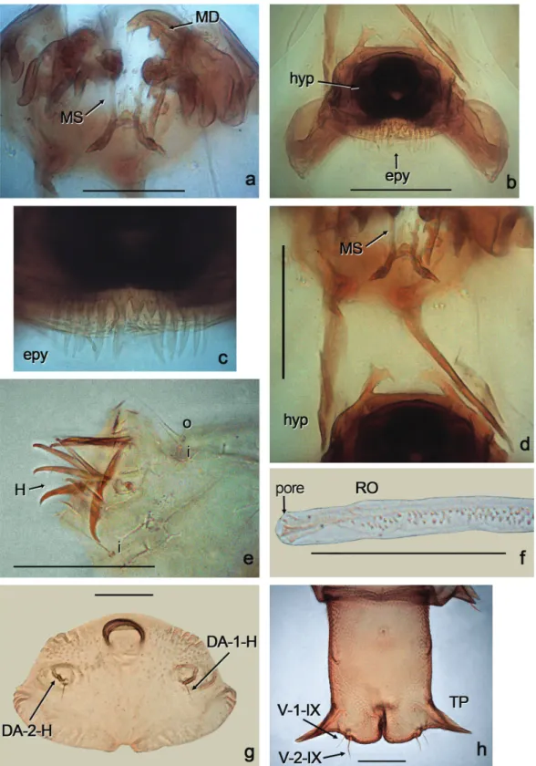
redescriptionfemalepupa (Figs. 2a-i, 3f-h, 4a-c). General coloration of exuvium pale brown, except anterior portion of cephalotorax brown. Total
length 3.80–4.08 (3.95, n=5) mm. Exuvium with flagellum appressed against lateral margin of face. Dorsal apotome (Figs. 2b-d, 3g) 2.66 X broader
than long, apex rounded, surface covered with small
rounded tubercles, anterior margin slightly concave
with a notch, with 2 pairs of raised, wrinkled areas;
dorsal apotome sensilla (Figs. 2b-d, 3g) DA-1-H long, thin seta, DA-2-H campaniform sensillum; posterior margin rounded with stout, dorsomedial, rounded tubercle; DAL 0.10 mm; DAW 0.38 mm;
DAW/DAL 3.80; mouthparts with mandible, lacinia absent; palpus extending posterior to posterolateral margin of labium; labium divided medially by longitudinal suture. Cephalothorax (Fig. 2a)
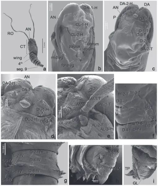
Figure2a-i - Dasyhelea mediomunda Minaya, pupa (SEM). a-h female pupa, i male pupa; a, entire pupa (lateroventral view); b, cephalothorax (ventral view); c, cephalotorax (dorsal view);
surface with small rounded tubercles, length 1.40-1.77 (1.60, n=5) mm, width 0.775-0.925 (0.862, n=5) mm. Cephalothoracic sensilla as follows: three dorsolateral cephalic sclerite sensilla (Figs. 2d-e) DL-1-H medium-sized, thin seta, DL-2-H stout, short setae, DL-3-H campaniform sensillum; three anterolateral sensilla (Fig. 2e), AL-1-T
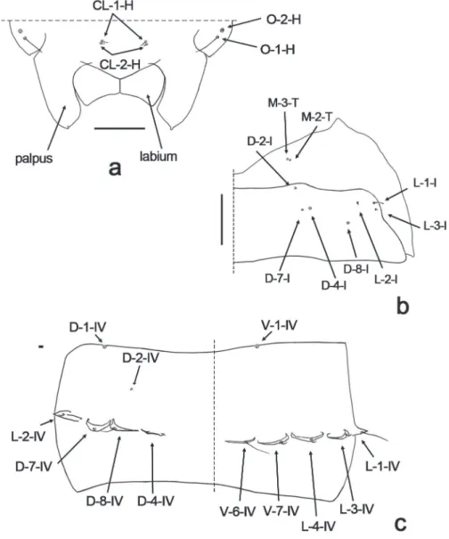
long, thin seta, 2-T medium-sized, thin, AL-3-T short, stout setae; two anteromedial sensilla (Fig. 2e), AM-1-T long, thin seta, AM-2-T short, thin seta; two clypeal/labrals (Figs. 2b, 4a); CL-1-H medium-sized, thin seta, CL-2-H short seta; two ocular sensilla, O-1-H long, thin, seta, O-2-H campaniform sensillum (Fig. 4a). Respiratory
organ (Figs. 2a, e) brown, slender, as long as total length of pupa, annulated on entire extension, with 3–4 apical and 22–24 lateral pores; RO length 3.20-3.75 (3.34, n=4) mm, RO width 0.03-0.06 (0.04, n=4) mm; without pedicel. Dorsals (Fig. 2c): D-1-T short, thin seta, D-3-T campaniform sensillum, D-2-T minute setae, supraalar (SA-2-T) campaniform sensillum. Metathoracics (Figs. 2f, 4b): M-2-T-M-3-T campaniform sensilla. Abdominal segments covered with small spinules. First abdominal segment (Figs. 2f, 4b) with sensilla as follows: D-2-I peg; D-4-I, D-7-I campaniform sensilla, D-8-I minute seta; L-1-I long thin seta, L-2-I, L-3-I short, thin setae. Second abdominal segment similar to the first one. Segment 4 with sensillar pattern (Fig. 2g) as follows: D-2-IVshort seta; D-4-IV, D-7-IV campaniform sensilla, D-8-IV medium-sized, thin seta, all located on flattened tubercles; L-1-IV medium-sized, thin seta, L-2-IV, L-3-IV-L-4-IV short, thin setae, all located on triangular tubercles; V-5-IV long, thin seta, V-6-IV short, stout seta, both on flattened tubercles. Segment 9 (Figs. 2h, 3h)1.50 X longer than wide, ventral surface with many spinules; length 0.30-0.35 (0.31, n=5) mm, width 0.20-0.29 (0.24, n=5) mm. Terminal process (Figs. 2h, 3h) triangular, divergent, tip pointed, base wide two setae, one long, thin seta on small rounded base, other medium-sized, stout seta on rounded tubercle; length 0.10-0.12 (0.11 n=5) mm.
Male pupa. Similar to female with usual sexual differences. Total length 3.51-4.27 (4.00, n=3) mm. Exuvium pale brown, except cephalotorax brown. Dorsal apotome with DAL 0.105 mm; DAW 0.335 mm, DAW/DAL 3.38. Respiratory organ (Fig. 3f), RO length 3.15-3.90 (3.53, n=4) mm, RO width 0.02-0.05 (0.03, n=4); without pedicel. Cephalotorax: length 1.57-1.80 (1.73, n=4) mm, width 0.92-0.97 (0.94, n=4) mm. Segment 4 as in Fig. 4c. Segment 9 (Fig. 2i) length 0-34-0.37 (0.35, n=4) mm, width 0.22-0.29 (0.25, n=4) mm; terminal process length 0.08-0.16 (0.11, n=4) mm.
Distribution: Peru (Lima), Chile (Valparaíso, Nu-ble), Argentina (Neuquén, Río Negro and Chubut).
DisCussion
The larva of Dasyheleamediomunda is very simi-lar to that of D. bahamensis by virtue of the short head capsule, the hypostoma bearing the medial portion smooth, the maxillary palpus short and button-like, the lateral arms of epipharynx stout and lacking teeth, and the caudal segment lacking hooklets. However, the larva of D. bahamensis dif-fers by having the palatum with one pair of sensilla campaniformia, the scopae absent; the mandible with three teeth, and the hypostoma having five-six teeth. The pupa is very similar to those of D. bahamensis and D. cincta slender and annulated respiratory organ, which is twice as long as the to-tal length of pupa. The pupae of D. bahamensis and D. cincta differ by the number of pores of the respiratory organ (D. bahamensis has 6 apical and 17 lateral pores and D. cincta bears 8-9 and 14-16, respectively).
Finally, the immatures of D. mediomunda could be also compared with D. paracincta. In the original description, Borkent (1991) mentioned only the head capsule length of the larva and the coloration and number of pores of the respiratory organ of the pupa (pale yellow with apex darkly pigmented, and 11-13 apical and 12-18 lateral pores respectively).
Ronderos et al. (2003) in the original description of the larva and pupa of D. bahamensis, incorrectly mentioned 5 dorsoposteromarginal setae (d.p.m.) and 4 ventroposteromarginal setae (v.p.m.). Díaz et al. (2009) in the redescription of pupa of D. cincta also incorrectly mentioned 4 d.p.m. and 3 v.p.m.
BionoMiCs
Immatures of Dasyhelea mediomunda were col-lected together with Culicoideslacustris Ronderos in a pond connected to a wetland (“mallin”), fed by groundwater, with intermittent flooding regime, without fishes in the semi-arid Patagonian Steppe (Epele and Archangelsky 2012). Larvae and pu-pae were collected in December on a sunny day, between 2:00-2:30 pm, air temperature was 20°C, water temperature was 17°C, pH 7.5. In the lab-oratory, with a temperature of 18-21°C, two lar-vae lasted five days to reach to pupal stage. Pupae completed their development in seven days. Lar-vae were motionless or only exhibited slow move-ments; pupae exhibited circular, slow abdominal movements.
aCKnoWleDGMents
We are grateful to Nélida Caligaris for technical as -sistance, to Dr. Pablo Pessacq for help in the collec-tion and rearing in the laboratory, and to Dr. Miguel Archangelsky for critical reading of the manuscript. This work was supported by National Research Council of Argentina (CONICET, Argentina). This is contribution number 108 of the LIESA.
resuMo
A larva de quarto ínstar de Dasyhelea mediomunda Minaya é descrita pela primeira vez e a descrição completa da pupa é fornecida, através da utilização de microscópio de contraste de fases e microscopia eletrônica de varredura. Os espécimes estudados foram coletados em um “mallin” conectado à zonas úmidas na estepa patagônia, Província de Chubut, Argentina.
palavras-chave: Argentina, Dasyhelea mediomunda, estágios imaturos, estepa patagônia.
reFerenCes
borkent a. 1991. The Ceratopogonidae Diptera of the
Galapagos islands ecuador with a discussion of their phylogenetic relationships and zoogeographic origins. Entomol Scand 22: 97-122.
borkent a. 2014. The pupae of the Biting Midges of the world (Diptera: Ceratopogonidae), with a generic key and analysis of the phylogenetic relationships between Genera. zootaxa 3879: 1-327.
borkent a. 2015. World species of biting midges (Diptera: Ceratopogonidae). Last update February 9, 2015. http:// www.inhs.uiuc.edu/research/FLYTREE/Ceratopogonidae-Catalog.pdf, 236 p.
borkent a and SPinelli gr. 2000. Catalog of the New World biting midges south of the United States of America (Diptera: Ceratopogonidae). Contrib Entomol Internat 4: 1-107.
borkent a and SPinelli gr. 2007. Neotropical Cerato-pogonidae (Diptera: Insecta). In: Adis J et al. (Eds), Aquat-ic Biodiversity in Latin AmerAquat-ica (ABLA). Vol. 4. Pensoft, Sofia-Moscow, 198 p.
borkent a and wirtH ww. 1997. World species of biting
midges (Diptera: Ceratopogonidae). Bull Am Mus Nat Hist 233: 1-257.
díaz f, ronderoS mm, SPinelli gr, ferreira-kePPler rl and torreiraS SRS. 2013. A new species of Dasy-helea Kieffer (Diptera: Ceratopogonidae) from Brazilian Amazonia. zootaxa 3686: 85-93.
díaz f, SPinelli gr and ronderoS mm. 2009. Biting Midges of the Dasyhelea cincta group from Patagonia (Diptera, Ceratopogonidae). Dtsch entomol z 56: 149-156.
díaz f, torreiraS SRS, SPinelli gr and ronderoS mm. 2014. A new species of Dasyhelea from Brazilian Amazonas and the description of the male of D. pau-listana (Diptera: Ceratopogonidae). Acta Entomol Mus Nat Pragae 54: 715-728.
ePele lb and arcHangelSky m. 2012. Spatial Variations
in Water Beetle Communities in Arid and Semi-Arid Pa-tagonian Wetlands and Their Value as Environmental Indi-cators. zool Stud 51: 1418-1431.
minaya g. 1978. Dasyheleamediomunda, sp. n. (Diptera: Ceratopogonidae) de la costa central del Perú. Rev Peru Entomol 21: 79-81.
ronderoS mm, díaz f and Sarmiento P. 2008. A new
method using acid to clean and a technique for preparation of eggs of biting midges (Diptera: Ceratopogonidae) for Scanning Electron Microscope. Trans Am Entomol Soc 134: 471-476.
ronderoS mm, SPinelli gr, Huerta H and díaz f. 2003.
Inmature stages of two Neotropical species of Dasyhelea Kieffer, 1911. (Diptera: Ceratopogonidae). Trans Am Entomol Soc 129: 295-308.
ronderoS mm, SPinelli gr and Sarmiento P. 2000. Preparation and Mounting of Biting Midges of the Genus Culicoides Latreille (Diptera: Ceratopogonidae) to be observed with a Scanning Electron Microscope. Trans Am Entomol Soc 126: 125-132.
de Dasyheleapenthesileae Macfie, y nuevas citas para el género Stilobezzia Kieffer. Limnobios 2: 403-411.
SPinelli gr and marino Pi. 2009. Estado actual del conocimiento de la familia Ceratopogonidae en la Patagonia (Diptera: Nematocera). Rev Soc Entomol Argent 68: 201-208.
SPinelli gr and ronderoS mm. 2011. Orden Diptera.
Familia Ceratopogonidae. En: Massaferro J (Comp.). Guía de Insectos Acuáticos del Parque Nacional Nahuel Huapi.
Larvas y Pupas. 1er ed., Río Negro Administración de Parques Nacionales, 2011 Fundación Flores, p. 118-127.
SPinelli gr and wirtH ww. 1984. Las especies
Neotropicales del género Dasyhelea, grupo cincta (Diptera: Ceratopogonidae). Limnobios 2: 586-608.
waugH wt and wirtH ww. 1976. A revision of the genus