Braz. J. of Develop.,Curitiba, v. 6, n.5, p.27034-27050 may. 2020. ISSN 2525-8761
Biodegradative capacity of Bacillus megaterium and Ralstonia
solanacearum on biodegradation of P(3HB) films in simulated soil
Capacidade biodegradativa de Bacillus Megaterium e Ralstonia
solanacearum na biodegradação de filmes de P(3HB) em solo simulado
DOI:10.34117/bjdv6n5-233
Recebimento dos originais: 13/04/2020 Aceitação para publicação: 13/05/2020
Matheus Marques Torres
Bacharel em Biotecnologia pela Universidade Federal de Pelotas Instituição: Universidade de São Paulo
Endereço: Laboratório de Bioprodutos, Instituto de Ciências Biomédicas, Avenida Professor Lineu Prestes, 1374, CEP: 05508-000, São Paulo-SP- Brasil
E-mail: matheus_mmt@hotmail.com
Camila Rios Piecha
Bacharela em Biotecnologia pela Universidade Federal de Pelotas Instituição: Universidade Federal de Pelotas
Endereço: Campus Capão do Leão s/n, prédio 19 -CEP: 96010-900-Pelotas-RS-Brasil, E-mail: camilapiecha@gmail.com
Karine Laste Macagnan
Doutora em Ciências pela Universidade Federal de Pelotas Instituição: Universidade Federal de Pelotas
Endereço: Campus Capão do Leão s/n, prédio 19 -CEP: 96010-900-Pelotas-RS-Brasil, E-mail: karinemacagnan@hotmail.com
Mariane Igansi Alves
Doutora em Ciências pela Universidade Federal de Pelotas Instituição: Universidade Federal de Pelotas
Endereço: Campus Capão do Leão s/n, prédio 19 -CEP: 96010-900-Pelotas-RS-Brasil, E-mail: marianeigansialves@hotmail.com
Angelita da Silveira Moreira
Doutora em Ciências pela Universidade Federal de Pelotas Instituição: Universidade Federal de Pelotas
Endereço: Campus Capão do Leão s/n, prédio 19 -CEP: 96010-900-Pelotas-RS-Brasil, E-mail: angelitadasilveiramoreira@gmail.com
Braz. J. of Develop.,Curitiba, v. 6, n.5, p.27034-27050 may. 2020. ISSN 2525-8761
Luciana Bicca Dode
Doutora em Ciências pela Universidade Federal de Pelotas Instituição: Universidade Federal de Pelotas
Endereço: Campus Capão do Leão s/n, prédio 19-CEP: 96010-900-Pelotas-RS-Brasil, E-mail: lucianabicca@gmail.com
Cristiane Wienke Raubach
Doutora em Ciências (com ênfase em química) pela Universidade Federal de São Carlos (UFSCAR)
Instituição: Universidade Federal de Pelotas
Endereço: Campus Anglo - Rua Gomes Carneiro n 1 - Cep: 96010-610-Pelotas-RS-Brasil, E-mail: cricawr@gmail.com
Patrícia Silva Diaz
Doutora em Ciências pela Universidade Federal de Pelotas Instituição: Universidade Federal de Pelotas
Endereço: Campus Capão do Leão s/n, prédio 19-CEP: 96010-900-Pelotas-RS-Brasil, E-mail: bilicadiaz@yahoo.com.br
RESUMO
O poli (3-hidroxibutirato) é um bioplástico microbiano biodegradável que vem sendo utilizado como substituto aos plásticos petroquímicos. Ele é completamente degrado em um ambiente microbiológico que contenha microrganismos com capacidade biodegradativa, sendo importante sua identificação para compreensão do processo de biodegradação. Além disso, novas metodologias próximas a um microambiente natural aumentam a confiabilidade do experimento. Neste contexto, o presente estudo visa analisar a capacidade degradativa das linhagens brasileiras Ralstonia solanacearum RS e Bacillus megaterium CN3 na biodegradação do P(3HB) sintetizado por Ralstonia solanacearum RS e pelo P(3HB) comercial. O experimento foi conduzido em casa de vegetação e a produção de P(3HB) foi realizada em um biorreator antes da extração. Os filmes foram enterrados no solo e removidos em intervalos de 20, 40, 60, 80 e 100 dias e analisados por porcentagem de biodegradação e diferenças nas características macro e microestruturais. Após 100 dias do experimento, o filme de P(3HB) mais degradado foi o produzido por R. solanacearum RS, sendo completamente degradado (100%) em solo não estéril. Além disso, B. megaterium CN3 provou-se ser um microrganismo com potencial degradativo, degradando 88% do P(3HB) RS em solo estéril. A análise macroscópica mostrou modificações na superfície ao longo do tempo, incluindo rachaduras, buracos, perda gradual de massa e alterações de cor. A análise microscópica demonstrou o aumento no tamanho dos poros e rachaduras, confirmando as etapas da biodegradação. Portanto, o experimento demonstrou ser capaz de simular condições viáveis para o crescimento de microrganismos e avaliar a capacidade de biodegradação de bactérias.
Palavras Chaves: Biodegradação, Poli (3-hidroxibutirato), Bioplástico, Ralstonia solanacearum, Bacillus megaterium.
ABSTRACT
Poly(3-hydroxybutyrate) is a microbial biodegradable bioplastic that has been used as a substitute for petrochemical-based plastics. It complete degradates in a microbiological
Braz. J. of Develop.,Curitiba, v. 6, n.5, p.27034-27050 may. 2020. ISSN 2525-8761 environment containing microorganisms with biodegradative capacity, being its identification important to the comprehension of the biodegradation process. In addition, new methodologies that are closer to a natural microenvironment increase the reliability of the experiment. In this context, the present study analyses the degradation capacity of the Brazilian strains Ralstonia
solanacearum RS and Bacillus megaterium CN3 on biodegradation of P(3HB) synthesized by Ralstonia solanacearum RS and commercial P(3HB). It was conducted under greenhouse
conditions and P(3HB) production was performed in a bioreactor before extraction. Films were buried in the soil and removed at 20, 40, 60, 80, and 100 days intervals and analyzed by biodegradation percentage and differences in macro and microstructural characteristics. After 100 days of the experiment, the most degraded P(3HB) was the one produced by R.
solanacearum RS, completely degraded (100%) in non-sterile soil. In addition, B. megaterium
CN3 proved to be a potential degradative microorganism, degrading 88% of P(3HB)RS in sterile soil. The macroscopic analysis showed surface modifications over time, including cracks, holes, gradual mass loss and color changes. Microscopic analysis demonstrates the increase in the pore size and cracks confirming the steps of biodegradation. Therefore, the experiment proved to be able to simulate viable conditions for the growth of microorganisms and to evaluated the biodegradation capacity of bacteria.
Key Words: Biodegradation, Poly(3-hydroxybutyrate), Bioplastic, Ralstonia solanacearum, Bacillus megaterium.
1 INTRODUCTION
Plastics derived from petrochemical sources are highly consumed in today's society due to its wide range of uses, from the manufacture of surgical instruments to the production of disposable packages. However, due to their mechanical properties of integrity and durability, they present prolonged degradation time (Alves et al., 2017). In order to minimize the environmental impacts caused by these plastics, several European Union countries such as the Netherlands and Belgium have already adopted policies to discourage their use, the same is reported for the cities of Ladakh in India, Dhaka in Bangladesh and South Australia, where its use is prohibited (Chanprateep, 2010). In 2016, France also banned the use of plastic bags and should carry out the transition from the use of plastic utensils, such as cups, plates, and cutlery to materials that are at least composed of 50% biodegradable materials such as corn or potato starch, cellulose fibers or bioplastics by 2020 and 60% by January 1st 2025 (France, 2016).
To replace these plastics, biodegradable polyesters such as polyhydroxyalkanoates (PHA)s, with wide production worldwide, are strong candidates. One of the most researched PHAs is the poly(3-hydroxybutyrate) [P(3HB)], a biocompatible, biodegradable and thermoplastic material that can be used in several industrial areas such as the manufacture of disposable utensils, medical materials and drug delivery (Alves et al., 2017; Raza et al. 2018).
Braz. J. of Develop.,Curitiba, v. 6, n.5, p.27034-27050 may. 2020. ISSN 2525-8761 Those, in a microbiologically active environment, shown effectively degradation in months compared to conventional plastics, which takes years (Boyandin et al., 2011; Altaee et al., 2016; Alves et al., 2017), resulting as final products carbon dioxide (CO2), water (H2O) in O2
presence and methane (CH4) under anaerobic conditions (Wang et al., 2013; Wang et al.,
2014).
The complete biodegradation of a material is a very attractive feature development of ecological plastic material for industrial use. Due to the high microbial biodiversity in the soil, the literature describes several methodologies to simulate P(3HB) biodegradation under different environmental conditions (Boyandin et al., 2011; Anstey et al., 2014). Analyses are reported in soil (Altaee et al., 2016; Araújo et al., 2015); composted soil (Casarin et al., 2013; Rosa et al., 2002); composted and humid soil based on poultry manure and organic materials of plant origin (Pellicano et al., 2009); and soil simulating landfill (Weng et al., 2013)
Recently, Emadian et al. (2017) reviewed numerous bacteria and fungi from soil, river, seawater and activated sludge and reported them as responsible for the release of depolymerases that hydrolyzes ester bonds of the polymer into water-soluble monomers and oligomers. Some bacteria such as Streptomyces, Bacillus, Pseudomonas, and Leptothrix, also possess the ability of synthesis and biodegradation of P(3HB). In this context, the aim of the present work was to analyze the degradation capacity of Brazilian strains Ralstonia
solanacearum RS and Bacillus megaterium CN3 on biodegradation of P(3HB) films
synthesized by Ralstonia solanacearum RS and commercial P(3HB), conducted in a greenhouse, a procedure not described yet.
2 MATERIALS AND METHODS
2.1 P(3HB) SAMPLES PRODUCTION
P(3HB)RS was produced at Biopolymers Laboratory of the Federal University of Pelotas by submerged fermentation of Ralstonia solanacearum RS, isolated from the local biodiversity.
Multiplicative cultures were transferred to 500 mL Erlenmeyer containing 200 mL of liquid yeast malt (YM) medium composed of (in g.L-1) yeast extract (Kasvi®), 2.7; malt extract (Kasvi®), 2.7 and peptone (Kasvi®), 4.5, (Jeanes, 1974), incubated in an orbital shaker at 32 °C (R. solanacearum) at 150 rpm for 24 hours for cell growth. On polymer production phase, the inoculum (DO600 =10) was transferred to the F4 mineral medium, composed of (in
Braz. J. of Develop.,Curitiba, v. 6, n.5, p.27034-27050 may. 2020. ISSN 2525-8761 element solution (MgSO4 0.2 g; CaCl2, 0.01 g; Na2MoO4, 0.005 g; ZnSO4, 0.1 g; FeCl3, 0.05
g), in Erlenmeyers of 500 mL of capacity, containing 200 mL of the final volume. Incubating in an orbital shaker at 32 °C and 200 rpm for 72 hours. Subsequently, the fermented broth was centrifuged at 10,000 x g for 15 min at 4 °C and the pellet resuspended in 0.89% saline solution and centrifuged again under the same conditions, being oven-dried at 56 °C until constant weight. The bioplastic extraction was performed as described by Macagnan et al. (2017).
Commercial P(3HB) (Biocycle®) was supplied as powder by PHB Industrial Usina da Pedra-Açúcar e Álcool (Serrana, SP, Brazil).
Films were obtained by solubilizing the powder in chloroform 1:40 (w/v) at 58 °C under stirring. The solution was transferred to a 9 cm diameter semi-open petri dish for slow evaporation of the solvent and formation of the film. P(3HB) specimens of approximately 3.5 cm2 were oven-dried at 56°C, weighed and individualised into polyester fabric envelopes measuring 5 x 6 cm.
2.2 BIODEGRADATION ASSAY
To the biodegradation experiment, plastic seedling trays (6.5 x 6.5 x 6.0 cm) with
individualised cells were used containing approximately 130 g of commercial soil. The experiment was carried out in a greenhouse with weekly monitoring of maximum and minimum temperature with the employment of a thermohygrometer (SH 122 - J. Prolab - Brazil).
The commercial soil used, contained 25% of clay and 5.4% of organic residues (pH 5.8) with a cation exchange capacity of 31.5 cmolc/dm3 and water holding capacity of 24%.
The moisture content was measured by withdrawing 5 g of soil samples during the periods of removal of the specimens, which were oven-dried at 56 °C until constant weight and analyzed by gravimetry.
Different treatments were used: (T1) soil sterilized by two cycles of 45 min in an autoclave at 121 °C (negative control); (T2) soil sterilized and added to R. solanacearum RS, (DO600 = 6.0); (T3) soil sterilized and added to B. megaterium, (DO600 = 5.8); (T4) natural soil
with no processes of sterilization; The treatments T1 to T4 were carried out from April to August 2016. The inoculums in T2 and T3 were made according to the cell growth phase described for P(3HB) production, with a temperature of 36 °C for B. megaterium. After that, 10 mL of the respective inocula were applied in 1 cm depth. After the experiments, the soils were sterilized prior to proper disposal.
Braz. J. of Develop.,Curitiba, v. 6, n.5, p.27034-27050 may. 2020. ISSN 2525-8761 2.3 ANALYSIS OF THE TEST SAMPLES
2.3.1 Percentage of biodegradation
The samples were collected at 20, 40, 60, 80 and 100 days, carefully washed with distilled water and oven-dried at 56 °C for 24 hours. The mass loss was analysed by gravimetry and the biodegradation by the percentage of mass loss according to equation 1 (Yew et al., 2006).
Equation 1
Where W1 is the initial mass of the film and W2 is the mass of the film after the period
of biodegradation.
2.3.2 Macro and microstructural characterization
Photographs to macrostructural analysis were performed using a digital camera (LG K8, South Korea) of 8 megapixels and 3264 x 2448 pixels of resolution.
The microstructural characterization was performed by scanning electron microscopy (Carl Zeiss Supra 35-V, Germany), conducted at room temperature with a voltage of 10 kV with the samples being previously metallized with gold layer overlay. Samples were analyzed at time 0 and after 60 days of biodegradation.
2.3.3 Statistical analysis
All averages were calculated from the triplicates of each treatment and the statistical analyses were performed using Tukey test with a significance level of 5% using the program Statistix 8 and the means with standard deviations were generated by the program GraphPad Prism 7.
3 RESULTS AND DISCUSSION
3.1 PERCENTAGE OF BIODEGRADATION
Microorganism biodegradation mechanism consists of 3 stages: first, the deposition of biofilms for bacterial adhesion and establishment of colonies on the surface (Capitelli et al., 2006); second, the biofilms serve as a surfactant facilitating ionic exchanges between hydrophobic and hydrophilic phases (Warscheid and Braams, 2000) and the third stage, where
Braz. J. of Develop.,Curitiba, v. 6, n.5, p.27034-27050 may. 2020. ISSN 2525-8761 the polymeric degradation is related to the excretion of degradative enzymes by the bacterial colonies, which assimilates the polymer as a source of carbon and nitrogen (Pelmont, 2005). In soil, under the tested conditions, all the bioplastics presented degradation, which increased according to the time elapsed as shown in figure 1.
P(3HB)RS presented the highest biodegradation degree in all treatments which was different to that observed in commercial P(3HB). The most effective biodegradation results were shown in treatments T3 and T4 with soil sterilized and inoculated with B. megaterium CN3 (88%) and natural unsterilized soil (100%) respectively. Similar results to those obtained in T3 and T4 have been described by Bucci et al. (2007) that obtained 100% in 90 days, Altaee et al. (2016), with 56% in 42 days and Weng et al. (2011), with 79.9% after 110 days.
The biodegradability capacity of P(3HB)-producing bacteria was observed in T3 (88%) in 100 days for P(3HB)RS, with the presence of B. megaterium CN3, which shown much higher biodegradation when compared to inoculated soil with Ralstonia solanacearum RS (T2-33%) and sterilized soil (T1-37%). The bacterium Bacillus megaterium possibly followed the steps of the microbial degradation mechanism more effectively, since the soil is its natural environment and researches demonstrates that it can aid the development of plants in general (Mendes, 2003). This fact can be attributed to the ability of B. megaterium CN3 to synthesize and excrete a large amount of the depolymerase enzyme involved in the bacterial pathway responsible for the breakdown of P(3HB) monomers (Grigull et al., 2015). Tansengco and Tokiwa (1998) analyzed the biodegradation capacity of Bacillus sp. in vitro incubated at 56 ºC. It was observed after 2 weeks 14.3% of mass loss of the films Bacillus gelatini was evaluated by Anh et al. (2010), achieving more than 50% PHB biodegradation after 30 days of incubation in vitro. These results are in agreement with those obtained in the present study. In T2 (sterilized soil with R. solanacearum RS) comparing to T3, the lower biodegradation rates were probably due to the low adhesion of the inoculated bacteria to the bioplastic film or the low production of functional and effective depolymerases to degrade it.
Ralstonia solanacearum has been known as a phytopathogenic bacterium (Denny, 2007) and
has its effective growth only in the infectious process (Genin and Denny, 2012). Despite that, Boyandin et al. (2013) described the genus Ralstonia as a potentially P(3HB) degrader. Similar results were presented in T1 (negative control) where the sterilization process extinguished the native microorganisms of the soil, producing an unfavorable environment to microbiological biodegradation. To both treatments, the probable mechanism of degradation is related to the abiotic conditions, such as hydrolysis, which comprises a degradation process
Braz. J. of Develop.,Curitiba, v. 6, n.5, p.27034-27050 may. 2020. ISSN 2525-8761 where the breakage of ester bonds occurs (Rosa and Filho, 2002). Similar results have been presented by Bonartesev et al. (2012) for the PHB synthesized by Azotobacter chroococcum 7b in the study of hydrolytic degradation, which obtained 20% degradation in 91 days.
Biodegradative capacity of microorganisms have been studied in in vitro systems by the measurement of the halo formed around colonies of microorganisms consuming bioplastic as the sole source of carbon in solid medium (Kumaravel et al., 2010; Volova et al., 2010) and by inoculation of bacteria with P(3HB) films in liquid medium (Colak and Guner, 2004; Woolnough, et al., 2008). The present study was conducted under simulated conditions in a greenhouse, although not described in the literature, proved to be a valid method to describe the biodegradation and catabolic capacity of the bacteria tested.
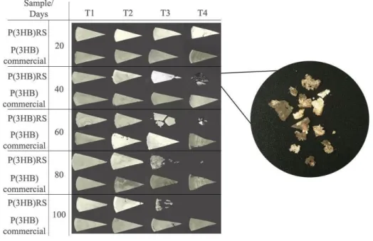
Figure 1. Percentage of biodegradation of P(3HB)RS and commercial P(3HB) in 20, 40, 60, 80 and 100 days in the treatments. (T1) sterilized soil; (T2) sterilized soil with R. solanacearum RS; (T3) sterilized soil with B.
megaterium CN3; (T4) natural soil;
The difference between the biodegradation of commercial P(3HB) and P(3HB)RS may be related to the molar mass thereof. According to Gu (2003), the molecular weight, composition of the polyester and the dominant specie of bacteria can interfere in the surface erosion. The average weight (Mw) of P(3HB)RS of 2.9x10⁵ Da (Macagnan et al., 2017) being approximately two times lower in comparison to commercial P(3HB), which ranges from
Braz. J. of Develop.,Curitiba, v. 6, n.5, p.27034-27050 may. 2020. ISSN 2525-8761 5.6x10⁵ Da (Machado et al., 2010) to 6.0x10⁵ Da (Faria and Martins-Franchetti, 2010). This is an extremely important factor directly affecting the mechanical strength of the bioplastic as well as the expansion capacity, the hydrolysis and consequently the biodegradation rate (Montoro et al., 2010). This, therefore, explains the faster biodegradation of P(3HB)RS.
It is also reported in the literature that depolymerisation, consisting of the cleavage of polymer molecules progressively reducing their molar mass (Lucas et al., 2008) is one step of biodegradation process. Lim et al. (2005) demonstrated the decrease in molar mass of P(3HB) (Fluka) film from 1.0x10⁵ Da to 8.3x10⁴ Da and 9.1x10⁴ Da in mangrove and after biodegradation for 112 days in tropical forest soil. Volova et al. (2017) also determined the molar mass changes of P(3HB) films synthesized by Cupriavidus eutrophus B10646 after 28 days of soil biodegradation from 6.2x10⁵ to 5.9x10⁵ Da.
3.2 SOIL TEMPERATURE AND MOISTURE
The experiments were performed from April to August 2016, a period of lower temperature average as shown in figure 2. Similar temperatures were recorded in the experiment developed by Weng et al. (2013) on the biodegradation of P(3HB) in soil and by Lim et al. (2005) on biodegradation in a forest and mangrove soils. Volova et al. (1998) report that higher temperatures tend to favour biodegradation, however, they should not exceed 35 °C, the optimum temperature being 28 °C. In another study, Volova et al. (2017) found that temperatures near 21 °C tended to decrease the degradation level of PHAs, whereas at 28 °C it was higher. However, the results presented in this study demonstrated a higher rate of mass loss in average values of temperature of 18 °C. Therefore, temperature were adequate for survival and reproduction of microorganisms according to figure 1.
Superior soil moisture is presented in the experiments conducted by Boyandin et al. (2011) that analysed the biodegradation of P(3HB) in root zones of Siberian larch (Larix
sibirica L.), obtaining 18-28% of humidity and under a drooping birch (Betula pendula L.),
obtaining values between 11- 23%. In addition, Lim et al. (2005) reported values of humidity around 72% with bioplastic buried under mangrove soil. The moisture values recorded in the present study may differ from the values presented in the literature due to the use of a greenhouse in opposition to open field. Despite this, even with moisture values lower than those reported in the literature, similar percentages of biodegradation were observed. For example, Lim et al. (2005) reported losses of 73.5% of P(3HB) buried in forest soil for 112 days and Boyandin et al. (2011) reports loss of mass in 78% of P(3HB). This evidences the
Braz. J. of Develop.,Curitiba, v. 6, n.5, p.27034-27050 may. 2020. ISSN 2525-8761 validity of the degradation experiment conducted in the greenhouse by providing adequate data and simulating natural disposal conditions. Although previous research has reported that low levels of moisture tend to limit the growth and activity of microorganisms (Briassoulis, 2007; Kapanen et al., 2008; Accinelli et al., 2012), in the present study, moisture content was not a limiting factor.
Figure 2. Average soil moisture and temperatures during the 100 days of the greenhouse experiment.
3.3 MACRO AND MICRO STRUCTURAL CHARACTERISATION
Physical modifications in the surface morphology of P(3HB)RS films were observed by macrostructural analysis (figure 3) which includes cracks, holes, gradual mass loss and color changes, which increased according to the time of biodegradation. As for commercial P(3HB), the physical surface modifications observed were a few cracks and color changes. A correlation between the results of percentage biodegradation presented in figure 1 and the physical changes in the surface of the bioplastics can be observed, which can be attributed to the fragmentation and assimilation of the bioplastics by the microorganisms.
The physical modifications in the surface morphology of the films of P(3HB)RS were higher than those presented by the commercial P(3HB), as already observed in the T4 with the beginning of fissures at 20 days of biodegradation.
Similar results are reported by Weng et al. (2013) for the biodegradation of P(3HB) in a landfill. After 30 days the fragmentation of the polymers were macroscopically visualized, which increased until 150 days of the experiment, and at 90 days complete biodegradation of one of the samples was observed, which was also detected in T4 for P(3HB)RS at 100 days of biodegradation. Weng et al. (2011) evaluated the biodegradation of P(3HB) under simulated conditions of the intense aerobic composting process and a decrease in the size of the films
Braz. J. of Develop.,Curitiba, v. 6, n.5, p.27034-27050 may. 2020. ISSN 2525-8761 from 10 days of the process was observed. Altaee et al. (2016) perceived the presence of holes and loss of mass in the biodegradation of P(3HB) in fertile garden soil.
Figure 3. Macroscopic analysis of P(3HB)RS and commercial P(3HB) films at 20, 40, 60, 80 and 100 days in the treatments. (T1) sterilized soil; (T2) sterilized soil with R. solanacearum RS; (T3) sterilized soil
with B. megaterium CN3; (T4) natural soil.
The samples present in Figure 3 shows the changes in coloration of bioplastics through the appearance of dark blurred areas that may have been caused by deposition of extracellular material excreted by microorganisms, water accumulation, penetration into the matrix of and/or excretion of lipophilic microbial pigments, especially the highlighted sample (Araújo et al., 2015). Pellicano et al. (2009) reported the detection of dark brown and reddish pigments in the biodegradation assay of PHBV/Ecoflex®/cassava starch and PHBV/Ecoflex® blends, respectively. Altaee et al. (2016) also reports the color change of bioplastics after the process of biodegradation of P(3HB) in fertile garden soil.
Bioplastics films micro-morphology modifications over time were more noticeable in T4, as presented in figure 4. It can be seen that the bioplastic samples surface at the initial time have porosities and it was observed that P(3HB)RS had higher porosity than commercial PHB, being more susceptible to biodegradation due to its larger surface contact area.
For P(3HB)RS the formation of large round cavities occurs in 20 days evidencing the bacterial accumulation on the surface. After the 40 day period, there are cracks in the surface
Braz. J. of Develop.,Curitiba, v. 6, n.5, p.27034-27050 may. 2020. ISSN 2525-8761 of the polymer that were observed even in higher number after 60 days, fractures increases as much as the number of hyphae and the appearance of structures derived from a microorganism (Yew et al., 2006; Ong and Sudesh, 2016). Thereafter, the accumulation of biomass-derived material in continuous growth up to 60 days is evident. The results of pores and biological material deposited on the surface are consistent with Corrêa et al. (2008) in that the same conditions were found after five months of biodegradation in soil. As for commercial P(3HB), the appearance of pores were visible from the 40 days, increasing continuously up to 60 and possibly further, also evidencing the accumulation of colonies of microorganisms on surface.
Since there are different degrees of degradation between the polymers, the micrographs could evidence the establishment of a soil biodegradation process, since they cover most of the steps at different times of degradation.
The biodegradation process begins when the polymeric material is placed in a microbiologically active environment, in this study it starts when the film was buried. After the adaptation of the native microbiota with the material, the appearance of pores and cavities initiate, as evidenced by commercial P(3HB) in 40 days, and P(3HB) RS in 20 days. In this step, it is important to note that the pores are formed due to enzymatic degradation and assimilation of the polymeric material by the colonies of adhered microorganisms (Pelmont, 2005), and the porosity of the material continues to increase until the surface is completely deformed.
After the bacterial degradation process, and most likely due to excessive deformation of the surface, the number of microorganisms like fungi begin to adhere more intrinsically to the polymer. The presence of hyphae could also exist on the surface of the film in 20 days, but with its integrity not yet fully committed, after washing with distilled water, the structure could have been easily removed. The presence of hyphae that increase continuously with time (as evidenced at 40 and 60 days of P(3HB)RS on the right corner) may indicate internal cavities and relative increase of the contact surface. Finally, the biodegradation of the polymer material at its most advanced stage is associated with the physical and visible breakage of the polymer, with cracks derived from the mechanical strength of the hyphae and complete disintegration of the uniformity of the polymer surface, as shown in the 60 day micrograph of the P(3HB)RS. According to the micrographs and the process described above, P(3HB)RS is in a more advanced stage of biodegradation since by the difference of the mass loss, it is possible to prove the difference in the rates of degradation.
Braz. J. of Develop.,Curitiba, v. 6, n.5, p.27034-27050 may. 2020. ISSN 2525-8761 Figure 4. Scanning electron micrograph of the surface of P(3HB)RS and commercial P(3HB) films at 0,
20, 40 and 60 days of biodegradation assay in T4 (natural soil).
The treatments T1 and T3 that contained sterilized soil and B. megaterium CN3 respectively (data not show), presented a significant difference on the number of pores of the polymer surface, evidencing the biodegradation as a result of bacterial adhesion end enzyme excretion and the biodegradative capacity of this bacterium. T1 and T2 (sterilized soil with R.
solanacearum) presented differences in relation to the samples at the initial time, but it was
Braz. J. of Develop.,Curitiba, v. 6, n.5, p.27034-27050 may. 2020. ISSN 2525-8761
4 CONCLUSION
Biodegradation was observed in both P(3HB)s in all treatments analyzed, with the highest percentage of biodegradation being P(3HB)RS. It has been proven that the type of bacteria used interferes with the percentage of biodegradation, as observed in the Bacillus
megaterium CN3 treatment, which presented biodegradation capacity on the biodegradation
of P(3HB)RS. Moisture positively influenced the biodegradation process of P(3HB)s and the lower temperatures present in this study did not influence the microbial growth. The microscopy and macroscopy analyses proved the presence of microorganisms and the biodegradation process which as confirmed by the color change and the presence of cracks and hyphaes. Therefore, the greenhouse experimental methodology tested in this study was able to verify the biodegradation capacity of biologically active microorganisms and to simulate a microenvironment with the natural steps of biodegradation.
ACKNOWLEDGMENTS
The authors would like to thank CNPQ and CAPES for financial support. The CEME-Sul in Federal University of Rio Grande by Technical support by user facility of MEV. Professor Eugênia Jacira Bolacel Braga, from the Department of Botany of the Federal University of Pelotas, for the loan of the greenhouse for the realization of the experiment, PHB Industrial S/A for supplying the polymer.
REFERENCES
Accinelli C, Saccà M, Mencarelli M, Vicari A. (2012) Deterioration of bioplastic carrier bags in the environment and assessment of a new recycling alternative. Chemosphere. 89(2):136-143. https://doi.org/10.1016/j.chemosphere.2012.05.028
Altaee N, El-Hiti G, Fahdil A, Sudesh K, Yousif E. (2016) Biodegradation of different formulations of polyhydroxybutyrate films in soil. Springerplus. 5(1): 762. https://doi.org/ 10.1186/s40064-016-2480-2
Anh T T V, Toan N T K, Huy N Q. (2010) Degradation of poly(3-hydroxybutyrate) (PHB) by
Bacillus gelatini isolated from Vietnam. Tap chi Sinh hoc. 32(3): 72-77.
https://doi.org/10.15625/0866-7160/v32n3.710
Anstey A, Muniyasamy S, Reddy M, Misra M, Mohanty A. (2014) Processability and Biodegradability Evaluation of Composites from Poly(butylene succinate) (PBS) Bioplastic and Biofuel Co-products from Ontario. J Polym Environ. 22(2):209-218. https://doi.org/10.1007/s10924-013-0633-8
Braz. J. of Develop.,Curitiba, v. 6, n.5, p.27034-27050 may. 2020. ISSN 2525-8761 Araújo R, Conceição I, Carvalho L, Alves T, Barbosa R. (2015) Influência da argila vermiculita brasileira na biodegradação de filmes de PHB. Polímeros. 25(5):483-491.http://dx.doi.org/10.1590/0104-1428.2031
Bonartsev A, Boskhomodgiev A, Iordanskii A, Bonartseva G, Rebrov A, Makhina T et al. (2012) Hydrolytic Degradation of Poly(3-hydroxybutyrate), Polylactide and their Derivatives: Kinetics, Crystallinity, and Surface Morphology. Mol Cryst Liq Cryst. 556(1):288-300. https://doi.org/10.1080/15421406.2012.635982
Boyandin, A. N. et al. (2013) Microbial degradation of polyhydroxyalkanoates in tropical soils. International Biodeterioration & Biodegradation. 83:77–84. https://doi.org/10.1016/j.ibiod.2013.04.014
Boyandin A, Prudnikova S, Filipenko M, Khrapov E, Vasil’ev A, Volova T. (2011) Biodegradation of polyhydroxyalkanoates by soil microbial communities of different structures and detection of PHA degrading microorganisms. Appl Biochem Microbiol. 48(1):28-36. http://dx.doi.org/10.1590/0104-1428.2031.
Bucci D, Tavares L, Sell I (2007) Biodegradation and physical evaluation of PHB packaging.
Polym Test. 26(7):908-915.https://doi.org/10.1016/j.polymertesting.2007.06.013
Cappitelli F, Principi P, Sorlini C. (2006) Biodeterioration of modern materials in contemporary collections: can biotechnology help?. Trends Biotechnol. 24(8):350-354. http://dx.doi.org/:10.1016/j.tibtech.2006.06.001
Casarin S, Agnelli J, Malmonge S, Rosário F. (2013) Blendas PHB/copoliésteres biodegradáveis: biodegradação em solo. Polímeros. 23(1):115-122. http://dx.doi.org/10.1590/S0104-14282013005000003
Colak A, Güner S. (2004) Polyhydroxyalkanoate degrading hydrolase-like activities by Pseudomonas sp. isolated from soil. Int Biodeterior Biodegradation. 53(2):103-109. http://dx.doi.org/ 10.1016/j.ibiod.2003.10.006
Corrêa M, Rezende M, Rosa D, Agnelli J, Nascente P. (2008) Surface composition and morphology of poly(3-hydroxybutyrate) exposed to biodegradation. Polym Test. 27(4):447-452. https://doi.org/10.1016/j.polymertesting.2008.01.007
Emadian S, Onay T, Demirel B. (2007) Biodegradation of bioplastics in natural environments.
Waste Manage. 59:526-536. https://doi.org/10.1016/j.wasman.2016.10.006
Faria A, Martins-Franchetti S. (2010) Biodegradação de filmes de polipropileno (PP), poli(3-hidroxibutirato) (PHB) e blenda de PP/PHB por microrganismos das águas do Rio Atibaia.
Polímeros. 20(2):141-147. http://dx.doi.org/10.1590/S0104-14282010005000024.
Genin S, Denny T. (2012) Pathogenomics of the Ralstonia solanacearum Species Complex.
Annu Rev Phytopathol. 50(1):67-89.
Braz. J. of Develop.,Curitiba, v. 6, n.5, p.27034-27050 may. 2020. ISSN 2525-8761 Grigull V, Mazur L, Garcia M, Schneider A, Pezzin A. (2015) Estudo Da Degradação De Blendas De Poli(Hidroxibutirato-Co-Hidroxivalerato)/Poli(L-Ácido Lático) Em Diferentes Condições Ambientais. Engevista. 17(4). https://doi.org/10.22409/engevista.v17i4.773 Gu J. (2003) Microbiological deterioration and degradation of synthetic polymeric materials: recent research advances. Int Biodeterior Biodegradation. 52(2):69-91. https://doi.org/10.1016/S0964-8305(02)00177-4
Kapanen A, Schettini E, Vox G, Itävaara M (2008) Performance and Environmental Impact of Biodegradable Films in Agriculture: A Field Study on Protected Cultivation. J Polym
Environ. 16(2):109-122. https://doi.org/10.1007/s10924-008-0091-x
Kumaravel S, Hema R, Lakshmi R (2010) Production of Polyhydroxybutyrate (Bioplastic) and its Biodegradation by Pseudomonas Lemoignei and Aspergillus Niger. E- J Chem. 7(s1):S536-S542. https://doi.org/ 10.1155/2010/148547
Lim S, Gan S, Tan I. (2005) Degradation of medium-chain-length polyhydroxyalkanoates in tropical forest and mangrove soils. Appl Biochem Biotechnol. 126(1):23-33. https://doi.org/10.1007/s12010-005-0003-7
Lucas N, Bienaime C, Belloy C, Queneudec M, Silvestre F, Nava-Saucedo J. (2008) Polymer biodegradation: Mechanisms and estimation techniques – A review. Chemosphere. 73(4):429-442. https://doi.org/10.1016/j.chemosphere.2008.06.064
Macagnan K, Rodrigues A, Alves M, Furlan L, Kesserlingh S, Moura A et al. (2017) Simplified recovery process of Ralstonia solanacearum-synthesized polyhydroxyalkanoates via chemical extraction complemented by liquid-liquid phase separation. Quím Nova. 40(2):125-130. https://doi.org/10.21577/0100-4042.20160162
Machado M, Pereira N, Miranda L, Terence M, Pradella J. (2010) Estudo das propriedades mecânicas e térmicas do polímero Poli-3-hidroxibutirato (PHB) e de compósitos PHB/pó de madeira. Polímeros. 20(1):65-71. https://doi.org10.11606/T.87.2008.tde-21012009-115422 Mendes I, Reis Junior F. (2003) Microrganismos e disponibilidade de fósforo (P) nos solos: uma análise crítica. Planaltina: Embrapa Cerrados. Embrapa Cerrados, ISSN 15175111. https://www.infoteca.cnptia.embrapa.br/bitstream/doc/568171/1/doc85.pdf. Accessed 10 february 2018.
Montoro S, Shigue C, Sordi M, Santos A, Ré M. (2000) Estudo cinético da redução da massa molar do poli(3-hidroxibutirato-co-3-hidroxivalerato) (PHBHV). Polímeros. 20(1):19-24. https://doi.org/10.1590/S0104-14282010005000005
Ong S, Sudesh K. (2016) Effects of polyhydroxyalkanoate degradation on soil microbial
community. Polym Degrad Stab. 131:9-19.
https://doi.org/10.1016/j.polymdegradstab.2016.06.024
Pellicano M, Pachekoski W, Agnelli J. (2009) Influência da adição de amido de mandioca na biodegradação da blenda polimérica PHBV/Ecoflex®. Polímeros. 19(3):212-217. https://doi.org/10.1590/S0104-14282009000300009
Braz. J. of Develop.,Curitiba, v. 6, n.5, p.27034-27050 may. 2020. ISSN 2525-8761 Pelmont J. (2005) Biodégradations et métabolismes. Les Ulis: EDP sciences. Edp Sciences, Les Ulis Cedex A, France
Rosa D, Chui Q, Pantano Filho R, Agnelli J. (2012) Avaliação da Biodegradação de Poli-beta-(Hidroxibutirato), Poli-beta-(Hidroxibutirato-co-valerato) e Poli-épsilon-(caprolactona) em Solo Compostado. Polímeros. 12(4):311-317. https://doi.org/10.1590/S0104-14282002000400015
Tansengco M, Tokiwa Y. (1997) Thermophilic microbial degradation of polyethylene succinate. World J Microbiol Biotechnol. 14(1):133-138. https://doi.org/10.1023/A:1008897121993
Volova T, Belyaeva O, Plotnikov V, Puzyr A. (1998) Studies of Biodegradation of Microbial Polyhydroxyalkanoates. Appl Biochem Microbiol. 34(5):488-492.
Volova T, Boyandin A, Vasiliev A, Karpov V, Prudnikova S, Mishukova O et al. (2010) Biodegradation of polyhydroxyalkanoates (PHAs) in tropical coastal waters and identification of PHA-degrading bacteria. Polym Degrad Stab. 95(12):2350-2359. https://doi.org/10.1016/j.polymdegradstab.2010.08.023
Volova T, Prudnikova S, Vinogradova O, Syrvacheva D, Shishatskaya E. (2017) Microbial Degradation of Polyhydroxyalkanoates with Different Chemical Compositions and Th7eir Biodegradability. Microb Ecol. 73(2):353-367. https://doi.org10.1007/s00248-016-0852-3 Wang Z, Lin X, An J, Ren C, Yan X. (2013) Biodegradation of Polyhydroxybutyrate Film by Pseudomonas mendocina DS04-T. Polym Plast Technol Eng. 52(2):195-199.https://doi.org/10.1080/03602559.2012.735738
Warscheid T, Braams J. (2000) Biodeterioration of stone: a review. Int Biodeterior
Biodegradation. 46(4):343-368. https://doi.org/10.1016/S0964-8305(00)00109-8
Weng Y, Wang L, Zhang M, Wang X, Wang Y. (2013) Biodegradation behavior of P(3HB,4HB)/PLA blends in real soil environments. Polym Test. 32(1):60-70. https://doi.org/10.1016/j.polymertesting.2012.09.014
Weng Y, Wang X, Wang Y. (2011) Biodegradation behavior of PHAs with different chemical structures under controlled composting conditions. Polym Test. 30(4):372-380. https://doi.org/10.1016/j.polymertesting.2011.02.001
Woolnough C, Charlton T, Yee L, Sarris M, Foster L. (2008) Surface changes in polyhydroxyalkanoate films during biodegradation and biofouling. Polym. Int. 57(9):1042-1051. https://doi.org/10.1002/pi.2444
Yew S, Tang H, Sudesh K. (2006) Photocatalytic activity and biodegradation of polyhydroxybutyrate films containing titanium dioxide. Polym Degrad Stab. 91(8):1800-1807. https://doi.org/10.1016/j.polymdegradstab.2005.11.011