Analysis of rapid maxillary expansion using
Cone-Beam Computed Tomography
Gerson Luiz Ulema Ribeiro*, Arno Locks**, Juliana Pereira***, Maurício Brunetto***
Whenever a maxillary arch is diagnosed as skeletally atresic the treatment of choice is usually maxillary orthopedic expansion, involving separation of the midpalatal suture. Basically, this suture used to be assessed with the aid of a maxillary occlusal radiograph, which limited its posteroanterior evaluation. Similarly, quantifying this atresia in cephalometric x-rays always posed an obstacle for clinicians owing to considerable superimposition of facial structures. With the advent of computed tomography, this technology has revolutionized diagnostic methods in dentistry because it provides high dimensional accuracy of the facial structures and a reliable method for quantifying the behavior of the maxillary halves, tooth inclination, bone formation at the suture in the three planes of space, as well as alveolar bone resorption and other conse-quences of palatal expansion.
Abstract
Keywords: Diagnosis. Radiographic images. Rapid maxillary expansion. Cone-Beam Computed Tomography.
* M.Sc. and Ph.D. in Orthodontics, Rio de Janeiro Federal University (UFRJ). Professor, Graduate and Postgraduate courses, UFSC. Diplomate, Brazilian Board of Orthodontics and Facial Orthopedics.
** M.Sc. and Ph.D. in Orthodontics, Rio de Janeiro Federal University (UFRJ). Postdoctoral research, University of Aarhus, Denmark. Professor, Graduate and Postgraduate courses, UFSC. Diplomate, Brazilian Board of Orthodontics and Facial Orthopedics.
*** Specialist in Orthodontics, UFSC. M.Sc. Candidate in Orthodontics, UFSC.
introduction
Recovery of transverse maxillary discrepancy seems to be essential for the proper treatment of various types of malocclusion. Several authors have investigated possible methods to expand the maxillary arch through different means. Propo-nents of rapid maxillary expansion (RME) argue that this method causes minimum tooth move-ment and maximum skeletal displacemove-ment. Con-versely, advocates of slow expansion believe that this method produces less tissue resistance in neighboring maxillary structures while enhancing
bone formation in the intermaxillary suture, and that these two factors help to minimize postex-pansion relapse.12,13
Some authors have advocated the separation of the midpalatal suture to expand narrow max-illary arches.11,15,20 Moreover, Graber,7 in 1972,
Given the diversity of structures comprised in the craniofacial complex various therapeutic resources have emerged which are capable of modifying the position or morphology of these components. Lateral maxillary atresia is a very common condition in different malocclusions. This transverse deficiency, caused by genetic and/ or functional4 factors, may involve only the
pos-terior dental segments, imparting excessive lin-gual tipping to these segments,6 but it may also
be associated with a skeletally compromised maxilla, which gives it a sicatréo appearance.6,14
When this happens, the maxilla presents with a narrow6 and gothic palate.14 To remedy this
situ-ation, an expansion is required which is capable of effecting maximum orthopedic movement of the maxillary bones while maintaining the integ-rity of the tissues and reducing the resulting tooth inclination.1,4,12,13,15,17,25 Rapid maxillary
expan-sion (RME) meets these requirements, restoring the transverse dimensions of this bone structure and corresponding dental arch14,25 by opening the
midpalatal suture in conjunction with orthopedic reactions in other facial sutures and slight move-ments in the posterosuperior segmove-ments.8
Numerous studies have been conducted to in-vestigate the changes caused in the maxillary bones and midpalatal suture as a result of rapid maxillary expansion. Histological experiments on animals showed new bone formation in the suture zone after palate splitting.5,10,28 Radiographic studies in humans
showed ossification in the region after expansion. However, the length of time that the palatal suture takes to restore its normal structure in humans is still the subject of considerable controversy. The vast majority of authors recommend that retention be performed with the appliance itself, after palate splitting, for a period of three months.2,8,10,16,18,19,22
The ability to measure these changes allows orthodontists to predict the effects of orthopedic treatment. Invasive techniques such as metal im-plants provide accurate information but are too aggressive for routine use. Histological control of
tissue reactions is possible only in animal studies or autopsy material.27
Several authors have studied the skeletal and dental changes resulting from opening the mid-palatal suture but the literature is still incon-clusive regarding dimensional changes in dental arches and maxillary displacement as a whole, and whether or not these changes are transient.4,11,12,30
According to Sato et al,23 posteroanterior
ceph-alometric radiography provides an assessment of the transverse dimensions of the face by broaden-ing the scope and thus facilitatbroaden-ing the diagnosis of crossbites and orthopedic changes inherent in the rapid opening of the midpalatal suture. Because it is an image in two dimensions, radiographic overlays of anatomical structures hamper the pre-cise location of cephalometric landmarks, which are instrumental for diagnosing and assessing the maxilla before or after any intervention, notably in the maxillary middle third.9
Assessment of frontal radiographs shows that the maxillary bones are displaced laterally with the fulcrum located close to the frontomaxillary suture while lower skeletal expansion progresses. The maxillary central incisors usually move mesi-ally and, in general, undergo uprighting after ap-pliance stabilization. Such movement aids in clos-ing the wide median diastema produced by the orthopedic effects of the appliance. As these teeth are uprighted, part of the arch length benefits obtained with the expansion is lost. The occlusal radiograph shows that the intermaxillary suture experiences a non-parallel opening accompanied by a further, V-shaped expansion, greater in the anterior than in the posterior region.30
In frontal view, a pyramid appears in the re-gion of this suture, whose base is turned inferiorly. Thus, real bone mass gain occurs with a conse-quent increase in arch perimeter.4,10,11
The occlusal view showed that in the antero-posterior direction the opening of the suture would be twice as large in the incisor than in the molar region, allowing the visualization of a new triangle with the base facing the anterior region. Apparently, the amount of opening varies with each individual. By comparing the opening of the intermaxillary suture with the dental effects it was found that the amount of suture separation would be equal to or less than the amount of expansion in the dental arch.10
The advent of Cone-Beam Computed Tomog-raphy (CBCT) has made possible three-dimen-sional assessment. Today, it is increasingly applied in dentistry mainly because it is more affordable and entails lower radiation exposure.9
To compare the biological effects of radiation on various parts of the body, effective equivalent dose is used, which yields a comparison of the bio-logical effects of different types of ionizing radia-tion and allows adjustments to be made in the vol-ume and radiosensitivity of irradiated tissue. The unit of measure used is the sievert (Sv).9,24
The effective equivalent dose in conven-tional radiographic examinations, comprising 3 maxillary periapical radiographs (5 µSv), 3 mandibular periapical radiographs to assess the bone tissue available in the mandibular symphy-sis (5 µSv), 1 upper occlusal radiograph (4 µSv), 1 panoramic radiograph (7 µSv), 1 posteroan-terior cephalometric radiograph (7 µSv), 1 lat-eral cephalometric radiograph (7 µSv), results in a total of 42 µSv.9,24 Using a Cone-Beam CT
scanner such as the i-CAT, radiation exposure is approximately 30-100 µSv for examining both the maxilla and mandible, which represents a reduction of 1/6 in patient radiation exposure compared to a conventional medical CT scan-ner (helical). Cone-Beam CT radiation dose is similar to the radiation dose used in the periapi-cal examination of the entire mouth, equivalent to approximately 4-15 times the dose of a pan-oramic X-ray.9
Moreover, compared to conventional radiog-raphy, the potential of CT to provide additional information is much higher. Additionally, with Cone-Beam CT, professionals can obtain recon-structions of all conventional dental radiographs in addition to the unique information provided by multiplanar and 3D reconstructions.9
As new knowledge is generated by three-di-mensional views of the skull and face, Cone-Beam CT is expected to change concepts and shift para-digms, redefining goals and treatment plans in or-thodontics. This would facilitate the diagnosis of maxillary atresia and maxillary behavior in terms of expansion procedures, thus allowing for quanti-fication of the actual skeletal gains in dealing with two different activation protocols. CT will there-fore contribute to diagnosis to the extent that it will be decisive in establishing the best protocol expansion to be used in treatment planning.9
diScuSSion
FIGURE 1 - Three-dimensional occlusal reconstruction of the maxilla from a CT scan, showing the closed midpalatal suture.
FIGURE 2 - Three-dimensional occlusal reconstruction of the maxilla from a CT scan, showing the open midpalatal suture.
central incisors returned spontaneously to their original position. Control over this now purely orthodontic movement is linked to the memory of stretched gingival fibers, which rapidly move, first the crowns, then the roots, closer to each other.
Total maxillary occlusal x-rays are the routine diagnostic tool used in orthodontic practice to ver-ify and document suture separation. Cone-Beam computed tomography enables more accurate re-sult evaluation and improved quantification. One can observe a triangular, radiolucent area with its base facing the anterior nasal spine, a region where bone strength is reduced (Figs 2 and 3). At the same time that CT confirms the orthopedic splitting of the maxilla, it subsequently records midpalatal suture reorganization, which occurs during the retention phase, when the appliance is kept in the mouth (Fig 4). The fixed expander should only be replaced by a removable retention plate after complete tomographic restructuring, which takes on average 3-4 months.29
It seems indisputable that, even though the predominant effect is of an orthopedic nature,
orthodontic effect, represented by the flaring of the posterior teeth and alveolar process, is an integral part of rapid maxillary expansion. It is known to practitioners who deal with orthopedic expansion that hand in hand with the gradual opening of the midpalatal suture, the force delivered by the ex-pander causes periodontal ligament compression, lateral tipping of the alveolar process and subse-quent flaring of the posterior teeth. These chang-es reprchang-esent the orthodontic effect of RME. But before these forces induce classical orthodontic movement with osteoclastic histological changes in the periodontium, the maxillary bones are split due to orthopedic effects (Figs 2 and 3).
A
A
B
B
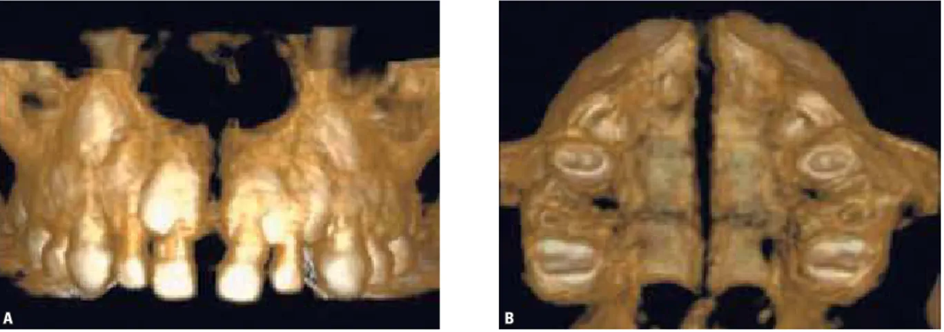
FIGURE 3 - Three-dimensional occlusal reconstruction of the maxilla from a CT scan, showing the open midpalatal suture: (A) posteroanterior view; (B) oc-clusal view.
FIGURE 4 - Three-dimensional occlusal reconstruction of the maxilla from a CT scan, showing the suture reorganization process: (A) posteroanterior view; (B) occlusal view.
concLuSionS
It could be argued that nowadays orthope-dic maxillary expansion is part and parcel of a coherent therapeutic approach in orthodon-tic pracorthodon-tice, provided that maxillary atresia is present. The lateral repositioning of the max-illa and increased basal bone, which can be accurately observed in Cone-Beam computed
tomography confirms the marked morphologi-cal changes that occur in the upper arch and nasomaxillary structure.
such as age. These variables will establish the orthodontic planning and treatment best suited for each case.
Cone-Beam Computed Tomography is a groundbreaking diagnostic method in dentistry as it provides high dimensional accuracy of the
facial structures and a reliable method for quan-tifying the behavior of the maxillary halves, dental tipping, bone formation at the suture in the three planes of space, as well as alveo-lar bone resorption and other consequences of palatal expansion.
1. Bell RA. A review of maxillary expansion in relation to rate of expansion and patient’s age. Am J Orthod. 1982 Jan;81(1):32-7. 2. Belli SJ. Long term anteroposterior, transverse and vertical
skeletal changes following rapid maxillary expansion in adults [thesis]. Columbus (Ohio): The Ohio State University; 1992. 3. Biederman W. A hygienic appliance for rapid expansion.
J Pract Orthod. 1968 Feb;2(2):67-70.
4. Bishara SE, Staley RN. Maxillary expansion: clinical implications. Am J Orthod Dentofacial Orthop. 1987 Jan;91(1):3-14. 5. Cleall JF, Bayne DI, Posen JM, Subtelny JD. Expansion of the
midpalatal suture in the monkey. Angle Orthod. 1965 Jan;35:23-35. 6. Dipaolo RJ. Thoughts on palatal expansion. J Clin Orthod.
1970 Sep;4(9):493-7.
7. Graber TM. Orthodontics principles and practice. 3rd ed. Philadelphia: WB Saunders; 1972, 953p.
8. Ekström C, Henrikson CO, Jensen R. Mineralization in the midpalatal suture after orthodontic expansion. Am J Orthod. 1977 Apr;71(4):449-55.
9. Garib DG, Raymundo R Jr, Raymundo MV, Raymundo DV, Ferreira SN. Tomograia computadorizada de feixe cônico (cone beam): entendendo este novo método de diagnóstico por imagem com promissora aplicabilidade na Ortodontia. Rev Dental Press Ortod Ortop Facial. 2007 mar-abr;12(2):139-56.
10. Haas AJ. Rapid expansion of the maxillary dental arch and nasal cavity by opening the midpalatal suture. Angle Orthod. 1961 Apr;31(2):73-90.
11. Haas AJ. The treatment of maxillary deiciency by opening the midpalatal suture. Angle Orthod. 1965 Jul:35(3):200-17. 12. Haas AJ. Palatal expansion: just the beginning of dentofacial
orthopedics. Am J Orthod. 1970 Mar;57(3):219-55.
13. Herold JS. Maxillary expansion: a retrospective study of three methods of expansion and their long-term sequelae. Br J Orthod. 1989 Aug;16(3):195-200.
14. Hershey HG, Stewart BL,Warren DW. Changes in nasal airway resistance associated with rapid maxillary expansion. Am J Orthod. 1976 Mar;69(3):274-84.
15. Isaacson RJ, Ingram AH. Forces produced by rapid maxillary expansion. II. Forces present during treatment. Angle Orthod. 1964 Oct;34(4):261-70.
16. Inoue N, Oyama K, Ishiguro K, Azuma M, Ozaki T. Radiographic observation of rapid expansion of human maxilla. Bull Tokyo Med Dent Univ. 1970 Sep;17(3):249-61.
17. Goddard CL. Discussion: separation of the superior maxilla at the symphysis. Dental Cosmos. 1893 Sep;35(9):882-2.
referenceS
18. Melsen B. A histological study of the inluence of sutural morphology and skeletal maturation on rapid palatal expansion in children. Trans Eur Orthod Soc. 1972:499-507.
19. Moss JP. Rapid expansion of the maxillary arch. Part II. J Clin Orthod. 1968 May;2(5):215-23.
20. Murphy JJ. A histological study of craniofacial sutures held in long retention following rapid palatal expansion in rhesus monkeys [thesis]. Ohio: The Ohio State University; 1975. 21. Ribeiro GLU, Retamoso LB, Moschetti AB, Mei RMS, Camargo
ES, Tanaka OM. Palatal expansion with six bands: an alternative for young adults. Rev Clín Pesq Odontol. 2009 jan-abr; 5(1):61-6. 22. Sandikçioglu M, Hazar S. Skeletal and dental changes after
maxillary expansion in the mixed dentition. Am J Orthod Dentofacial Orthop. 1997 Mar;111(3):321-7.
23. Sato K, Vigorito JW, Carvalho LS. Avaliação cefalométrica da disjunção rápida da sutura palatina mediana através da telerradiograia em norma frontal. Ortodontia.1986 jan-dez;19(1/2):44-51.
24. Scarfe WC, Farman AG, Sukovic P. Clinical applications of cone-beam computed tomography in dental practice. J Can Dent Assoc. 2006 Feb;72(1):75-80.
25. Silva Filho OG, Valladares Neto J, Rodrigues AR. Early correction of posterior crossbite: biomechanical characteristics of the appliances. J Pedod. 1989 Spring;13(3):195-221. 26. Silva Filho OG, Boas MC, Capelozza Filho L. Rapid
maxillary expansion in the primary and mixed dentitions: a cephalometric evaluation. Am J Orthod Dentofacial Orthop. 1991 Aug;100(2):171-9.
27. Souza MMG. Comportamento radiográico, histológico e histométrico da sutura palatina mediana de primatas adultos (Cebus apella) submetidos à expansão maxilar [tese]. Rio de Janeiro (RJ): Universidade Federal do Rio de Janeiro; 1992. 28. Starnbach H, Bayne D, Cleall J, Subtelny JD. Facioskeletal
changes resulting from rapid maxillary expansion. Angle Orthod. 1966 Apr;36(2):152-64.
29. Orlando T, Bruno O, Gerson R. Detalhes singulares nos procedimentos operacionais da disjunção palatal. Rev Dental Press Ortod Ortop Facial. 2004 jul-ago;9(4):98-107. 30. Wertz RA. Skeletal and dental changes accompanying rapid
midpalatal suture opening. Am J Orthod. 1970 Jul;58(1):41-66.
contact address
Gerson Luiz Ulema Ribeiro Rua Max Colin, 1356
CEP: 89.204-635 – Joinville / SC, Brazil E-mail: [email protected]
Submitted: July 2010