Hippocampus discovery
First steps
Eliasz Engelhardt1
ABSTRACT. The first steps of the discovery, and the main discoverers, of the hippocampus are outlined. Arantius was the first to describe a structure he named “hippocampus” or “white silkworm”. Despite numerous controversies and alternate designations, the term hippocampus has prevailed until this day as the most widely used term. Duvernoy provided an illustration of the hippocampus and surrounding structures, considered the first by most authors, which appeared more than one and a half century after Arantius’ description. Some authors have identified other drawings and texts which they claim predate Duvernoy’s depiction, in studies by Vesalius, Varolio, Willis, and Eustachio, albeit unconvincingly. Considering the definition of the hippocampal formation as comprising the hippocampus proper, dentate gyrus and subiculum, Arantius and Duvernoy apparently described the gross anatomy of this complex. The pioneering studies of Arantius and Duvernoy revealed a relatively small hidden formation that would become one of the most valued brain structures. Key words: hippocampus, silkworm, discovery, Arantius, Duvernoy, history.
O DESCOBRIMENTO DO HIPOCAMPO: PRIMEIROS PASSOS
RESUMO. Os primeiros passos no descobrimento e os principais descobridores do hipocampo são aqui rastreados. Arantius foi o primeiro a descrever uma estrutura que designou “hipocampo” ou “bicho-da-seda branco”. Apesar de muitas controvérsias e denominações alternativas, o termo hipocampo prevaleceu até os dias atuais como o mais amplamente utilizado. Duvernoy apresentou uma ilustração do hipocampo e estruturas próximas, considerada a primeira, de acordo a maioria dos autores, que apareceu mais de um e meio século depois da descrição de Arantius. Alguns autores identificaram outras figuras e textos, que supostamente antedataram os de Duvernoy, nas obras de Vesalius, Varolio, Willis e Eustachio, mas não de modo inequívoco. Considerando a definição da formação hipocampal como compreendendo a hipocampo propriamente dito, giro denteado e subículo, Arantius e Duvernoy aparentemente descreveram a anatomia macroscópica deste complexo. Os estudos pioneiros de Arantius e Duvernoy revelaram uma formação relativamente pequena e ocultada que se tornaria uma das estruturas cerebrais mais valorizadas.
Palavras-chave: hipocampo, bicho-da-seda, descobrimento, Arantius, Duvernoy, história.
INTRODUCTION
T
he hippocampus may be regarded as one of the most studied structures in the brain. Its anatomy was irst described over four centuries ago, but its function remained unclear until the beginning of the modern neurosciences era.1,2 Its function (e.g.,mem-ory processing) may be afected in various neurological and neuropsychiatric disorders such as Alzheimer’s disease, temporal lobe epilepsy, stroke, among others.1-3 Functional
and structural imaging of the hippocampus
has become an important surrogate marker for deining clinical states.2,3 he structure
may be regarded as a complex that comprises, despite lack of consensus, the hippocampus proper, dentate gyrus and subiculum – the hippocampal formation, where many also include subicular related regions and the ento-rhinal cortex – the hippocampal region, which pertains to the hippocampal system, part of the limbic network.4,5
A brief history is provided tracing the irst steps of the discovery, and main discoverers,
1Full Professor (retired), Cognitive and Behavioral Neurology Unit - Institute of Neurology Deolindo Couto (INDC)/Center for Alzheimer Disease (CDA) - Institute of Psychiatry - Federal University of Rio de Janeiro (UFRJ), Rio de Janeiro-RJ, Brazil
Eliasz Engelhardt. Avenida N.S. de Copacabana 749/708 – 22050-002 Rio de Janeiro RJ – Brasil. E-mail: [email protected]
Disclosure: The authors report no conflicts of interest.
Received December 12, 2015. Accepted in final form February 16, 2016.
restricted now to gross anatomical features of the hip-pocampus, identiied at this time as the hippocampal formation as deined above.
THE FIRST DESCRIPTION OF THE HIPPOCAMPUS
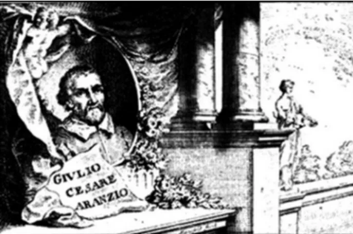
he irst description and denomination of the struc-ture, practically undisputed, is credited to Giulio Cesare Aranzio (Arantius) (Julius Caesar Arantius [Bononiensis]) (c. 1530-1589), an Italian anatomist and surgeon, and pupil of Vesalius1,6-8 (Figure 1). He described and named
the anatomical formation in a study of the human brain in the 1st issue of Anatomicarum Observationum Liber
(Book of Anatomical Observations), which appeared together with the 3rd revised edition of De Humano Foetu
Liber (Book on the Human Fetus) and the 1st version
of De Tumoribus Secundum Locus Afectus Liber (Book on Tumors According the Afected Site), published in 1587, compiled together into a single volume.1,2,6,9 he
Anatomicarum Observationum Liber, concerning the ventricles, choroid plexus and hippocampus, contains ive chapters: Chapter I and II describe the ventricles, choroid plexus, and the formation and storage of animal spirits; Chapter III, the main one, provides a description of the hippocampus or silkworm (vermis bombycinus) (caterpillar of the Bombyx mori moth) and its intraven-tricular location (inferior or temporal horn) (“ventricle of the hippocampus”); Chapter IV describes the proce-dure to reach the target structures, while in Chapter V he commented briely on the ventricles, including the hippocampal one, and the animal spirits produced there. Chapter III was featured in translated and commented form in papers by Lewis’10 and Tilney,11 whereas
Chap-ters I, III, and IV appeared as selections in the paper of Walther,12 presented here as excerpts from the texts in
their original and translated forms (Box 1).
Figure 1. Giulio Cesare Aranzio, from Bologna (illustration from Bram-billa, 1781).8
Box 1. Excerpts from Arantius`’ Anatomicarum Observationum Liber
(Chapters I and III)9 (translation checked against those of Lewis, Tilney,
and Walther).10,11,12
Arantius, regarding the ventricles, choroid plexus, and hippocampus, divided his text into five Chapters.
De cerebri ventriculis ab Hipocampo dominatis. Cap. I.
Praeter iam perspectos in cerebri susbstantia sinus, quos ventrícu-los appellare consueuimus,...duos insignes alios sinus, aut cauitates in peritioribus cerebri partibus recoditas,...qui a superiorum sinuum, aut uentriculorum magnitudine non admodum, recedunt;...Resident bi sub duobus illis uentriculis anterioribus,...quasi in subiecto nauigii alicuius abdito cubículo, latent, ad anterioraque, versus frontem pro-tendunt, tertioque, uel communi sinui,...quemadmodum & duo superi-ores,...atque in illum uelut cerebri centrum concurrunt.
Chapter I. On the cerebral ventricles named after the Hippocampus.
“In addition to the previously recognized sinuses (cavities) in the brain matter, which are commonly called ventricles…two other prominent sinuses or cavities are found, in hidden parts of the brain, laying deep-ly buried,…considerabdeep-ly smaller compared to the higher sinuses, or ventricles...Seated under the two anterior ventricles,…like a hidden chamber (cavity), undetected, and stretching out anteriorly, the third or common sinus (ventricle),...and as the two superior,…meet together in the center of the brain.”
De plexibus Choroidibus per eosdem sinus distributis. Cap. II.
Chapter II. On the Choroid plexus and their distribution in these cavities.
De cerebri particulis Hippocampum referentibus. Caput III.
Horum ventriculorum basi, quae intro ad medium respicit, candida insurgens supereminet, & quasi adnascitur substantia, quae ab in-feriori superficie, velut additamentum extollitur, psalloidique corpori, seu testudini est continua, ac per longitudinem, in anteriora, versus frontem protenditur inequalique, ac flexuosa figura predita est, quae Hippocampi, hoc est marini equuli effigie refert, vel potius, bombycini vermis candidi spinalis medullae initium hinc inde amplexantis, forma indicant, de cuius usu alibi dicemus: huius particula caput referens tertio vocato ventriculo proxima est, reflexum vero corpus in caudam abiens, ad anterior proteditur; quocirca ad superiorum differentiam, Hippocampi, vel Bombycini vermis ventrículos appellare libuit...
Chapter III. On those parts of the brain which constitute the hip-pocampus.
“At the base of the ventricles, a white growth rises up, as an expan-sion of matter that originates from the lower surface, like an attached elevation that faces the midline, continuous with the psalloid body [lyra] or tortoise [vault, fornix], and extends frontally along its length, displaying an unequal flexuous (curving, bending) shape, with resem-blance to a seahorse, the Hippocampus, or rather, to a white silkworm, which embraces the beginning of the medulla spinalis. Regarding the structure, it may be said from other experience: the part of the head is closely related to the third ventricle, the reflected (bent) body contin-ues as a tail, extending anteriorly; therefore, different from the supe-rior, it is pleased (preferred) to call it ventricles of the Hippocampus or of the silkworm (caterpillar of the Bombyx)…“
De ratione administrationis. Cap. IIII.
Chapter IIII. On the rationale of the procedure.
Quae in animalis spiritus generatione conueniant rationi. Cap. V.
THE FIRST ILLUSTRATION OF THE HIPPOCAMPUS
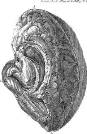
According to most authors, the irst drawing of the human hippocampus was provided by Johann [Johannes] Georg Duvernoy (Johannes Georgius Duvernoi) (1691-1759), a German anatomist and bota-nist.13,14 He wrote a short essay, De Sinibus Cerebri (On
sinuses [ventricles, cavities] of the brain), published in 1729 in the Commentarii Academiae Scientiarum Imperialis Petropolitanae (Commentaries of the Impe-rial Academy of Sciences of St. Petersburg), where he presented the text divided into four paragraphs (§1-§4), also denominating the structure as hippocampus or silkworm. It was illustrated with a drawing, Plate XII, of a right human dissected hemisphere, depicting the hippocampus and neighboring structures.15
his material was included as summarized descriptions and some original and translated excerpts (Box 2), as well as a igure with explanations (Figure 2).
Box 2. Excerpts from Duvernoy’s De sinibus cerebri (paragraphs
1-4).10,15,17
Duvernoy divided his text into four paragraphs.
§.1. Contains a citation of Galen’s comments on the ventricles, and a statement that Arantius was the first to observe and describe their structure in Anatomicarum Observationum Liber, and quoted Chap-ters I and III.
§.2. Deals further with the ventricles and the adjacent related struc-tures (corpus striatum, medulla oblongata and choroid plexus), and reiterates the finding of:
..ea fere hactenus neglecta fuisse: nam si sinum bombycinum oculi vidissent, miror quomodo res notatum digníssima praefato sinui in-clusa, quae ab Arantio detecta & titulo Hippocampi, vel Bombycini vermis descripta est, usque adeo obscure exposita fit.
“…hidden deeper cavities, like the neglected silkworm sinus (silk-worm ventricle), detected by Arantius and described under the name of Hippocampus or silkworm, which so far obscure became revealed.”
He commented also about the absence of an illustration.
§.3. Deals with the illustration he provided, describing the main axes of one hemisphere, detailing the characteristics and location of the pellucid septum (speculi lucidi), again mentioning Galen’s findings.
§.4. He described his procedure, ventricle exposure, and finding of the adjacent and internal structures (corpus striatum, part of the medulla oblongata, choroid plexus, fornix), as well as the silkworm, which he described, with apparent admiration:
In hoc facto particulam observa, qua in toto cerebro propter albedi-nem occaecantem fabricaeve elegantiam, pulchrior haud datur, puta Vermem bombycinum seu caput Hippocampi Arantius [C],...in cujus exteriore superfície, spiralium circum volutionum exsculpta sunt vestigia[ff]. Ad haec, quum tereti & ovali figura, usque ad duorum fere pollicum longitudinem, gaudeat, eo toto itinere ad vermis bombycini crassioris efigiem non nihil accedit, vel ad efigiem cornu arietini,….
“Thus, a small elegant structure of a dazzling whiteness may be ob-served, possibly nothing more beautiful in the whole brain as the silk-worm, or the head of the Hippocampus of Arantius [C],…in which the outer surface, appears with spiral winding carved tracks [ff] [prob-ably digitations]. For this purpose, the round and oval form, up to about two thumbs [inches] in length, appreciate, nothing resembles the coarse shape of the silkworm, like the figure of a ram’s horn,…”
He described the fornix (body, legs and arms) again, and its relation-ship with the hippocampus.
Finally, there is the explanation of the figure (his Plate XII), which depicts the drawing of a dissected right hemisphere, showing the hippocampus and neighboring structures (Figure 2).
A.A. Two apices of the hemisphere, with the sinus (ventricle) in the middle a.b.
B.B.B. Medullary limbus surrounding the sinus.
a.b. Sinus resembling the commonly known human ear coil [?].
c. sinus, resembling ear lobe, in posterior apex of the hemisphere [?].
d.e. Sinus of the silkworm or hippocampus of Arantius.C. Hippocampus or silkworm of Arantius.
ff. Circumvoluted spirals of the silkworm.
g. Curvature and slender part of the silkworm.
h. Part of the fornix.
COMMENTARIES
he pioneering description of Arantius, and much later Duvernoy’s depiction of the hippocampus, revealed a relatively small formation that would become one of the most valued brain structures. It should be noted that up to this point, outstanding anatomical researchers such as Galen, Vesalius, and Willis, had practically overlooked this formation, located deep among numerous other structures in the brain.10,12,16
Arantius was the irst to describe this structure, pro-truding from the loor of the inferior (temporal) horn of the lateral ventricle, which he denominated hippocam-pus, as to his mind it bore resemblance to a seahorse (or hippocampus, Greek: hippocampus [hippos=horse, kampus=sea monster]) or rather, to a white silkworm (bombycini vermis candidi) (white caterpillar of the Bom-byx).1,6,7,9,10 He provided a summarized explanation of the
technical procedure, where the dissection was performed with the aid of a bone knife and hands used to reach the deep structures, suggesting that he examined the brain through the exposed lateral ventricle, inspecting the temporal extension, thereby locating the hippocampus with its three parts – head, body, and tail. No illustration of the structure was presented. hus, his perception of a seahorse or of a silkworm remained rather unclear.1
Many controversies arose concerning the description and denomination, as well as alternative designations.6,10,11
However, the term hippocampus has endured until the present day, being the most widely used in the literature.1
Other terms emerged designating the structure or its component parts, which will be reviewed on another occasion.7,10,16,17
Duvernoy endorsed Arantius’ description of the ven-tricles and the structures therein, and additionally pro-vided an illustration of the hippocampus and surround-ing formations,15 regarded as the irst by most authors.
It must be stressed that it appeared more than one and a half century after Arantius’ description.
However, some authors have identiied other illus-trations and texts, which they claim predate Duvernoy’s depictions listed in chronological order of publication as follows: Andrea Vesalius (Andreas Vesalius) (1514-1564) in the 1543 edition of the De Humani Corporis Fabrica
(On the Fabric of the Human Body), presented drawings and text that might be identiied as pertaining to the hip-pocampus. he structure allegedly illustrated, though not unmistakably, was not described or named.7 It should be
noted that Vesalius clearly depicted and labeled only the fornix or tortoise, in a horizontally (axially) sectioned brain (e.g. Fifth Figure: S, T, V. Superior corporis instar for-nicis seu testudinis extructi superfícies,… (“S, T, V. Upper
body surface shaped like a fornix [vault] or tortoise…”).
X, X…, corpori testudinem referenti continua. (“X, X….the reported body of the tortoise continues.”),18 without any
reference to the hippocampus.
Costanzo Varolio (Constantius Varolius) (1543-1575), in his book De nervis opticis (On the optic nerves), pub-lished in 1573, supposedly presented a “rough sketch of the hippocampus”, but without any reference to this structure.10,17 he two plates displayed in this book,19
as far as can be seen, do not allow this formation to be distinguished.
homas Willis (homae Willis) (1621-1675), in the
Cerebri Anatome (Brain Anatomy) of 1664, Chapter X, Figure VII, neglected the structure as he described and depicted a dissected ovine (sheep) brain. However, a formation considered recognizable as the hippocampus was identiied,10 labeled as D.D. Corporis callosi margo,
qui caudicem medullarem prope Cerebellum amplexabatur.
(“D.D. Margin of the corpus callosum, which embraces the medullary stem close to the Cerebellum.”), closely related to C.C. Fornicis brachia, qui caudicem medullarem e regione glandulae pinealis amplexabantur. (“C.C. Arms [bra-chia] of the Fornix, which embraces the medullary stem and the pineal gland.”).20 he illustration displays a
dis-torted and unclear anatomy of the dissected brain. he recognition of the hippocampus is not convincing. he apparently same dissected brain is presented redrawn in his Anima Brutorum (he Soul of Beasts) in the 1672 edition, Plate V, with modiications, and changes in the labels,10,21 where the alleged structure now becomes
unrecognizable.
Bartolomeo Eustachio (Bartholomeus Eustachius) (c. 1510-1574) showed in the Tabulae Anatomicae
(Anatomical Plates), probably commissioned in 1552, but irst published by Giovanni Maria Lancisi (1654-1720) only in 1714, almost one and a half century after Eustachio’s death, a dissection that displayed a struc-ture presumed to be the hippocampus.10 However, this
dissection (Plate XVII, Figure V, legend on pp 43-44), depicts the median fornix and the posterior pillars seemingly fusing in an indistinct way and designated by him as the cornua (horns), labeled only in the 1717 edition (Plate VI, igure 5… Insuper fornicem in situ [… In addition to the fornix in situ], cujusprincipium [whose body], b. cornua verò [the real horns], c, c...(b=body of the fornix, c, c=horns).22,23 here is no mention of the
hippocampus. he distinction between the posterior pil-lars and the horns is not at all clear, as they appear as a single structure, constituting more an illustration of the fornix only.
irst drawing of the structure. If not the irst, it may be stated that it was a good depiction10 and the best and
most representative at the time.
As originally described, and remains so in the present day, the name “hippocampus” applies to the entire ven-tricular protrusion. Considering here one of the
hippo-campal formation deinitions, comprising the hippocam-pus proper, dentate gyrus and subiculum, as described above, Arantius and Duvernoy apparently described the gross anatomy of this complex. Further identiication of the component structures occurred later, and will be the focus of another study at a later date.
REFERENCES
1. Bir SC, Ambekar S, Kukreja S, Nanda A. Julius Caesar Arantius (Giulio Cesare Aranzi) (1530–1589) and the hippocampus of the human brain: history behind the discovery. J Neurosurg 2015;122:971-975. 2. Duvernoy H, Cattin F, Risold P-Y. The Human Hippocampus. 4th ed.
Berlin: Springer-Verlag, 2013.
3. Bartsch T. The Clinical Neurobiology of the Hippocampus. An Integrative View. Oxford: Oxford University Press, 2012.
4. Andersen P, Morris R, Amaral D, Bliss T, O’Keefe J. The hippocampal formation. In: The Hippocampus Book. Andersen P, Morris R, Amaral D, Bliss T, O’Keefe J editors. Oxford: Oxford University Press, 2007, pp 3-6. 5. Angevine Jr J. The development of the hippocampal region. In: The
Hippocampus. Volume I: Structure and Development. Isaacson RL, Pribram KH editors. New York: Plenum Press, 1975, pp 61-94. 6. Judaš M, Pletikos M. A note on the sea-horse in the human brain.
Translat Neurosci 2010;1(4):335-337.
7. Swanson LW. Neuroanatomical Terminology. Oxford: Oxford University Press. 2015, pp 296-299.
8. Brambilla GA. Storia delle scoperte fisico-medico-anatomico-chirur-giche fatte dagli uomini illustri italiani. Tom 2. Giulio Cesare Aranzio. Milano: Nel Imperial Monistero di s. Ambrogio Maggiore, 1781, pp188-198. [Retrieved from: https://books.google.com.br/books/ download/Storia_delle_scoperte_fisico_medico_anat.pdf?id=j2mmU-NVuw3gC&hl=ptBR&capid=AFLRE72pDS09gbo8EWsRy2gnLQCiAI fQZWDhXUWlBh9acxJVm4GbOomUxPnf5nBdjahfrFH6J2u9mdzpY jeNP_bLWmaSFY5Q&continue=https://books.google.com.br/books/ download/Storia_delle_scoperte_fisico_medico_anat.pdf%3Fid%3Dj2 mmUNVuw3gC%26hl%3Dpt-BR%26output%3Dpdf]
9. Arantius JC. De Humano Foetu Liber tertio editus, ac recognitus. Anatomicarum Observationum Liber, ac de Tumoribus Secundum Locos Affectos Liber. Venetiis: Apud Iacobum Brechtanum, 1578, pp 41-46. [Retrieved from: http://gallica.bnf.fr/ark:/12148/bpt6k606047/f2.image] Lewis FT. The significance of the term hippocampus. J Comp Neurol 1923;35:213-230.
10. Tilney F. The hippocampus and its relations to the corpus callosum. J Nerv Ment Dis 1939;89:433-513.
11. Walther O. Hippocampal terminology: concepts, misconceptions, origins. Endeavour 2002; 26(2):41-44.
12. Hirsch A. “Duvernoy, Johann Georg”. In: Allgemeine Deutsche Biographie 13. (1877), 2016. [Retrived from http://data.deutsche-biographie.de/rest/
sfz12158.pdf]
14. Mazolinni RG. Schemes and Models of the Thinking Machine (1662-1762). In: The Enchanted Loom: History of Neuroscience P. Corsi (editor),. Oxford: Oxford University Press 1991, pp 68-143, 198-200. [Retrieved from: https://www.academia.edu/11367646/_Schemes_
and_Models_of_the_Thinking_Machine_1662-1762_._In_P._Corsi_ editor_The_Enchanted_Loom_Chapters_in_the_History_of_Neurosci-ence._Oxford_Oxford_University_Press_1991_pp._68-143_198-200] 15. Duvernoy JG. De Sinibus Cerebri. Comm Acad Sci Imp Petropolitanae
1729;4:112-116. [Retrieved from: http://books.googleusercontent.com/ books/content?req=AKW5Qafqc_X_lQ8kIW-ReJhb81bZJ-16zsLAZJSoH LojCO7-KFTdnSAGa6HISzFaxZjdC2Qg9tOzFz9IYB8gXogb8BVdOrkUS- Bmz2B54tsTq-F23XgkfGgdRKWiegZs891rCVaretg89uVnbPfMvVpzlp-PwsWNxmL6oaILdU6j9wwYQLT_c0bHUm1SV6YQ2VVr84J74PIjlwR_ NFyw1XN0DptadUfA1EAQSu1Y51lXlIYaAwiVuPk2voh3vwtI1x__LlbbBng-SJUPTjrDXNro095DqPrJZ9GZlNSk9W7R-59b3p9D8_MsHU]
16. Hill A. The Hippocampus. Philosoph Trans Roy Soc B 1893;389-427. [Retrieved from: http://rstb.royalsocietypublishing.org/content/royptb/ 184/389.full.pdf]
17. Olry R. Métaphores zoologiques au sein des ventricules latéraux du cerveau, ou l’imagination au service de la linguistique. Hist Sci Méd 1991;25(3):221-224. [Retrieved from: http://www.biusante.parisdescartes. fr/sfhm/hsm/HSMx1991x025x003/HSMx1991x025x003x0221.pdf]. 18. Vesalius A. De Humani Corporis Fabrica Libri Septem. Basel: Oporinus. 1543. [Retrieved from: http://www.bvh.univ-tours.fr/B372615206_ 47294/B372615206_47294.pdf]
19. Varolio C. De nervis opticis. Padua: Paulum & Antonium, Meittus fratres, 1573. [Retrieved from: http://www.archive.org/details/constantiivaroliO Ovaro]
20. Willis T. Cerebri Anatome, cui Accessit Nervorum Descriptio et Usus. Londini: typis J. Flesher, impensis J. Martyn et J. Allestry, 1664. [Retrieved from: http://www.biusante.parisdescartes.fr/histoire/medica/resultats/? cote=05344x01&do=pdf]
21. Willis T. De Anima Brutorum quae Hominis Vitalis ac Sensitiva est, exerci-tationes duae. Londini: Prostant apud Gulielm. Wells, & Rob. Scot, 1672. [Retrieved from: http://www.biusante.parisdescartes.fr/histoire/medica/ resultats/?cote=05342&do=pdf]
22. Eustachio B. Tabulae Anatomicae. Lancisio JM illust & ed. Romae: Ex Officina typographica Francisci Gonzagae. 1714. [Retrieved from: http://books.googleusercontent.com/books/content?req=AKW5Q adgactdW1cpIQyZRjiRC-fJWcN-7DRht4wA5B8AzADcEKt7OuYL-piBpUkhvMoYNj_9Apsfl3WmouHmdYNwZANPEwig8D5o9DQSt6 sC7eDs8WIvsaXixSux9j-SNOXZjSyeIkpXDDbw-OOx1S4yKw2Xw- pOH4JaIadCYkthYT-Kmn2MaMLA3-WQN8CMCDLXckox_HlNNy- 588k9UYuVMic7qW1teA07zfej0veooRhBYioCBAEJy4x3dRhAQxp-WRAXEj4N66sd54dJUnDQYPnu4OyOOVLi7HhVNiw8tSKN4qPu8TvCQ] 23. Eustachio B. Tabulae anatomicae. Lancisio JM illust & ed. Colloniae