An interview with
How to cite this section: Hwang HS. An interview with Hyeon-Shik Hwang. Dental Press J Orthod. 2016 Jan-Feb;21(1):24-33. DOI: http://dx.doi.org/10.1590/2177-6709.21.1.024-033.int
Submitted: October 19, 2015 - Revised and accepted: November 03, 2015
Hyeon-Shik Hwang
It gives me great pleasure to interview Dr. Hyeon-Shik Hwang, an innovative orthodontist who has developed many creative techniques over his career. Dr. Hwang was born in Korea and received his DDS and PhD degrees from Yonsei University in Seoul. He is professor and chairman of the Department of Orthodontics at Chonnam National University School of Dentistry, Gwangju, Korea. Dr. Hwang, as a faculty at the university hospital, has maintained a successful clinical practice for more than 25 years. He has treated many adult patients focusing on esthetics and periodontal health and has developed many clinical techniques to improve the efectiveness and eiciency of treatment to the beneit of both the patient and practitioner. Dr. Hwang is also in-terested in the evaluation of facial asymmetry two- and three-dimensionally. As one of the early adopters of cone-beam volume imaging, he has given special emphasis on the management of surgical cases. He is married to Jung-Un Park with whom he has two sons. His favorite hobbies are photography and listening to music. When I was presented to him in a congress, it was a great pleasure meeting someone who I already admired for his singular work. Later on, his humbleness and knowledge made me marvel at him even more. I hope that all readers of Dental Press Journal of Orthodontics also enjoy the teachings from this brilliant Korean orthodontist! Guilherme Thiesen – interview coordinator
» DDS and PhD, Yonsei University, Seoul, Korea.
» Professor and Chairman, Chonnam National University, School of Dentistry, Department of Orthodontics, Gwangju, Korea.
» Director of Korean Adult Orthodontic Research Institute. » President of Korean Society of Surgery-irst Orthodontics. » Over 30 books and book chapters published.
» Over 130 papers published.
» Former Editor-in-Chief of the Korean Journal of Orthodontics (KJO). » Reviewer of various of scientiic journals, including the American Journal of
Orthodontics and Dentofacial Orthopedics (AJODO) and the Angle Orthodontist (AO).
DOI: http://dx.doi.org/10.1590/2177-6709.21.1.024-033.int
É uma grande satisfação poder entrevistar, pelo Dental Press Journal of Orthodontics, o Dr. Hyeon-Shik Hwang, um ortodontista inovador que tem desenvolvido muitas técnicas criativas ao longo de sua carreira. Dr. Hwang nasceu na Coreia do Sul e recebeu seus títulos de DDS e PhD pela Yonsei University, em Seul. Ele é professor e chefe do departamento de Ortodontia da Faculdade de Odontologia da Chonnam National University, em Gwangju, Coreia do Sul. Dr. Hwang é membro do corpo docente do hospital universitário, e tem mantido uma prática clínica bem-sucedida por mais de 25 anos. Ele tem tratado muitos pacientes adultos, com foco na estética e saúde periodontal, e desenvolveu técnicas clínicas para melhorar a eicácia e eiciência do tratamento, beneician-do tanto pacientes quanto proissionais. Dr. Hwang tem interesse cientíico especial na avaliação bidimensional e tridimensional de assimetrias faciais. Como um dos pioneiros a adotar a tomograia volumétrica de feixe cônico, acabou dando ênfase especial para o tratamento de casos cirúrgicos. Ele é casado com Jung-Un Park, com quem tem dois ilhos. Seus hobbies favoritos são a fotograia e a música. Quando fui apresentado a ele em um congresso, foi um grande prazer conhecer alguém que eu já respeitava, por seu trabalho singular. Mais tarde, sua humildade e inteligência me izeram admirá-lo ainda mais. Espero que todos os leitores do Dental Press Journal of Orthodontics também possam desfrutar dos ensinamentos desse brilhante ortodontista coreano!
Your knowledge about craniofacial asymmetries is singular, and your papers on 2D and 3D evalu-ation of these deformities are classic. How much do you think CBCT images have contributed to the diagnosis of dentofacial asymmetry? What was not possible to be properly analyzed in the past, with two-dimensional images? (Guilherme Thiesen)
Needless to say, CBCT has contributed very much to the diagnosis of dentofacial asymmetry. One of the main advantages of CBCT in the diagnosis of facial asymmetry is that frontal cephalograms can be generated in a standardized and reproducible manner.1,2 Although 3D image is available, it is believed that frontal cepha-lograms are better than 3D image for determining the presence and the degree of facial asymmetry. However, conventional radiographs have the inherent limitations of enlarging and distorting the image, and this can lead to misdiagnosis. It is because all cephalograms are obtained in perspective projection. In contrast, cephalograms can be generated in ‘parallel’ projection using CBCT volume image. Clear and accurate cephalograms without distor-tion and/or enlargement can be generated.3 We call this CG ceph (CBCT-generated cephalograms). In addition to accurate diagnosis, this imaging allows superimposi-tion of frontal cephalograms to be obtained. In the past, we had diiculty with the superimposition of frontal cephalograms due to a diference in head posture. Using computer algorithm, reorientation of second volume im-age into the same position, as in the initial volume imim-age, became possible. As a result, two CG cephs can be gen-erated in the same posture and angle. Standardized and reproducible cephalograms can be generated simply from CBCT volume images. Not only accurate diagnosis, but also precise treatment evaluation became possible with the help of this unique imaging modality, CG ceph.
How do you see the current status of CBCT use in orthodontic diagnosis and treatment planning by Asian clinicians as well as around the world? Do you think that published guidelines and clinical recom-mendations from the European SEDENTEXCT and the American Academy of Oral and Maxillofacial Radiology, as well as routine aspects like software and exam costs, the time consumed in diagnosis and even lack of training, have been inluencing the way orthodontists deal with 3D imaging? (Alexan-dre Trindade Simões da Motta)
Whether routine use of CBCT in orthodontic di-agnosis and treatment planning is justiied is one of the hottest issues in today’s Orthodontics. If somebody asks me about it, my answer would be: “It depends on the practitioner.” Many other clinicians will say it depends on the “case.” But my answer is that it depends on the “practitioner.” As we all know, CBCT examinations are justiied only when the beneits outweigh the risks. Here, the beneits from CBCT scan in orthodontic pa-tients are quite diferent according to the practitioner. The ability and knowledge to use scan data varies from practitioner to practitioner. If he or she has little knowl-edge on the use of CBCT scan data (e.g., just see the volume image, and sometimes observe MPR image), a routine taking of CBCT in orthodontic cases might be blamed as unethical.
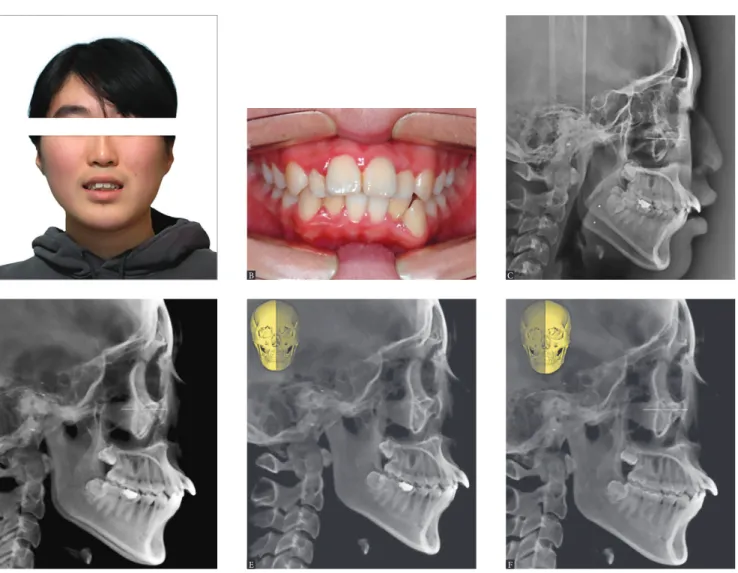
On the other hand, it should be respected if a de-cision of CBCT taking was made by a practitioner based on the risk-benefit analysis. I routinely take CBCT in my orthodontic practice. It is because I am certain that the benefits always outweigh the risks in my practice. A number of images can be generated simply once one single scan of CBCT is obtained. As an example, all cephalograms are made on the ba-sis of CBCT volume data.4 While cephalograms can be generated with perspective projection geometry for comparison with previous cephalograms taken with conventional cephalometer, cephalograms can be made also with parallel projection without any magnification and distortion. Accurate diagnosis is possible with CBCT-generated cephaolgrams (CG ceph). Uncertainty with perspective projection can be explained clearly with parallel projection of CG ceph. In addition, right side and left side can be generated separately, forming a unique image called ‘half ceph.’ Right and left difference can be evaluated clearly and simply by using a pair of half cephs (Fig 1).
Figure 1 - Generation of cephalograms from cone-beam volume image. A 20-year-old lady came to us complaining of upper anterior protrusion (A and B). Cepha-lometric radiograph showed a significant difference in mandibular outline between the right and left side (C). Is this a real asymmetry? If so, which one is right or left? These questions cannot be answered with conventional lateral ceph, whereas they can be answered with cone-beam CT (CBCT). CBCT-generated cephalograms (CG Ceph) revealed no significant asymmetry (D). In order to differentiate right and left side clearly, right and left side half cephs were generated separately. It was revealed that the pattern of Class II high angle is more evident on the right side. Everything is clear with the help of half ceph generation (E and F).
However, it should be noted that this treatment evaluation during treatment is only possible when we have initial data. During the early stages of my 3D imaging, CT/CBCT was scanned in selected cases. Nevertheless, I had a difficulty with progress assessment during treatment of patients with no ini-tial scan data. First, it happened in part of my pa-tients, but it has become always the case with in-creasing ability on the use of CBCT scan data.
It is obvious that treatment quality is improved with the help of CBCT imaging. This is due to the fact that imaging is used not only for diagnosis, but also for progress evaluation during treatment. Un-like other dental treatments, progress evaluation during treatment is quite important in Orthodon-tics, and this is only possible with comparison with ‘initial’ data. There is an obvious trend that the ben-efits outweigh the risks (Fig 3).
D
B
E
C
What are the great indings from your papers on craniofacial asymmetry that an orthodontist should keep in mind when analyzing a patient with this kind of disharmony? (Guilherme Thiesen)
My decision in diagnosis and treatment planning is based on the philosophy of minimally invasive Den-tistry, and there is no exception for patients with fa-cial asymmetry. As an example of chin deviation, how much deviation needs to be corrected? Based on men-ton deviation, two degrees or four degrees? All sub-jects with menton deviation over 4 degrees should be treated? Treatment decision needs to be based not only on the degree of asymmetry, but also on patient’s per-ception of asymmetry. In some cases, patient’s percep-tion comes irst. If asymmetry afects dental occlusion
and oral health signiicantly, it should be corrected. However, if not, treatment decision needs to be based on patient’s perception.
For accurate diagnosis and treatment planning, more material is used in general. Considering the nature of asymmetry in the human body, more diagnostic ma-terial reveals more asymmetry. However, it should be noted that the use of more material is not for more treat-ment, it is for accurate diagnosis and understanding of the nature of asymmetry. This is really important in a patient with mild asymmetry. Some patients unduly worry about their asymmetry which is within normal limit. Just a verbal explanation such as “you do not need correction” does not work well in this kind of patient. If the patient does not understand the nature of his or Figure 3 - Risk-benefit analysis on the use of CBCT in orthodontic practice. If a medical CT is used to obtain 3D image, the benefit usually does not outweigh the risk (A). However, the CBCT has reduced the risk considerably, so that the benefit may or may not outweigh the risk depending on the cases (B). With the development of computer technology, the benefit has outweighed the risk. However, this analysis might work only for practitioners who have a basic knowledge and ability to use CBCT scan data appropriately (C).
Figure 2 - Construction of composite cepha-lograms (composite ceph) from cone-beam volume image. In order to visualize condylar position in cephalograms, overall head and seg-mental view of the condyle area were captured in the 3D image program. The segmental view was created by removing overlapping area us-ing the clippus-ing and sculpt functions of the pro-gram (A). The captured images were imported into Photoshop, and color was inverted for vis-ibility (B). Frontal cephalograms with highlighted condyles were generated by overlapping the two images, overall and segmental (C). After treat-ment, composite ceph can also be generated in the same way as in initial composite ceph (D). If we make an animated GIF using the two imag-es, before and after composite cephs, treatment changes can be evaluated precisely and dynam-ically: this animated GIF (C and D) is available at http://goo.gl/sQ7R2B
A
C
B
D
A B C
Beneit
Beneit
Risk Risk
her asymmetry, he or she might go and see other cli-nician, such as a plastic surgeon. For the management of this kind of patient, CBCT 3D analysis is crucial. The detailed explanation of the exact nature of their asymmetry could alleviate patients’ concerns. It should be stressed that recently-developed 3D analysis is not for the practitioner, but for the patient.
You have developed, by cluster analyses, a clas-siication system for facial asymmetries, admit-tedly a great challenge for clinical Orthodontics. This classiication establishes four groups (based on menton deviation and ramal length diferences) and advocates treatment protocols for each one of them. Do you think that every type of facial asym-metry can be included in one of these groups? Which group is the most prevalent? Which group presents the easiest and the toughest treatment for the clinician? (Guilherme Thiesen)
Patients with facial asymmetry can be classiied into four groups: RM, M, RA, and B types, as de-scribed in Figure 4.5,6 This classiication is really use-ful in orthodontic practice because the causes of facial asymmetry can be identiied easily. Moreover, the clas-siication is possible just by using frontal cephalograms. Only two variables, menton deviation and ramus length
diferences, are needed. From this classiication, accu-rate diagnosis can be made and a proper treatment plan can be established according to the type (Fig 4).
Can all subjects with facial asymmetry be included in one of these groups? My answer is yes and no, more accurately ‘yes’ in orthodontic practice and ‘no’ for oral surgeon’s practice which is dealing with craniofacial deformities. Nearly all asymmetry patients are classi-ied into one of these four groups. On the other hand, it needs to be realized that all patients concerning asym-metry are not included into one of these groups. Ac-cording to unpublished data, 91% of orthodontic pa-tients who visited a university hospital and had frontal cephalograms taken for diagnostic purposes were classi-ied into four groups. The remaining patients (9%) were diagnosed as being within normal limits.5
Considering the causes of each type of asymmetry, RM type is the toughest case in terms of treatment. The prognosis is questionable because the cause of this asymmetry is condylar growth diference between the right and let side. On the other hand, M type of asym-metry is the easiest case. Once any prematurity which may result in functional shit of mandible is removed, a balanced facial growth can be expected. While RM type is the toughest asymmetry, this is the most preva-lent type of asymmetry in orthodontic clinic.5,6
Figure 4 - Facial asymmetry can be classi-fied into four groups: RM (Ramus Menton), M (Menton), RA (Ramus Angle), and B (Bulkiness). These groups are based on menton deviation and ramal length differences on frontal cepha-lograms. This classification allows the clinician to determine the cause of a given asymmetry and to formulate a proper treatment strategy for facial asymmetry patients.
Menton deviation
Ramus length diference (Rt / Lt)
yes
yes
Type
Causes
Ramus Menton type
Asummetric condylar growth
Growth modiication Removal of prematuriy
Camoulage treatment
Orthognathic surgery
Restoration of
bilateral mastication Explanation
Plastic surgery Functional
shit of mandible Unilateralmastication Not speciic Menton type Ramus Angle type Bulkiness type
Treatment
yes no
no
Do you indicate surgery-irst orthognathic treat-ment for all your patients with marked skeletal dis-harmony in the three planes of space?
(Telma Martins de Araújo)
The surgery-irst (SF) approach demands more care-ful surgical planning and stronger collaboration be-tween skilled orthodontists and surgeons in order to accurately predict post-surgical tooth movement and surgical movement. Therefore, previous advocates of this approach recommend only using the SF approach for mild to moderate skeletal discrepancies. However, the scope of this approach has been expanding with ad-vances in 3D imaging technology and virtual orthodon-tic and surgical simulation.7
Another important issue is that unstable occlusion is inevitable ater surgery in the SF approach. This could lead to surgical instability and interfere with subsequent orth-odontic treatment. At the early stages of my SF practice, only selected cases with tripod or at least bilateral contact in the state of surgical occlusion were treated by using the SF approach. However, presently, unstable occlusion can be managed properly with the use of continued splint wear with stepwise modiication, as illustrated in Figure 5. Un-wanted mandibular shiting can be prevented by using sur-gical splint continuously. More and nearly all patients can beneit from the SF approach (Fig 5).
In general, the advantage of surgery-irst ortho-dontics is to avoid temporary deterioration in pa-tient’s appearance during the pre-surgical phase. In your experience, this is the key point of indica-tion of this technique? What are the main diicul-ties that you have had in the inalization of these cases? (Maria Perpétua Mota Freitas)
It is certain that patients appreciate immediate im-provement in facial appearance with the help of surgery-irst (SF) orthodontics. However, the most important thing is that orthodontic tooth movement is easier and more physiologically favorable ater surgical elimination of skeletal disharmony. It is because the direction of tooth movement for decompensation is not against sot tissue pressure. This is a beneit not only to the patient, but also to the practitioner. We have no diiculties even during the inalization phase.
On the other hand, maintaining the condylar position during surgery is absolutely essential. In the conventional three-stage surgical orthodontic approach, minor changes in condylar position oten go unnoticed because pre-sur-gical orthodontics enables stable occlusion to be routinely obtained ater surgery. However, in the SF approach, even minor changes in the condylar position can cause unwant-ed shiting of the mandible due to the relatively unstable occlusion. Therefore, it is essential to monitor changes
in condylar position before and ater surgery. If a signii-cant change is detected in post-surgery CBCT imaging, it is necessary to have the patient wearing a removable splint with continuous adjustment until stable occlusion is achieved and stable condylar position is obtained through the remodeling process.
Considering the orthodontic phase after the sur-gery-irst procedure, what would be your major concerns about the way patients face treatment? In other words, do you think the surgery-irst crite-ria promote a diferent psychological impact from conventional orthodontic-surgical treatment, thus inluencing patients’ overall compliance (appoint-ment non-attendance rate, hygiene, appliance breakage) and expectations?
(Alexandre Trindade Simões da Motta)
The conventional three-stage surgical orthodontic ap-proach, which includes pre-surgical orthodontics, surgery, and post-surgical orthodontics, has been well established as the gold standard in most cases. However, one of the drawbacks is the long pre-surgical treatment time that typically worsens facial appearance and exacerbates mal-occlusion. During the pre-surgical orthodontic period, all tooth movement is against sot tissue pressure. The teeth are leveled to a lat occlusal plane, relative to their own arches. Although the resulting occlusion facilitates proper positioning of the jaws, patients will experience discom-fort throughout treatment of pre-surgical stage. Treatment does not improve quality of life, but rather deteriorates it, at least before surgery. In addition, patients become in-creasingly anxious about surgery under general anesthesia as the date of the surgery approaches. On the other hand, everything is completely the opposite in the surgery-irst approach. I incorporated it into my practice in 2009. The last six years of experience have demonstrated greater pa-tient satisfaction using the SF approach in surgical ortho-dontics. All of my patients appreciate treatment which is SF approach. Overall patients’ expectations for SF orthodon-tics are beyond imagination, although their compliances are similar to those in conventional surgical orthodontics.
Still on surgery-irst orthodontics, how do you work with Spee curve in the treatment of asymme-tries, knowing that this is a limiting factor for man-dibular positioning during surgery?(Maria Perpé-tua Mota Freitas)
In the surgery-irst approach, occlusion cannot be used as a guide for surgical movement, and the surgeon is limited by tooth position in correcting skeletal discrepancy. This is really true in severe asymmetry cases which show deep Spee curve and dental compensation in the transverse dimension. Unlike surgeons, orthodontists can aford to simulate post-surgical orthodontic treatment. Accurate post-post-surgical tooth movement and surgical movement can be predicted, even in patients with severe skeletal discrepancy. Figure 6 shows an example of skeletal Class III with severe asymmetry treated by means of the SF approach. Although the patient showed severe asymmetry with deep Spee curve on one side, this did not act as a limiting factor for mandibular positioning dur-ing surgery. It is believed that nearly all patients can beneit from SF orthodontic treatment (Fig 6).
Recently, the use of mini-implants is becoming a routine in orthodontic treatment. This is also your routine, or do you have some restriction or speciic indications for these accessories? (Maria Perpétua Mota Freitas)
Not any appliance can be used as a routine in orthodontic treatment, and mini-implants are no exception. It should be realized that mini-implant is not an orthodontic appliance, but merely an anchorage device. It is certain that there is a trend towards overuse of this device. Although the devices are very efective as a sure anchorage, they should be used only when necessary. If we overuse some devices habitually, we may misuse them. Indiscriminate use of this anchorage device can cause an imbalance in force system which can be a source of malpractice.
What is your experience with intrusion of posterior teeth using skeletal anchorage (mini-implants or mini-plates) to correct anterior open bite maloc-clusions in order to avoid orthognathic surgery?
(Telma Martins de Araújo)
In the last AAO Congress held in San Francisco, USA, you gave a lecture on the use of the mini-tubes appliance (MTA) for alignment of anterior teeth. What are the indications and contraindica-tions of this technique? What are the diferences in using it in the buccal or in the lingual surfaces of anterior teeth? (Telma Martins de Araújo)
The mini-tube appliance (MTA), a round tube with diameter of 0.018-in and length of 3 mm, has been de-signed especially for anterior teeth alignment.8 It was originally developed to be used in young adults seeking a rapid improvement in their anterior esthetics. With the combined use of light NiTi wire and interproximal
stripping, rapid alignment can be obtained within a very short period of time by using the MTA. Before the in-troduction of MTA, many patients with uneven ante-rior teeth received ‘instant orthodontics’ which is not orthodontic treatment, but ceramic veneer treatment by some of cosmetic dentists.9
While the MTA was used mostly in young adults during the early stages of MTA development, the scope of the MTA has expanded to all age groups of patients who need low-proile appliances and/or light force ap-plication. However, the appliances are used only in non-extraction cases because retraction of anterior teeth can-not be carried out by the MTA. On the other hand, Figure 6 - A case example of skeletal Class III malocclusion with severe asymmetry treated with surgery-first approach. Although the patient showed a severe asym-metry and concomitant dental compensation in the transverse dimension, surgical occlusion could be obtained after simulation of post-surgical orthodontic tooth movement. After elimination of skeletal disharmony by two-jaw surgery — that included maxillary advancement and differential mandibular set-back using a surgery-first approach —, overall alignment and leveling by fixed orthodontic treatment was obtained so rapidly. Please note that Spee curve on the left side was relieved at the early stage of post-surgical orthodontic treatment. A, initial; B, surgical occlusion; C, two months into fixed treatment; D, seven months into fixed treatment.
A
C B
Figure 7 - A new appliance for rapid alignment. Mini-tube appliance (MTA), a round tube with diameter of 0.018-in and length of 3mm, has been especially de-signed for anterior teeth alignment. With the combined use of light NiTi wire and interproximal stripping, alignment can be obtained so rapidly. For this reason, MTA has been suggested as a sure alternative to ceramic veneers which require a considerable amount of tooth reduction.
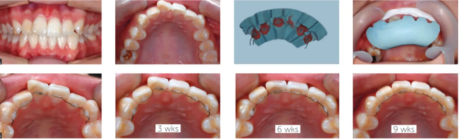
Figure 8 - Lingual application of tube appliance (MTA). The tubes can be placed also in the lingual surface. However, one potential problem for mini-tube is that a wire cannot be inserted easily into the mini-tube, particularly in case of crowding. To overcome this limitation, a unique indirect bonding technique, named Indirect Bonding with Wire, has been developed. The tubes are attached to the models with an active wire, usually 012 NiTi. Not only the tubes, but also the wire can be transferred into the mouth using the indirect bonding tray. A) Indirect bonding; B) treatment progress.
A
B
3 wks 6 wks
3 wks 6 wks
3 wks
3 wks 6 wks 9 wks
some orthodontists prefer to use the MTA for initial alignment of anterior teeth even in extraction cases due to the characteristic of rapid alignment. It is interesting to note that anterior teeth are aligned with good gingi-val line leveling with the MTA, even without the use of rectangular wire (Fig 7).
With regards to labial or lingual application, it usu-ally depends on the patient’s demand. If a patient wants alignment with esthetic manner, the appliances are placed on the lingual surfaces. Sometimes, the MTAs are applied to lingual surfaces regardless of patient’s de-sire. Also in this case, the appliances can be placed with-out any discomfort to the patient because the thickness of the appliance is very small, only 0.65 mm. One par-ticular advantage of this unique appliance is that the ap-pliance can be used as a retainer as it is ater alignment, indicating no additional work for the retainer (Fig 8).10,11
REFERENCES
1. Hwang HS, Lee KH, Park JY, Kang BC, Park JW, Lee JS. Development of posteroanterior cephalometric analysis for the diagnosis of facial asymmetry. J Korean Dent Assoc. 2004;42:219-31.
2. Hwang HS, Hwang CH, Lee KH, Kang BC. Maxillofacial 3-dimensional image analysis for the diagnosis of facial asymmetry. Am J Orthod Dentofacial Orthop. 2006 Dec;130(6):779-85.
3. Sun MK, Uhm GS, Cho JH, Hwang HS. Use of Head Posture Aligner to improve accuracy of frontal cephalograms generated from cone-beam CT scans. Korean J Orthod. 2009 Oct;39(5):289-299.
4. Hwang HS, Lee KM, Uhm GS, Cho JH, McNamara JA Jr. Use of Reference Ear Plug to improve accuracy of lateral cephalograms generated from cone-beam computed tomography scans. Korean J Orthod. 2013 Apr;43(2):54-61. 5. Hwang HS. A new classiication of facial asymmetry. In: McNamara JA, editor.
Early orthodontic treatment: is the beneit worth the burden? Ann Arbor: Center for Human Growth and Development, The University of Michigan; 2007. p. 269-94. Craniofacial Growth Series, vol. 44.
6. Hwang HS, Youn IS, Lee KH, Lim HJ. Classiication of facial asymmetry by cluster analysis. Am J Orthod Dentofacial Orthop. 2007 Sep;132(3):279.e1-6.
7. Hwang HS, Oh MH, Oh HK, Oh HS. Surgery-irst approach in correcting skeletal Class III malocclusion with mandibular asymmetry. Am J Orthod Dentofacial Orthop. In press.
8. Hwang HS, Jeon HR, Kim SP, Kim WS, Lee GH. A new orthodontic appliance for rapid anterior alignment in adults; Mini-Tube Appliance (MTA). J Korean Dent Assoc. 2011;49:334-45.
9. Christensen GJ. Are veneers conservative treatment? J Am Dent Assoc. 2006 Dec;137(12):1721-3.
10. Hwang HS. Indirect bonding techniques in orthodontics. In: Hardin JF, editor. Clark’s Clinical Dentistry. Chicago: Mosby-Year Book; 1998. p. 1-19, vol 2, chap. 23C.
11. Hwang HS. Lingual Mini-Tube Appliance (MTA). In: Park YC, editor. Contemporary lingual orthodontics. Seoul: DaehanNarae; 2015. chap. 16, p. 299-316.
Guilherme Thiesen
» Professor, Universidade do Sul de Santa Catarina (UNISUL), Department of Orthodontics,
Florianópolis, Santa Catarina, Brazil.
» PhD in Dentistry, Universidade Luterana do Brasil (ULBRA), Canoas, Rio Grande do Sul, Brazil. » MSc in Orthodontics and Facial Orthopedics,
Pontifícia Universidade Católica do Rio Grande do Sul (PUCRS), Porto Alegre, Rio Grande do Sul, Brazil. » Diplomate by the Brazilian Board of Orthodontics
and Dentofacial Orthopedics (BBO).
Telma Martins de Araújo
» Full professor, Universidade Federal da Bahia (UFBA), Department of Orthodontics, Salvador, Bahia, Brazil. » Coordinator of UFBA José Édimo Soares Center
of Orthodontics and Facial Orthopedics, Salvador, Bahia, Brazil.
» MSc and PhD in Orthodontics, Universidade Federal do Rio de Janeiro (UFRJ), Rio de Janeiro, Rio de Janeiro, Brazil.
» Associate editor, Dental Press Journal of Orthodontics.
» Former chairman of the Brazilian Board of Orthodontics and Facial Orthopedics (BBO).
Maria Perpétua Mota Freitas
» Adjunct professor, Universidade Luterana do Brasil (ULBRA), Canoas, Rio Grande do Sul, Brazil.
» PhD in Dentistry, Pontifícia Universidade Católica do Rio Grande do Sul (PUCRS), Porto Alegre, Rio Grande do Sul, Brazil.
» MSc in Orthodontics and Facial Orthopedics,
Pontifícia Universidade Católica do Rio Grande do Sul (PUCRS), Porto Alegre, Rio Grande do Sul, Brazil. » Scientific editor of Stomatos Journal.
Alexandre Trindade Simões da Motta
» Adjunct professor, Universidade Federal
Fluminense (UFF), School of Dentistry, Niterói, Rio de Janeiro, Brazil.
» Chairman of the graduate program in Orthodontics, Universidade Federal Fluminense (UFF), School of Dentistry, Niterói, Rio de Janeiro, Brazil.
» Specialist, MSc and PhD in Orthodontics,
Universidade do Estado do Rio de Janeiro (UERJ), Rio de Janeiro, Rio de Janeiro, Brazil.