BASIC RESEARCH
The therapeutic effect of a pulsed electromagnetic
field on the reproductive patterns of male Wistar rats
exposed to a 2.45-GHz microwave field
Sanjay Kumar, Kavindra Kumar Kesari, Jitendra Behari
Bioelectromagnetics Laboratory, School of Environmental Sciences. Jawaharlal Nehru University/New Delhi, India.
INTRODUCTION:Environmental exposure to man-made electromagnetic fields has been steadily increasing with the growing demand for electronic items that are operational at various frequencies. Testicular function is particularly susceptible to radiation emitted by electromagnetic fields.
OBJECTIVES:This study aimed to examine the therapeutic effects of a pulsed electromagnetic field (100 Hz) on the reproductive systems of male Wistar rats (70 days old).
METHODS: The experiments were divided into five groups: microwave sham, microwave exposure (2.45 GHz), pulsed electromagnetic field sham, pulsed electromagnetic field (100 Hz) exposure, and microwave/pulsed electromagnetic field exposure. The animals were exposed for 2 hours/day for 60 days. After exposure, the animals were sacrificed, their sperm was used for creatine and caspase assays, and their serum was used for melatonin and testosterone assays.
RESULTS: The results showed significant increases in caspase and creatine kinase and significant decreases in testosterone and melatonin in the exposed groups. This finding emphasizes that reactive oxygen species (a potential inducer of cancer) are the primary cause of DNA damage. However, pulsed electromagnetic field exposure relieves the effect of microwave exposure by inducing Faraday currents.
CONCLUSIONS: Electromagnetic fields are recognized as hazards that affect testicular function by generating reactive oxygen species and reduce the bioavailability of androgen to maturing spermatozoa. Thus, microwave exposure adversely affects male fertility, whereas pulsed electromagnetic field therapy is a non-invasive, simple technique that can be used as a scavenger agent to combat oxidative stress.
KEYWORDS: Microwave; Caspases; Creatine kinase; Testosterone; Infertility.
Kumar S, Kesari KK, Behari J. The therapeutic effect of a pulsed electromagnetic field on the reproductive patterns of male Wistar rats exposed to a 2.45-GHz microwave field. Clinics. 2011;66(7):1237-1245.
Received for publication onDecember 10, 2010;First review completed onJanuary 12, 2011;Accepted for publication onMarch 1, 2011
E-mail: [email protected]
Tel.: 91 11-26704323
INTRODUCTION
The electromagnetic fields emitted from various sources (e.g., microwave ovens, mobile phones) have been reported to have causative effects on biological systems.1Electromagnetic fields have many effects on biological systems, but the testes are more sensitive to a variety of stresses, such as hyperther-mia, inflammation, radiation, and exposure, which lead to germ cell apoptosis.2,3 Exposure to various frequencies present in the environment can evoke a number of alterations at the cellular level, such as increased Ca2+efflux,4
reduced melatonin secretion,5disturbed antioxidant enzyme balance,6 and the induction of micronuclei.7 Microwave exposure
disrupts the seminiferous tubules and reduces the Leydig cell population and testosterone concentration in rats while increasing the LH level.8It is well established that testosterone is essential for spermatogenesis and the formation of spermatozoa.9
During spermatogenesis, apoptosis plays a critical role in adjusting the appropriate number of proliferating germ cells associated with the surrounding Sertoli cells. The regulation of apoptosis is based on the intracellular dominance of various proteins that induce or inhibit apoptosis, such as BAX, Bcl, caspase-3, and several key enzymes.10Apoptosis
is a cell death process that is characterized by blebbing, the loss of the cell membrane, asymmetry, cell detachment, cell shrinkage, nuclear fragmentation, chromatin condensation, and chromosomal DNA fragmentation, which are elimi-nated by phagocytes.11 Caspases are present as inactive precursors and are activated by caspase initiators through auto-active proteolysis.12The caspase 8 and 9 initiators and the caspase 3 effector are considered to be the main
phosphate, thus maintaining an immediately accessible energy reservoir in the cell.16Creatine phosphokinase, which is a key enzyme in transport and energy synthesis, supplies ATP to the sperm.17 Sperm are frequently associated with
elevated activities of certain key enzymes, including creatine kinase, which indicate male infertility.18-20
In most cases of infertility, high concentrations of reactive oxygen species (ROS) have been reported.21Kumar et al7also reported an increased level of ROS due to microwave exposure. Oxidative stress is a cellular condition of increased ROS concentrations that causes molecular damage and leads to various diseases.22,23 However, ROS are neutralized by
melatonin and its stimulatory effects on antioxidants. Melatonin is synthesized from serotonin in the pineal gland, and its level increases during the dark period and decreases during the light period. It has been reported that melatonin synthesis is suppressed by low-frequency EMF.24Brainard et al25have shown that melatonin synthesis and secretion from
the pineal gland are under the control of visible light. In this study, a pulsed electromagnetic field (PEMF) was used to relieve the effects caused by microwaves. Following the experiments of DiCarlo et al26on chick embryos exposed to radiation, the protection conferred by the magnetic fields appears to influence free radical scavenging. PEMF has a variety of biological effects, such as its effects on bone healing,27,28pain relief,29and the balance of the neuroendo-crine system (including hormone production and melatonin levels).30 PEMF has frequencies at the lower end of the electromagnetic spectrum (from 6 Hz to 500 Hz). In this study, we demonstrate that PEMF can be used as healing agent that activates the normal physiology of the body. PEMF activates normal metabolic processes, indirectly acts through the endocrine system, and controls the main cause of stress in the exposed system. We treated rats that had previously been exposed to 2.45 GHz radiation with PEMF. As biomarkers of this treatment, we chose the following parameters: melato-nin, creatine kinase activity, caspase assay, and testosterone.
MATERIALS AND METHODS
Animals
Seventy-day-old male Wister rats (190¡10 g) were used in
this study. The animals were maintained in accordance with national guidelines and protocols. The study was approved by the Institutional Animal Ethics Committee (IAEC-JNU/ 83/675-687; code no. 12/2008). A flowchart of the entire methodology is shown in Figure 1. The animals were housed in clean polypropylene cages and maintained at a controlled temperature with a constant 12-h/12-h light/dark schedule. The animals were fed a standardized normal diet (Tetragon Cheime Private Limited, Bangalore) and waterad libitum.
Animal Exposure
The rats were divided into five groups (Figure 1): Group I, microwave (MW) sham; Group II, MW exposure (2.45 GHz);
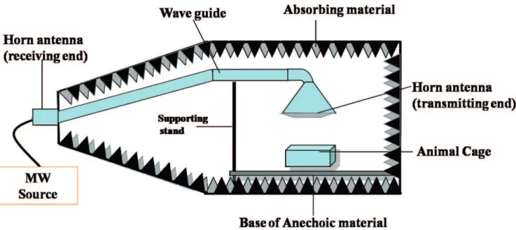
placed vertically such that all of the animals were irradiated homogeneously at a single power level. The rats were exposed to a 2.45-GHz radiation source at a 50-Hz modulation frequency (input, 1080 W; output, 700 W). A microwave oven (Haier India Co. Ltd, made by PRC (China); model no. HR-18MS1) was used as the source of exposure. Exposure was performed through the horn antenna for 2 h per day for 60 days (Figure 2).
Specific Absorption Rate (SAR) Calculation
The emitted power of the microwaves was measured by a power meter, which is a peak-sensitive device (RF power sensor (6900 series) and IFR 6960 B RF power meter; made by Aeroflex, Inc., Wichita, Kansas, USA). Every day, the cage was placed in the same position below the horn antenna, and the same numbers of rat positions were reshuffled. A similar experiment was performed with the MW sham but without energizing the system. A similar experimental setup was adopted previously by Paulraj and Behari31,32and Kesari and Behari.33A full description of the exposure setup has been discussed by Kesari et al.34 The SAR was calculated for the average size of the small animals by following the work of Durney et al.35For a plane-wave exposure with a random polarization and a power density of 0.21 mW/cm2, the SAR is 0.014 W/kg.
Materials
The caspase 3 assay kit (colorimetric; cat. no. CASP-3-C) was purchased from Sigma, USA. The creatine assay kit (cat no. K635-100) was purchased from BioVision Research Products (Mountain View, CA, USA), the ELISA melatonin kit (cat. no. E90908Ra) was purchased from Uscn Life Science Inc. (Wuhan, China), and the testosterone EIA kit (cat. no. 582701) was purchased from the Cayman Chemical Company (Ann Arbor, MI, USA). The remaining chemicals were purchased from Merck Chemicals, India.
Melatonin Assay
minimum detectable dose of rat melatonin is typically less than 1.02 pg/mL.
Creatine Kinase Assay
A CK kit was used. The seminal plasma was removed by washing the sperm sample with ice-cold imidazole buffer (0.15 M NaCl and 0.03 M imidazole (pH 7.0) at a ratio of 1515). The supernatant was decanted after centrifugation at
500g, and the pellet was re-suspended in a 0.1% Triton X-100 detergent solution by vortexing for 20 seconds. The sample was centrifuged again at 500g,and the CK activity of the supernatant was analyzed. In this assay, the creatine is enzymatically converted into sarcosine, which is then specifically oxidized to generate a product that converts a colorless probe to an intensely red product. This final product is detected colorimetrically (lmax= 570 nm). The CK
activity is expressed in international units/108spermatozoa. Measurement of Caspase 3 Activity
The activity of caspase 3 was also measured using an assay kit. The spermatozoa were centrifuged at 300g for 10 min at 4
˚
C. The pellet was then re-suspended in lysis buffer for 20 min and centrifuged at 20,000gfor 20 min at 4˚
C, and the supernatant was collected. The assays were conducted in 96-well plates. The colorimetric caspase 3 assay is based on the hydrolysis of the peptide substrate acetyl-Asp-Glu-Val-Asp p-nitroanilide (Ac-DEVD-pNA), which results in the release of the p-nitroaniline (pNA) moiety. To assess the specific contribution of caspase 3 activity, the Ac-DEVD-pNA substrate (2 mM) was added to each well according to the instructions of the manufacturer.The plates were incubated overnight at 37
˚
C to measure the caspase 3 activity. The absorbance was measured using a microplate reader (Spectromax M2) at 405 nm. The caspase 3activity is expressed inmmol of released pNA per min per ml of cell lysate at 37
˚
C.Testosterone Assay
A serum testosterone assay was performed using the testosterone EIA kit (Cayman, USA). The procedures for the assay were followed as described in the manual of the manufacturer. In brief, 50ml of the testosterone standard or serum, 50ml of the testosterone AChE tracer, and 50ml of the testosterone antiserum were added to the wells of an ELISA plate containing 100ml of EIA buffer. The sensitivity of the assay was 6 pg/ml. The optical density was read using a spectrophotometer (SpectroMax M2) that was sensitive at a wavelength range of 405-420 nm.
Pulsed electromagnetic field therapy
Immediately after exposure to the 2.45-GHz radiation, the Group-V animals (MW+PEMF) were exposed to PEMF for two hours daily. Three animals were exposed at a time. The rats were placed into a cage (28661726115 mm) ventilated
by one-cm-diameter holes. The animals were exposed in a mu-metal box at 100 Hz (Pulsed Magnetic Field Inventors Groups for Lawson Health Research, London, Ontario). The Mu box (38663336206 mm) was lined with opaque black
plastic and fitted with acrylic rods to transmit light into the enclosure (Figure 3). The box was made of 1.6-mm-thick mu-metal (Magnetic Shield, Bensenville, IL), with four rectangular Merritt-like configurations in the box. The coils
were 0.32 mm in diameter, had a thin layer of enamel insulation (Belden, St. Louis, MO), and were framed on rectangular plastic formers (30 cm617.8 cm) with 150 turns.
The four coils were spaced 9.7 cm apart from one another. The coils were connected in parallel pairs. Each pair was connected to one channel of a two-channel amplifier, and the connection leads were connected to a distribution unit that permitted individual coil-phase selection. The coils were electrically shielded with conductive silver paint and copper foil to minimize the introduction of an electric field. An in-house-built arbitrary function signal generator was configured to produce a 100 Hz (64-step) step. Prato et al36
also used the same setup. The wave shape of the PEMF in the time domain is shown in Figure 4.
Statistical analysis
All of the experimental results were compared with those of the sham exposure group and are expressed as means¡SEMs. The analyses were performed using GraphPad Prism software and a one-way analysis of variance.p,0.05 was considered to be significant. To test the effectiveness of the exposure, a multiple-range test was performed by using Fisher’s LSD test. The mean differences between the various experimental groups were found to be greater than the LSD values at a
Figure 2 -Schematic layout of the 2.45-GHz exposure device.
significance level of 0.05, which reveals that the experimental groups were significantly different from their controls and from one another.
RESULTS
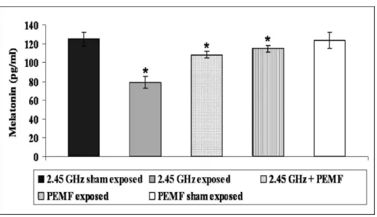
Melatonin
The average concentration of melatonin in the serum of the MW exposure group (78.82¡6.28 pg/ml) was
signifi-cantly less (p,0.003) than the average concentration of melatonin in the MW sham group (124¡7.29 pg/ml,
Figure 5). In contrast, the concentration of melatonin (108.29¡3.67) in the MW+ PEMF group was significantly
(p,0.01) decreased. The melatonin level in PEMF exposure group (114¡3.76) was significantly (p,0.05) less than that
of the PEMF sham group (123.22¡8.63). Creatine Kinase
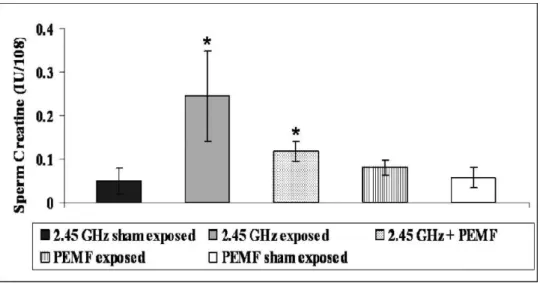
The critical role for CK in sperm energy transport was examined by measuring the ATP concentrations or ATP/ ADP ratios. The mean value of the CK level was higher (p,0.001) in the sperm from the MW exposure group (0.24¡0.10) than in the sperm from the MW sham group
(0.04¡0.03). A significant (p,0.001) decline was also observed in the MW + PEMF group (0.11¡0.02). The PEMF exposure group (0.08¡0.01) showed an insignificant increase (p.0.06) in the CK level compared with the PEMF sham group (0.05¡0.02, Figure 6).
Caspase 3 Activity
A statistically significant activation of caspase-3 was observed in the MW-induced animals (Figure 7). The sperm caspase activity showed a significant increase (p,0.003) in the MW exposure group (34.62¡1.98) compared with the
MW sham group (13.72¡0.81). After exposure to 2.45-GHz radiation, the animals were subjected to PEMF treatment (22.72¡1.74,p,0.001). A significant (p,0.049) decrease was found in the PEMF exposure group (15.62¡1.31) compared
with the PEMF sham (13.81¡1.48). Serum testosterone level
The serum testosterone level decreased significantly (p,0.002) in the MW exposure group (1.36¡0.54) compared with the MW sham group (5.41¡1.25). PEMF exposure
produced a significant recovery effect (p,0.008) on the serum testosterone level in the MW+PEMF exposure group (3.46¡0.74). However, significant (p= 0.05) changes were
observed in the PEMF exposure group (4.20¡0.69)
com-pared with the PEMF sham group (5.46¡1.21, Figure 8).
DISCUSSION
The interaction between EMF and biosystems is a wide-ranging phenomenon that is not essentially confined to emission from any particular source. In general, such exposure has already been identified as a stressor whose receptor mechanism at the cellular level is still unknown.
Figure 4 -A diagram showing the output pattern obtained from a 100-Hz sawtooth wave.
The thermal energy inside the body at room temperature is random, whereas the thermal energy from an external source is coherent (over a period of time exceeding 10 sec). It may be possible that a coherent signal interferes with the rhythmicity of the physiological process inside the body.
ROS generation in the testes was responsible for the possible toxic effects on the physiology of reproduction. However, cells have a defense mechanism [i.e., antioxidants (reduced glutathione, catalase, superoxide dismutase)] to fight against increased ROS production. Furthermore, melatonin stimulates the activity of several important endogenous antioxidants (GSH-Px, SOD) to combat the effect of reactive oxygen species.37Melatonin is exclusively synthesized and secreted at night and is an efficient endogenous free radical scavenger.
Decreased serum testosterone levels and increased crea-tine kinase activity were associated with reduced male fertility. Our data reflect an appreciable increase in sperm creatine kinase activity and a decline in the activities of
antioxidants and melatonin. In spermatozoa, CK-BB is localized to the mitochondria of the midpiece region.38 Creatine phosphate serves as a donor for the re-phosphor-ylation of adenosine diphosphate (ADP) into ATP, which supports flagellar dynein/adenosine triphosphate and sperm quality. The CK activity differences were reflected in the sperm ATP concentrations and ATP/ADP ratios.39 CK was associated with increased ROS and served as an indicator for oxidative stress.
Genotoxic stresses induced by irradiation trigger an arrest or delay in the G2 phase of the cell cycle to permit the repair of damaged DNA.40Cell cycle analysis by flow cytometry with propidium iodide has confirmed these results because EMF exposure induces the appearance of a sub-G1 apoptotic peak, which is characteristic of DNA fragmenta-tion in spermatozoa.7Activated caspases commit the cells with defective repair machinery to an apoptotic death by cleaving a number of substrates. The results of the caspase assay suggest that the apoptotic cell counts are significantly
Figure 6 -The five columns represent the CK activity distribution in the sperm fractions of the various groups. The results are expressed as IU/108spermatozoa¡standard deviation. All of the determinations were performed in triplicate.
increased. Therefore, apoptosis is considered to be involved in the impairment of spermatogenesis and the seminiferous tubules.
It is apparent from our data that a 2.45-GHz frequency increases caspase activity and decreases melatonin levels. These effects are indications of increased apoptosis and, hence, cancer promotion, which affects fertility. These effects were controlled by the administration of PEMF. Previously, our laboratory reported increased levels of PKC and ODC, changes in the levels of antioxidant enzymes (GPx, CAT, and SOD), micronucleus formation, and many other related changes.32,33The present work supports these findings and others.41Von Wilmsdorff et al42hypothesized
that brain behavioral modifications due to kainic acid treatment cause sex-specific responses via the hypothala-mic-pituitary-adrenal-axis. Such effects may be achieved through either a chemical or radiation-based treatment, which suggests that the effects are mediated by the higher brain centers and are transferred to the reproductive system, thereby affecting the testosterone and sperm counts through the endocrine system.
The exposure of a biological system to a microwave causes a weakly induced signal near the cellular boundary. Energy from a coherent signal penetrates the body, with amplification derived from noise via stochastic phenomena. Thus, the signal can circumvent the barrier height of the plasma membrane (,105 V/m), which may cause DNA
damage (strand breaks). This signal may also provide sufficient energy to exacerbate silent mutations existing in biological systems. ROS overproduction is inhibited by the application of a pulsed electromagnetic field (100 Hz, magnetic component), which provides a free electron and thus eliminates the effect of the stressor. Triplet states can be transformed into singlet states via spin-lattice relaxation, which originates in fluctuating local magnetic fields around the electron (because of its random motion).43
In fact, many medical devices used in medical therapies generate circulating currents that trigger cellular, hormonal, and behavioral responses. Wolsko et al44demonstrated that
PEMF relieves or eliminates pain through a mechanism mediated by Ca2+ion channels. Luo et al45
found that metal
ions are necessary for enzymatic activity, and, thus, pulsed electromagnetic field are able to influence the catalytic activities of enzymes. Pulsed electromagnetic therapy is believed to have beneficial effects on tissue growth and repair, probably via alterations in the cellular microenvironment.46A
low-level alternating electromagnetic field (,50 Hz) has also
been reported to have a healing effect on spinal cord injury.47
When a microwave field penetrates a biological body, it induces endogenous physiological processes. The major difference between the EMF generated from a 2.45-GHz source and that of the pulsed electromagnetic fields (100 Hz) is that the latter induces a circulating electric current in the tissue because of its constantly changing magnetic flux,29 thereby scavenging the free radical. Free radical formation also occurs at other microwave frequencies, which suggests that this phenomenon occurs generally. The destressor
Figure 8 -The effect of an electromagnetic field on testosterone production. The bars represent the mean values (¡SEMs). The statistical significance between the values of the sham and exposure groups is indicated by an asterisk (p,0.05).
Our results suggest that a 2.45-GHz exposure causes apoptosis during spermiogenesis or sperm maturation, and sperm caspase-3 activity seems to affect the physiology of reproduction. Several other parameters are affected by electromagnetic field exposure. All of these studies reveal that oxidative stress is a major mechanism affecting health, and microwave fields cause chronic stress via ROS over-production. Pulsed electromagnetic field therapy provides significant protection by controlling ROS production.
ACKNOWLEDGEMENTS
The authors would like to thank the Indian Council of Medical Research (ICMR) for its financial support.
REFERENCES
1. Hardell L, Sage C. Biological effects from electromagnetic field exposure and public exposure standards. Biomed Pharmacother. 2008;62:104-9, doi: 10.1016/j.biopha.2007.12.004.
2. Lue YH, Hikim AP, Swerdloff RS, Im P, Taing KS, Bui T, et al. Single exposure to heat induces stage-specific germ cell apoptosis in rats: role of intratesticular testosterone on stage specificity. Endocrinology. 1999;140:1709-17, doi: 10.1210/en.140.4.1709.
3. Richburg JH. The relevance of spontaneous- and chemically-induced alterations in testicular germ cell apoptosis to toxicology. Toxicol Lett. 2000;112-113:79-86, doi: 10.1016/S0378-4274(99)00253-2.
4. Kunjilwar KK, Behari J. Effect of amplitude-modulated radio frequency radiation on cholinergic system of developing rats. Brain Res 1993; 601:321-4, doi: 10.1016/0006-8993(93)91729-C.
5. Burch JB, Reif JS, Noonan CW, Ichinose T, Bachand AM, Koleber TL et al. Melatonin metabolite excretion among cellular telephone users. Int J Radiat Biol. 2002;78:1029-36, doi: 10.1080/09553000210166561. 6. Kesari KK, Behari J. Fifty-gigahertz microwave exposure effect of
radiations on rat brain. Appl Biochem Biotechnol. 2009;158:126-39, doi: 10.1007/s12010-008-8469-8.
7. Kumar S, Kesari KK, Behari J. Influence of microwave exposure on fertility of male rats. Fertil Steril 2010.
8. Hu PY, Chu XL, Li JY, Yang D, He P. [Effect of microwave contraception on human serum testosterone and luteinizing hormone]. Shengzhi Yu Biyun. 1985;5:32-4.
9. Steinberger E. Hormonal control of mammalian spermatogenesis. Physiol Rev. 1971;51:1-22.
10. Cayli S, Sakkas D, Vigue L, Demir R, Huszar G. Cellular maturity and apoptosis in human sperm: creatine kinase, caspase-3 and Bcl-XL levels in mature and diminished maturity sperm. Mol Hum Reprod. 2004;10:365-72, doi: 10.1093/molehr/gah050.
11. Allen RT, Hunter WJ, III, Agrawal DK. Morphological and biochemical characterization and analysis of apoptosis. J Pharmacol Toxicol Methods. 1997;37:215-28, doi: 10.1016/S1056-8719(97)00033-6.
12. Ceruti S, Beltrami E, Matarrese P, Mazzola A, Cattabeni F, Malorni W et al. A key role for caspase-2 and caspase-3 in the apoptosis induced by 2-chloro-29-deoxy-adenosine (cladribine) and 2-chloro-adenosine in human astrocytoma cells. Mol Pharmacol. 2003;63:1437-47, doi: 10. 1124/mol.63.6.1437.
13. Riedl SJ, Shi Y. Molecular mechanisms of caspase regulation during apoptosis. Nat Rev Mol Cell Biol. 2004;5:897-907, doi: 10.1038/ nrm1496.
14. Pommier Y, Sordet O, Antony S, Hayward RL, Kohn KW. Apoptosis defects and chemotherapy resistance: molecular interaction maps and networks. Oncogene. 2004;23:2934-49, doi: 10.1038/sj.onc.1207515. 15. Hallak J, Sharma RK, Pasqualotto FF, Ranganathan P, Thomas AJ, Jr.,
Agarwal A. Creatine kinase as an indicator of sperm quality and maturity in men with oligospermia. Urology. 2001;58:446-51, doi: 10. 1016/S0090-4295(01)01224-9.
16. Wallimann T, Hemmer W. Creatine kinase in non-muscle tissues and cells. Mol Cell Biochem. 1994;133-134:193-220, doi: 10.1007/BF01267955.
associated with increased creatine phosphokinase concentration and abnormal head morphology. Mol Reprod Dev. 1993;34:292-8, doi: 10. 1002/mrd.1080340309.
21. Moller P, Wallin H, Knudsen LE. Oxidative stress associated with exercise, psychological stress and life-style factors. Chem Biol Interact. 1996;102:17-36, doi: 10.1016/0009-2797(96)03729-5.
22. Adams ML, Little PJ, Bell B, Cicero TJ. Alcohol affects rat testicular interstitial fluid volume and testicular secretion of testosterone and beta-endorphin. J Pharmacol Exp Ther. 1991;258:1008-14.
23. Mahfouz R, Sharma R, Thiyagarajan A, Kale V, Gupta S, Sabanegh E et al. Semen characteristics and sperm DNA fragmentation in infertile men with low and high levels of seminal reactive oxygen species. Fertil Steril 2010.
24. Wilson BW, Anderson LE, Hilton DI, Phillips RD. Chronic exposure to 60-Hz electric fields: effects on pineal function in the rat. Bioelectromagnetics. 1981;2:371-80, doi: 10.1002/bem.2250020408.
25. Brainard GC, Gaddy JR, Barker FM, Hanifin JP, Rollag MD. Mechanisms in the eye that mediate the biological and therapeutic effects of light. In: Wetterberg L, editor. Light and Biological Rhythms in Man. Oxford: Pergamon, 1993:29-54.
26. DiCarlo AL, Hargis MT, Penafiel LM, Litovitz TA. Shortterm magnetic field exposures (60 Hz) induce protection against ultraviolet radiation damage. Int J Radait Biol. 1999;75:1541–49, doi: 10.1080/095530099139142. 27. Manjhi J, Mathur R, Behari J. Effect of low level capacitive-coupled pulsed electric field stimulation on mineral profile of weight-bearing bones in ovariectomized rats. J Biomed Mater Res B Appl Biomater. 2010;92:189-95.
28. Prakash D, Behari J. Synergistic role of hydroxyapatite nanoparticles and pulsed electromagnetic field therapy to prevent bone loss in rats following exposure to simulated microgravity. Int J Nanomedicine. 2009;4:133-44.
29. Fernandez MI, Watson PJ, Rowbotham DJ. Effect of pulsed magnetic field therapy on pain reported by human volunteers in a laboratory model of acute pain. Br J Anaesth. 2007;99:266-9, doi: 10.1093/bja/ aem129.
30. Shupak NM. Therapeutic uses of pulsed magnetic-field exposure: A review. Radio Science Bulletin. 2003;307:9-32.
31. Paulraj R, Behari J. The effect of low level continuous 2.45 GHz wave on brain enzymes of developing rat brain. Electromagn Biol Med. 2002;21:231-41, doi: 10.1081/JBC-120015993.
32. Paulraj R, Behari J. Single strand DNA breaks in rat brain cells exposed to microwave radiation. Mutat Res. 2006;596:76-80.
33. Kesari KK, Behari J. Effect of microwave at 2.45 GHz radiations on reproductive system of male rats. Toxicology and Environmental Chemistry. 2010;92:1135-47, doi: 10.1080/02772240903233637.
34. Kesari KK, Behari J, Kumar S. Mutagenic response of 2.45 GHz radiation exposure on rat brain. Int J Radiat Biol. 2010;86:334-43, doi: 10.3109/ 09553000903564059.
35. Durney CH, Iskander MF, Massoudi H, Johnson CC. An empirical formula for broad band SAR calculations of prolate spheroidal models of humans and animal. In: Osepchuk JM, editor. Biological Effects of Electromagnetic Radiation. New York: IEEE Press, 1984:85-90. 36. Prato FS, Desjardins-Holmes D, Keenliside LD, DeMoor JM, Robertson
JA, Stodilka RZ, Thomas AW. The detection threshold for extremely low frequency magnetic fields may be below 1000 nt-hz in mice. Bioelec-tromagnetics. 2011;(DOI 10.1002/bem.20661).
37. Reiter RJ, Tan DX, Osuna C, Gitto E. Actions of melatonin in the reduction of oxidative stress. A review. J Biomed Sci. 2000;7:444-58, doi: 10.1007/BF02253360.
38. Wallimann T, Moser H, Zurbriggen B, Wegmann G, Eppenberger HM. Creatine kinase isoenzymes in spermatozoa. J Muscle Res Cell Motil. 1986;7:25-34, doi: 10.1007/BF01756199.
39. Vigue C, Vigue L, Huszar G. Adenosine triphosphate (ATP) concentra-tions and ATP/adenosine diphosphate ratios in human sperm of normospermic, oligospermic, and asthenospermic specimens and in their swim-up fractions: lack of correlation between ATP parameters and sperm creatine kinase concentrations. J Androl. 1992;13:305-11. 40. Hemmati PG, Normand G, Gillissen B, Wendt J, Dorken B, Daniel PT.
41. Kumar S, Kesari KK, Behari J. Evaluation of genotoxic effects in male Wistar rats following microwave exposure. Indian J Exp Biol. 2010;48:586-92.
42. Von Wilmsdorff M, Sprick U, Bouvier ML, Schulz D, Schmitt A, Gaebel W. Sex-dependent behavioral effects and morphological changes in the hippocampus after prenatal invasive interventions in rats: implications for animal models of schizophrenia. Clinics. 2010;65:209-19.
43. Adey WR. Bioeffects of mobile communications fields: possible mechanism for cumulative dose. In: Kuster N, Balzano Q, Lin JC, editors. Mobile communications safety. London:Chapman & Hall, 1997:95-131.
44. Wolsko PM, Eisenberg DM, Simon LS, Davis RB, Walleczek J, Mayo-Smith M et al. Double-blind placebo-controlled trial of static magnets for
the treatment of osteoarthritis of the knee: results of a pilot study. Altern Ther Health Med. 2004;10:36-43.
45. Luo Q, Li SS, He C, He H, Yang L, Deng L. Pulse electromagnetic fields effects on serum E2 levels, chondrocyte apoptosis, and matrix metallo-proteinase-13 expression in ovariectomized rats. Rheumatol Int. 2009;29:927-35, doi: 10.1007/s00296-008-0782-6.
46. Lee EW, Maffulli N, Li CK, Chan KM. Pulsed magnetic and electromagnetic fields in experimental achilles tendonitis in the rat: a prospective randomized study. Arch Phys Med Rehabil. 1997;78:399-404, doi: 10.1016/S0003-9993(97)90232-X.