Cop
yright
© ABE&M t
odos os dir
eit
os r
eser
vados
.
Two cases of thyroid sarcoidosis
presentation as painful, recurrent
goiter in patients with Graves’ disease
Dois casos de sarcoidose na tireoide com apresentação na forma de bócios recorrentes dolorosos em pacientes com doença de Graves
Piotr Kmieć1, Marta Lewandowska1, Anna Dubaniewicz2, Krystyna
Mizan-Gross3, Artur Antolak4, Barbara Wołyniak1, Krzysztof Sworczak1
SUMMARY
Sarcoidosis rarely involves the thyroid gland. Pain in the thyroid gland area was only spora-dically reported in patients suffering from this disease. The aim of this paper is to report and discuss the cases of two female patients with Graves’ disease who presented painful, rapidly growing, recurrent goiters (after strumectomy in their early adult lives). Invasive treatment was applied and sarcoidosis was revealed histologically. The irst patient suffered from dysphagia and dyspnoea due to large goiter; skin lesions were present as well. Sarcoidosis was diagnosed in histological examination of the thyroid tissue specimens. Steroid treatment was ineffective; thus, the thyroid was removed. Two years later thyroid sarcoidosis recurred as a painful goiter and surgical treatment was applied once again. In the second case, thyroid ultrasound indings suggesting malignancy, and prompted the decision to perform thyroidectomy despite the fact that FNAB (ine needle aspiration biopsy) revealed cells indicative of a “granulomatous disease in the post-resection scar” and results of the thorax high-resolution computed tomography scan suggested pulmonary sarcoidosis. Pathological examination conirmed sarcoidosis. Ho-wever, a papillary cancer focus was also found. Arq Bras Endocrinol Metab. 2012;56(3):209-14
SUMÁRIO
A sarcoidose raramente envolve a glândula tireoide, e apenas esporadicamente foi relatada dor na região da glândula em pacientes que sofrem dessa doença. O objetivo deste trabalho é rela-tar e discutir os casos de duas mulheres que apresentavam bócios dolorosos, de rápido cresci-mento e recorrentes (após tireoidectomia na adolescência). Foi usado um tratacresci-mento invasivo e a sarcoidose foi revelada pelos achados histológicos. A primeira paciente sofria de disfagia e dispneia em decorrência de um grande bócio; lesões cutâneas também estavam presentes. A sarcoidose foi diagnosticada em um exame histológico das amostras de tecido da tireoide. O tratamento com esteroides foi ineicaz; foi feita assim a ressecção da glândula. Dois anos depois, houve recidiva da sarcoidose da tireoide como um bócio doloroso, e o tratamento cirúr-gico foi feito mais uma vez. No segundo caso, os resultados do ultrassom da tireoide sugeriam malignidade e levaram à decisão de se realizar a tireoidectomia, apesar de as células de PAAF indicarem uma doença granulomatosa na cicatriz pós-ressecção e os resultados da tomograia computadorizada de alta resolução de tórax sugerirem sarcoidose pulmonar. O exame histopa-tológico da glândula revelou sarcoidose. Entretanto, também foi encontrado um foco de câncer papilar. Arq Bras Endocrinol Metab. 2012;56(3):209-14
Correspondence to:
Piotr Kmie´c
Department of Endocrinology, Medical University of Gdansk, 7 Debinki Street
80-952 – Gdansk, Poland [email protected]
Received on 21/Nov/2011 Accepted on 18/Mar/2012
1 Department of Endocrinology and
Internal Medicine, Medical University of Gdansk, Gdansk, Poland
2 Department of Pneumology,
Medical University of Gdansk, Gdansk, Poland
3 Department of Nuclear Medicine,
Medical University of Gdansk, Gdansk, Poland
4 Department of Pathology, St.
Adalbert Specialist Hospital, Gdansk, Poland
INTRODUCTION
S
arcoidosis (SA) is a multisystemic, idiopathic disease that may affect almost all organs, most commonly lungs, the lymph nodes, liver, spleen and skin (1,2). InCop
yright
© ABE&M t
odos os dir
eit
os r
eser
vados
.
toimmune reaction in genetically predisposed individuals (3,4). As a result, noncaseating, epithelioid granulomas are formed. Presence of these formations in histological examination, along with clinical and radiological indings enables SA diagnosis as long as “granulomas of known causes and local sarcoid reactions” are excluded (5).
As has been extensively reported, sarcoidosis coexists with autoimmune thyroid disease (ATD), although the frequency of this association varies largely between stu dies, i.e., ATD percentage ranges from 1.9% to 16.6% of SA patients (610). The prevalence of antithyroid anti bodies (antiTPO: antithyroperoxidase antibodies, and antiTg: antithyroglobulin antibodies) and Hashimoto’s disease was higher in sarcoidosis patients than in age and gendermatched control subjects (11). Moreover, pri mary hypofunction of the gland was frequently reported (7,12,13). Although the incidence of hyperthyroidism is considered rare (14), one study showed a signiicantly higher prevalence of Graves’ disease in SA patients than in matched controls (11). On the one hand, HLA gene association between Graves’ disease and sarcoidosis has been established (15). On the other hand, Graves’ disease in SA patients may only be coincidental (14). Other thy roid disorders that accompany sarcoidosis include goiter, de Quervain’s thyroiditis and thyroid cancer (9,16).
Thyroid iniltration by noncaseating granulomas in the course of sarcoidosis diagnosed in other organs is rare. In a review paper from 1993, Valiati and cols. found 40 patients with thyroid sarcoidosis in the litera ture (17). Although in autopsy studies showed sarcoid granulomas in the gland in approximately 4% to 4.5% SA subjects, less than 1% of sarcoidosis patients pre sented signiicant clinical involvement of the thyroid (7,9,18,19). In these cases, iniltration was associated with hypo, eu or hyperthyroidism (9,16,20) and may be manifested as or imitate thyroiditis, nodular and dif fuse goiter, and thyroid cancer (2,9,21,22).
To our knowledge, painful thyroid sarcoidosis was reported only sporadically (2325). In this paper, we are going to present the cases of two female patients with Graves’ disease who suffered from painful, rapidly growing, recurrent goiter due to thyroid sarcoidosis.
CASE REPORTS
Patient AThe patient case was already partially reported in 2008 (26). A middleaged woman with several comorbidities (after strumectomy in her early adult life) was admitted
to our clinic due to rapidly growing, painful recurrent goiter (Table 1). She reported increasing, lower neck swelling leading to dysphagia, dyspnea and choking, which had started approximately 10 weeks prior to hospital admission. Upon physical examination, the left lobe of the thyroid was enlarged, and there were skin lesions above the gland, as well as on the left shoulder and calf (Table 1).
Laboratory indings revealed no signiicant abnor malities apart from increased erythrocyte sedimenta tion rate (ESR; Table 1).
Thyroid cytology by ine needle aspiration biopsy (FNAB) indicated de Quervain’s thyroiditis (Figure 1A). However, histological examination of the tissue material from the thyroid and affected skin suggested sarcoidosis (Figure 2A). Despite oral steroid therapy implementation, dyspnea and dysphagia became exa cerbated, and a new strumectomy was carried out. Af ter the surgery, symptoms subsided. The patient was prescribed a daily prednisone dose of 10 milligrams.
Nevertheless, twenty months later, the granuloma tous disease recurred in the thyroid lodge, presented as a painful movable tumour causing dyspnoea and stridor. Exudative, nodular skin lesions were present in the stru mectomy scar area and other body regions. Following ultrasound and thyroid FNAB, the patient was qualiied for another goiter resection. During hospitalization, a thorax highresolution computed tomography (HR CT) scan suggested stage 1 sarcoidosis in the lungs (the Xray image from two years earlier was normal).
Currently, our patient remains under outpatient cli nic care and receives a daily Lthyroxine dose of 125 micrograms.
Patient B
An 49 yearold female with Graves’ disease, who appeared for regular checkups in our outpatient clinic due to recur rent goiter after strumectomy in her early adult life, repor ted pain in the thyroid region that lasted for about four weeks. It radiated toward the pharynx and was accompanied by a pulling sensation (Table 1). Laboratory examinations reve aled no signiicant abnormalities (Table 1).
Cop
yright
© ABE&M t
odos os dir
eit
os r
eser
vados
.
Table 1. Results of clinical and additional examinations
Age at SA diagnosis Patient A52 Patient B49
Admission date 26.12.2006 24.11.2010
Primary thyroid disease Graves’ Graves’
Age at strumectomy 21 26
Co-morbidities Resistant hypertension, diabetes mellitus, obesity, HCV infection, post spinal column surgeries
None
Symptoms Rapidly growing, painful, recurrent goitre, dysphagia, dyspnea Pain of the thyroid region radiating toward the pharynx; pulling sensation
Signs Thyroid’s left lobe enlargement, edematous nodules and hard iniltration of the skin above the gland
Redness and hyperesthesia of the skin above the thyroid
Laboratory results TSH = 1.51 uU/mL, fT4 = 17.45 pmol/L, ESR = 37 mm, CRP < 5 mg/L TSH = 0.87 uU/mL, fT4 = 19,1 pmol/L, CRP < 5 mg/L
Thyroid gland ultrasound Heterogeneous thyroid tissue with altered echogenicity, volume of left lobe 6.4 mL, volume of right lobe 14.3 mL
Multiple, solid foci, up to 3 cm in diameter with luid-illed areas and calciications; neoplasm characteristics in the post-resection
scar; two pathologic supraclavicular lymph nodes, thyroid gland volume 102 mL
FNAB Histiocyte iniltration, multinucleated cells (Figure 1A) Histiocyte reaction with cells indicative of a granulomatous disease in strumectomy scar (Figure 1B)
Treatment Ineffective steroid therapy, consecutive partial thyroidectomy (02.2007)
Thyroidectomy (01.2011)
Lung involvement Stage 1 pulmonary sarcoidosis reported two years after thyroid sarcoidosis diagnosis in a HR-CT scan (01.2009)
Stage 2 pulmonary sarcoidosis found in a HR-CT scan (01.2011)
Remission/recurrence Jan., 2009. Thyroid sarcoidosis recurrence as painful goiter – second re-strumectomy (02.2009); currently in remission
Currently in remission
CRP: C-reactive protein; ESR: erythrocyte sedimentation rate; fT4: free thyroxine; HR-CT: high resolution computed tomography; TSH: thyroid-stimulating hormone.
Figure 1. FNAB specimens. HE x200. A. Multinucleated cells with a histiocyte granulomatosis (patient A); B. Histiocyte granolumatosis fragment with adjacent multinucleated cells (patient B). HE: hematoxylin-eosin staining.
A B
plasmatic cells were found. Furthermore, the results of the thorax HRCT scan suggested stage 2 pulmonary sarcoidosis. Despite thyroid FNAB and thorax CT re sults the patient was qualiied for surgery. In histolo gical examination, thyroid sarcoidosis was conirmed
Cop
yright
© ABE&M t
odos os dir
eit
os r
eser
vados
.
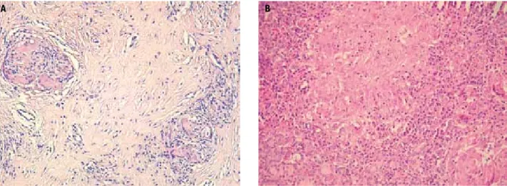
Figure 2. Thyroid sarcoidosis. HE x200. A. Diffused ibrosis between sarcoid granulomas (patient A); B. Sarcoid granuloma (patient B). HE: hematoxylin-eosin staining.
A B
Figure 3. Microfocus of papillary thyroid cancer (patient B). HE x400. HE: hematoxylin-eosin staining.
At present, the patient remains under care of our outpatient clinic. She receives a daily dose of 100 mi crograms of Lthyroxine.
REVIEW OF LITERATURE AND DISCUSSION
As mentioned above, thyroid sarcoidosis is rarely diag nosed in vivo, although noncaseating granulomas are found post-mortem in up to 4.5% of SA patients. In clinically signiicant thyroid sarcoidosis, abnormalities in respect to gland morphology (i.e., nodules, goiter, thyroiditis) and function (hypo and hyperthyroidism) may be isolated or combined. A different criterion for categorizing thyroid sarcoidosis patients is the involve ment of other organs; in this respect, concomitant SA
manifestation in the lungs is more frequent than isola ted thyroid iniltration.
Three main groups of patients with thyroid sarcoi dosis emerge from the literature review. First, patients in whom the disease manifested in the form of (possibly incidental) thyroid nodule or nodules – associated with or not with goiter (2,6,27,28). Functional disorders of the glands are not apparent.
Second, several reported cases of thyroid sarcoidosis involved patients who presented thyrotoxicosis (14,29 31). Concomitant Graves’ disease was found in three men and one woman, aged 23 to 30; all were resistant to antithyroid treatment and, therefore, underwent thyroidectomy (14,2931). Two other hyperthyroid patients, in a much greater extent, resembled the irst group of thyroid sarcoidosis subjects, since their main clinical abnormality was the multinodular goiter, and not thyrotoxicity (29,32).
Cop
yright
© ABE&M t
odos os dir
eit
os r
eser
vados
.
when evidence of systemic SA is absent (35,36). These reactions contrast with sarcoidosis accompanying a ma lignant tumour. The latter case is exempliied by one of our patients (patient B), and has also been reported by other authors (21,31,37).
To our knowledge, concomitant occurrence of pa pillary cancer, Graves’ disease and thyroid sarcoidosis, which we present here, had only been reported once before, by ZimmermannBelsing and cols. (31). Their patient, known to have sarcoidosis, underwent total thyroidectomy because of inadequate control of hyper thyroidism with thiamazole treatment in the course of Graves’ disease (the goiter gradually enlarged). Thyroid sarcoidosis and papillary cancer of the gland with me tastases to the lymph nodes were incidental indings in histological examination. In patient B (described here), ultrasound characteristics were disturbing, although FNAB result did not support the suspicion of cancer, and thorax CT scan suggested pulmonary sarcoidosis. In case of suspicion of malignancy, the approach should aim at clarifying diagnosis.
A case similar to ours in these aspects (i.e., malignancy suspicion and surgical treatment) was reported by Ma ría GonzálvezGasch and cols. (25). Their patient, who had undergone left hemithyroidectomy due to follicular cancer of the thyroid ifteen years earlier and had been diagnosed with sarcoidosis two years earlier, presented a painful nodule in the right lobe region of the thyroid. Due to the need to exclude malignancy (and the absen ce of inlammatory signs), the female patient reported by the Spanish authors underwent right hemithyroidec tomy. Thyroid sarcoidosis was diagnosed histologically.
Obviously, the three groups of patients with thyroid sarcoidosis reviewed above do not exhaust all possible manifestations of the disease. Our irst patient presen ted here may serve as an example. Initially, subacute thyroiditis was suspected in patient A, based on pain ful goiter, increased ESR (although, importantly, it did not exceed 40 mm/h) and result of FNAB cytology. Although tissue specimens revealed thyroid sarcoidosis, the exclusion of an underlying (i.e., sarcoid reaction) or accompanying malignancy could not have been made without thyroidectomy. A similar case was reported by Cooke – his patient also presented dysphagia and dysp nea due to large goiter, leading to suspicion of malig nancy, and was proven negative after surgery (38).
Yet, an other differential diagnosis (apart from thyroid sarcoidosis) should be considered in cases such as that of patient A or the one reported by Cooke, i.e.,
anaplastic cancer of the thyroid manifesting as subacute, granulomatous inlammation of the gland. Patients with this diagnosis were reported by MizanKalisiak and cols. as well as Rosen and cols. (39,40). In the irst instance, steroid, antibiotic and salicylate treatment were ineffec tive. The tissue specimens obtained during thyroid sur gery and postmortem examination of Cooke’s patient revealed anaplastic carcinoma of the gland. In this case, diagnosis was also based on histological examination of the gland (in samples collected during tracheostomy).
CONCLUSIONS
Similar to previous reports, other diseases were suspec ted in our patients before the diagnosis of sarcoidosis was determined by histology. Not only is thyroid sar coidosis a rare condition, but it also may manifest in many different forms. Furthermore, the diagnosis may only be conirmed if known causes of granulomas and, in particular, sarcoid reactions, are excluded.
In summary, it should be underscored that, in the vast majority of cases, sarcoid involvement of the thyroid remains clinically insigniicant. The emergence of gland disorders (change in function or morpholo gy) is not associated with pain. The process is mild and frequently leads to hypothyroidism. However, sporadi cally, sarcoidosis can cause pain and compression symp toms, and may coexist with thyroid cancer. One should also bear in mind that papillary thyroid carcinoma may cause a sarcoidtype reaction in the gland. Therefore, in patients with suspected thyroid sarcoidosis, thorough diagnosis and observation are necessary. Often, parti cularly due to the diagnostic dificulties, surgery is in dicated.
Funding: this research did not receive any speciic grant from any funding agency in the public, commercial or nonproit sector.
Disclosure: no potential conlict of interest relevant to this article was reported.
REFERENCES
1. Bell NH. Endocrine complications of sarcoidosis. Endocrinol Me-tab Clin North Am. 1991;20(3):645-54.
2. Ozkan Z, Oncel M, Kurt N, Kargi AB, Ozdemir N, Kaptanoglu L, et al. Sarcoidosis presenting as cold thyroid nodules: report of two cases. Surg Today. 2005;35(9):770-3.
3. Dubaniewicz A. Mycobacterium tuberculosis heat shock proteins and autoimmunity in sarcoidosis. Autoimmun Rev. 2010;9(6):419-24. 4. Rosen Y. Pathology of sarcoidosis. Semin Respir Crit Care Med.
Cop
yright
© ABE&M t
odos os dir
eit
os r
eser
vados
.
5. Statement on sarcoidosis. Joint Statement of the American Tho-racic Society (ATS), the European Respiratory Society (ERS) and the World Association of Sarcoidosis and Other Granulomatous Disorders (WASOG) adopted by the ATS Board of Directors and by the ERS Executive Committee, February 1999. Am J Respir Crit Care Med. 1999;160(2):736-55.
6. Cabibi D, Di Vita G, La Spada E,Tripodo C, Patti R, Montalto G. Thyroid sarcoidosis as a unique localization. Thyroid. 2006;16(11):1175-7. 7. Gentilucci UV, Picardi A, Maniini S, D’Avola D, Costantino S,
Po-zzilli P. Granulomatous thyroiditis: an unexpected inding leading to the diagnosis of sarcoidosis. Acta Biomed. 2004;75(1):69-73. 8. Isern V, Lora-Tamayo J, Capdevila O, Villabona C, Mañá J.
Sar-coidosis and autoimmune thyroid disease. A case series of ten patients. Sarcoidosis Vasc Diffuse Lung Dis. 2007;24(2):148-52. 9. Porter N, Beynon HL, Randeva HS. Endocrine and reproductive
manifestations of sarcoidosis. QJM. 2003;96(8):553-61.
10. Papadopoulos KI, Hörnblad Y, Liljebladh H, Hallengren B. High frequency of endocrine autoimmunity in patients with sarcoido-sis. Eur J Endocrinol. 1996;134(3):331-6.
11. Antonelli A, Fazzi P, Fallahi P, Ferrari SM, Ferrannini E. Prevalen-ce of hypothyroidism and Graves disease in sarcoidosis. Chest. 2006;130(2):526-32.
12. Ilias I, Panoutsopoulos G, Batsakis C, Nikolakakou D, Filippou N, Christakopoulou I. Thyroid function and autoimmunity in sarcoi-dosis: a case-control study. Croat Med J. 1998;39(4):404-6. 13. Nakamura H, Genma R, Mikami T, Kitahara A, Natsume H, Andoh
S, et al. High incidence of positive autoantibodies against thyroid peroxidase and thyroglobulin in patients with sarcoidosis. Clin Endocrinol (Oxf). 1997;46(4):467-72.
14. Yarman S, Kahraman H, Tanakol R, Kapran Y. Concomitant asso-ciation of thyroid sarcoidosis and Graves’ disease. Horm Res. 2003;59(1):43-6.
15. Muzaffar TH, Al-Ansari JM, Al-Humrani H. Brief review of sarcoi-dosis of the thyroid gland. Int J Med Sci. 2009;1(3):44-5. 16. Yamauchi M, Inoue D, Fukunaga Y, Kakudo K, Koshiyama H. A
case of sarcoid reaction associated with papillary thyroid carci-noma. Thyroid. 1997;7(6):901-3.
17. Mihailovic-Vucinic V, Sharma OP. Atlas of sarcoidosis: pathoge-nesis, diagnosis, and clinical features. Springer; 2005. p. 103-4. 18. Winnacker JL, Becker KL, Katz S. Endocrine aspects of
sarcoido-sis. New Engl J Med. 1968;278:483-92.
19. Sharma OP, Vucini V. Sarcoidosis of the thyroid and kidneys and cal-cium metabolism. Semin Respir Crit Care Med. 2002;23(6):579-88. 20. Gostiljac DM, Dordević PB, Maric-Zivković J, Canović F.
Sarcoi-dosis localized in endocrine glands. Med Pregl. 2005;58 Suppl 1:25-9.
21. Bruins NA, Oswald JE, Morreau H, Kievit J, Pavel S, Smelt AH. Papillary thyroid carcinoma in a patient with sarcoidosis treated with minocycline. Neth J Med. 2007;65(5):185-7.
22. Mizukami Y, Nonomura A, Michigishi T, Ohmura K, Matsubara S, Noguchi M. Sarcoidosis of the thyroid gland manifested initially as thyroid tumor. Pathol Res Pract. 1994;190(12):1201-5; discus-sion 1206-7.
23. Cilley RE, Thompson NW, Lloyd RV, Shapiro B. Sarcoidosis of the thyroid presenting as a painful nodule. Thyroidology. 1988;(1):61-2. 24. Johnson LW, Sehon JK, McDonald JC. Diagnosis and manage-ment of a painful thyroid nodule in a patient with systemic sar-coidosis. J La State Med Soc. 2000;152(3):125-7.
25. María Gonzálvez-Gasch A, Mora Rufete A, Moltó Marhuenda J, Martín-Hidalgo A. Thyroid sarcoidosis in a woman with previous thyroid cancer. Med Clin (Barc). 2002;119(8):316-7.
26. Mizan-Gross K, Lewczuk A, Wołyniak B, Antolak A, Sworczak K. Izolowana sarkoidoza tarczycy: opis przypadku. Endokrynol Pol. 2008;59(supl. A):133-4.
27. Lemerre D, Caron F, Delval O, Goujon JM, Hira M, Meurice JC, et al. Thyroid manifestations of sarcoidosis: a case report. Rev Pneu-mol Clin. 1999;55(6):393-6.
28. Warshawsky ME, Shanies HM, Rozo A. Sarcoidosis involving the thyroid and pleura. Sarcoidosis Vasc Diffuse Lung Dis. 1997;14(2):165-8.
29. Papi G, Briganti F, Artioli F, Cavazza A, Carapezzi C, Roggeri A, et al. Sarcoidosis of the thyroid gland associated with hyperthyroi-dism: review of the literature and report of two peculiar cases. J Endocrinol Invest. 2006;29(9):834-9.
30. Rodriguez MC, Rani D, Faas FH. Unusual clinical course of Gra-ves’ thyrotoxicosis and concomitant sarcoidosis: case report and review of literature. Endocr Pract. 2007;13(2):159-63.
31. Zimmermann-Belsing T, Christensen L, Hansen HS, Kirkegaard J, Blichert-Toft M, Feldt-Rasmussen U. A case of sarcoidosis and sarcoid granuloma, papillary carcinoma, and Graves’ disease in the thyroid gland. Thyroid. 1997;7(6):901-3.
32. Langsteger W, Lind P, Beham A, Költringer P, Eber O. Isolated thyroid gland sarcoidosis and hyperthyroidism. Schweiz Med Wochenschr. 1989;119(17):544-8.
33. Saydam L, Bozkurt MK, Kutluay L, Ozçelik T. Sarcoidosis of the thyroid gland initially diagnosed as malignancy. Otolaryngol Head Neck Surg. 2003;129(1):154-6.
34. van Assendelft AH, Kahlos T. Sarcoidosis of the thyroid gland. Sar-coidosis. 1985;2(2):154-6.
35. Komatsu M, Itoh N, Yazawa M, Kobayashi S, Inoue K, Kuroda T. Sarcoid reaction in thyroid diseases: report of a case of thyroid carcinoma demonstrating sarcoid reaction in regional lymph no-des. Endocr J. 1997;44(5):697-700.
36. Nag S. Sarcoidosis diagnosis and management. Motamedi MHK, editor. InTech, October, 2011.
37. Middleton WG, deSouza FM, Gardiner GW. Papillary carcino-ma of the thyroid associated with sarcoidosis. J Otolaryngol. 1985;14(4):241-4.
38. Cooke R. Sarcoidosis of thyroid. Proc R Soc Med. 1959;52(3):174-5. 39. Mizan-Kalisiak K, Sworczak K, Markuszewska A, Warnke D. Ana-plastic carcinoma of the thyroid gland with the clinical course suggesting subacute thyroiditis--personal observations. Pol Arch Med Wewn. 1988;79(5):278-82.