Cop
yright
© ABE&M t
odos os dir
eit
os r
eser
vados
.
Analysis of anti-Müllerian hormone
(
AMH
) and its receptor (
AMHR2
)
genes in patients with persistent
Müllerian duct syndrome
Pesquisa de mutações nos genes do hormônio antiMülleriano
(AMH) e do seu receptor (AMHR2) em pacientes com
síndrome de persistência dos ductos Müllerianos
Mirian Yumie Nishi1, Sorahia Domenice1, Andréa Trevas Maciel-Guerra2,
Alberto Zaba Neto1,Marcia Alessandra Cavalaro Pereira da Silva2, Elaine
Maria Frade Costa1, Gil Guerra-Junior2, Berenice Bilharinho de Mendonca1
ABSTRACT
Objective: To screen for mutations in AMH and AMHR2 genes in patients with persistent Mülle-rian duct syndrome (PMDS). Patients and method: Genomic DNA of eight patients with PMDS was obtained from peripheral blood leukocytes. Directed sequencing of the coding regions and the exon-intron boundaries of AMH and AMHR2 were performed. Results: The AMH mu-tations p.Arg95*, p.Arg123Trp, c.556-2A>G, and p.Arg502Leu were identiied in ive patients; and p.Gly323Ser and p.Arg407* in AMHR2 of two individuals. In silico analyses of the novel c.556-2A>G, p.Arg502Leu and p.Arg407* mutations predicted that they were harmful and were possible causes of the disease. Conclusion: A likely molecular etiology was found in the eight evaluated patients with PMDS. Four mutations in AMH and two in AMHR2 were identiied. Three of them are novel mutations, c.556-2A>G, and p.Arg502Leu in AMH; and p.Gly323Ser in
AMHR2. Arq Bras Endocrinol Metab. 2012;56(8):473-8
Keywords
AMH; AMHR2; anti-Müllerian hormone; hernia inguinalis; persistent Müllerian duct syndrome
RESUMO
Objetivo: Analisar os genes AMH e AMHR2 em indivíduos com síndrome de persistência dos ductos de Müller (SPDM). Pacientes e método: Amostras de DNA genômico de oito pacien-tes com SPDM foram obtidas de leucócitos de sangue periférico. Sequenciamento direto da região codiicadora e das áreas intrônicas próximas aos éxons dos genes AMH e AMHR foi realizado. Resultados: As mutações p.Arg95*, p.Arg123Trp, c.556-2A>G e p.Arg502Leu no gene
AMH foram identiicadas em cinco pacientes e as mutações p.Gly323Ser e p.Arg407* no gene
AMHR2, em dois indivíduos. As análises in silico das mutações c.556-2A>G, p.Arg502Leu e p.Arg407*, não descritas anteriormente na literatura, previram que elas são deletérias e pos-sivelmente a causa da doença. Conclusão: Uma provável etiologia molecular foi encontrada nos oito pacientes portadores de SPDM avaliados. No gene do AMH foram identiicadas quatro mutações e no AMHR2, duas mutações. Três das seis mutações encontradas são mutações no-vas, c.556-2A>G e p.Arg502Leu no gene AMH; e p.Gly323Ser no AMHR2. Arq Bras Endocrinol Metab. 2012;56(8):473-8
Descritores
AMH; AMHR2;hormônio anti-Mülleriano; hérnia inguinal; síndrome da persistência dos ductos Müllerianos
1 Unidade de Endocrinologia do
Desenvolvimento, Laboratório de Hormônios e Genética Molecular LIM42, Disciplina de Endocrinologia, Hospital das Clínicas, Faculdade de Medicina da Universidade de São Paulo (HC-FMUSP), São Paulo, SP, Brazil
2 Grupo Interdisciplinar de
Estudos da Determinação e Diferenciação do Sexo, Faculdade de Ciências Médicas, Universidade Estadual de Campinas (FCM-Unicamp), Campinas, SP, Brazil
Correspondence to:
Mirian Yumie Nishi
Unidade de Endocrinologia do Desenvolvimento, Laboratório de Hormônios e Genética Molecular LIM42, Disciplina de Endocrinologia e Metabologia, Hospital das Clínicas, Faculdade de Medicina da Universidade de São Paulo Av. Dr. Enéas de Carvalho Aguiar, 155, PAMB, 2o andar, Bloco 6
05403-900 – São Paulo, SP, Brazil [email protected]
Cop
yright
© ABE&M t
odos os dir
eit
os r
eser
vados
.
INTRODUCTION
N
ormal development of male internal and external genitalia during embryogenesis depends on the action of testicular hormones: anti-Müllerian hormone (AMH) and testosterone. AMH is secreted by Sertoli cells and acts on its receptor in the Müllerian ducts, de-termining their regression. Testosterone, secreted by testicular Leydig cells, acts on the androgen receptor in the Wolfian ducts inducing the formation of epidydimis, deferent ducts, and seminal vesicles. Testosterone is fur-ther reduced to dihydrotestosterone (DHT) by the en-zyme 5α-reductase 2, which acts on the androgen recep-tor of the prostate and external genitalia, determining their masculinization (1). The development of female internal genitalia in a male individuals may occur due to the incapacity of Sertoli cells to synthesize or secrete AMH, or due to alterations in type II AMH receptor.Patients with persistent Müllerian duct syndrome (PMDS) have normal male phenotype, and the presence of an uterus and Fallopian tubes is usually discovered during corrective surgery for inguinal hernia or crypt-orchid testis (2). Leydig cell function is preserved, but azoospermia is common, due to the malformation of the ductus deferens or agenesis of the epididymis (3). The testes are usually adhered to the uterine tubes, and may be located in the abdominal or inguinal region. Hernia
uteri inguinalis is a condition where the testes and
Mül-lerian derivatives are found within a hernia inguinalis, and when the testes descend through a single inguinal canal, this is known as a transverse testicular ectopia.
The anatomical abnormalities common to all patients with PMDS lie in the failure of the gubernaculum to anchor the testes in the base of the scrotum. Migration
ofthe gubernaculum to the scrotum creates a processus
vaginalis in which one or both testes, accompanied by
Müllerian derivatives, may cause hernias (4). Abnormal mobility of the testes facilitates their torsion and may lead to uni- or bilateral testicular degeneration (3).
The testes are not often connected to the ejacula-tory ducts due to aplasia of the epididymis and the up-per portion of the deferent duct or, secondarily, to the disassociation between the epididymis and testis caused by the presence of the Müllerian derivatives. The vas deferens is generally short and strongly associated with the uterine wall. Testicular histology is often normal, with the presence of spermatogonia, except in cases of long duration cryptorchidism. The presence of testicular germ cell tumors in adults patients, sometimes, may lead to an unexpected discovery of Müllerian derivatives (5).
PMDS is a heterogeneous disorder that is inherited in a sex-limited autosomal recessive manner. Occur-rences in several members of some families have already been described (6). The phenotype can be produced by a mutation in the gene that encodes anti-Müllerian hor-mone, or by a mutation in type II AMH receptor gene. These two forms result in the same phenotype and are referred to as PMDS type I and type II, respectively.
The AMH gene was cloned in 1986 by Cate and cols.
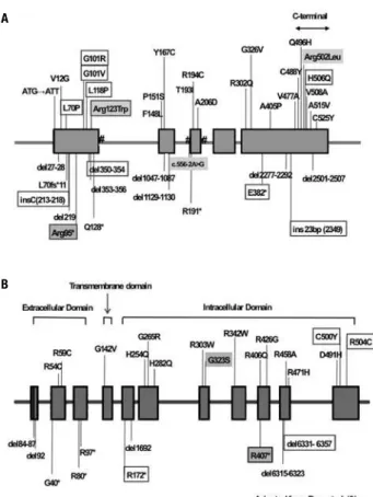
(7). AMH is a small gene containing ive exons located in chromosome 19p13.3 (8), and its protein product acts by means of its speciic type II receptor (AMHR2), a ser-ine/threonine kinase, member of the family of type II re-ceptors for TGF-b-related proteins (8). Several mutations in AMH have already been described (Figure 1) (9-11).
The AMHR2 gene, located in 12q13, was cloned
in 1994 by two different groups (12,13). AMHR2
contains 11 exons and more than 27 mutations have already been described in this gene (Figure 1) (5,9,14).
The aim of this study is to screen for mutations in
AMH and AMHR2 genes in patients with PMDS.
Figure 1. Schematic igure of AMH(A) and AMHR2(B) genes showing the location of the described mutations.
Missense mutations are shown above the genes. The boxes indicate recurrent mutations; gray background designates the mutations identiied in this study. #: shows the splicing site mutations.
A
Cop
yright
© ABE&M t
odos os dir
eit
os r
eser
vados
.
SUBJECTS AND METHODS
Patients
Eight unrelated patients with PMDS were selected. Five patients (patients 1, 2, 3, 4, and 5) were evaluated at the Unit of Endocrinology of Development at Hospital das Clínicas da Faculdade de Medicina da Universidade de São Paulo, and three patients (patients 6, 7 and 8) were evalu-ated in the Interdisciplinary Group for the Study of Sex Determination and Differentiation, Faculdade de Ciên-cias Médicas da Universidade Estadual de Campinas. Fifty ive normal males were used as the control group. This study was approved by the Ethical Committee of Hospital das Clínicas da Faculdade de Medicina da Universidade de São Paulo and Faculdade de Ciências Médicas da Univer-sidade Estadual de Campinas. Written informed consent was obtained from all participants of this study.
Clinical evaluation
The patients presented chronological age from four months to 37 years. Four patients reported having con-sanguineous parents. The main complaints were pres-ence of inguinal hernia and/or cryptorchidism. All sub-jects had male external genitalia and uni- or bilateral cryptorchidism. Four of them presented inguinal hernia. The presence of Müllerian derivatives was de monstrated by pelvic ultrasound and/or anatomopathological study. AMH levels were determined in three patients: 64.4 ng/mL in patient 7, and < 0.18 ng/mL in patient 8 (normal levels for their age: 33.8-110.8 ng/mL). and 4.6 ng/mL in patient 6 (normal levels for his age: 63.6-132.2). All of them had 46,XY karyotype.
Molecular evaluation
Genomic DNA samples were obtained from peri pheral blood leukocytes using the Salting Out technique (15). DNA was ampliied by PCR using speciic prim-ers for the AMH (ENSG00000104899) and AMHR2 (ENSG00000135409) genes (Table 1). All PCR re-actions were performed with 100-500 ng of genomic DNA, 200 mM of dNTP, 20 pmol of each primer, 0.5 units of Go Taq DNA polymerase (Promega Corpo-rations, Madison, WI, USA), 50 mM KCl, 1.5 mM MgCl2,and 10 mM Tris-HCl at pH 9.0, in a total vo lume of 25 mL. PCR conditions were: one cycle of 95oC for ive minutes, 35 cycles consisting of 97oC for
45 seconds; annealing temperatures (Table 1) for 45 seconds; 72oC for one or two minutes; and a inal cycle
of 72 oC for 10 minutes. Amplicons were sequenced
following the protocol of the ABI Prism BigDye Termi-nator Cycle Sequencing Ready Reaction Kit, and were placed in the ABI PRISM 3100 automatic sequencer (Life Technologies Corporation, CA, USA).
Cloning: The amplicon of AMH exon 1 was cloned
into pcDNA3.1/v5-His TOPO TA vector. The TOPO 10 E. coli were transformed with this vector using pcD-NA3.1/v5-His TOPO TA Expression Kit (Invitrogen, Life Technologies Corporation, CA, USA). Trans-formed E. coli were grown in selective medium (LB with 50 µg/mL ampicilin), the vector was extracted with QIAprep Spin Miniprep Kit (QIAGEN Inc., Valencia, CA, USA), and was sequenced as described above.
In silico analysis: The allelic variants identiied were
analyzed using the software BDGP Splice Site Predic-tion by Neural Network (http://www.fruitly.org/
Table 1. Primer sequences, annealing temperatures, and size of amplicons of AMH and AMHR2
Gene exon Forward primer (5’ – 3’) Reverse primer (5’ – 3’) Ann temp (oC) Size (bp)
AMH 1 1F – AAAcAccccAccTTccAcTc 1R – ccggcccAccTgAAggAA 60 530
AMH 2 2F – cAgggAcAgATcccAAAgAT 2R – TAcTgcAgAcccTgcAAcAA 60 288
AMH 3-4 3-4F – gTAgAgcggggcTgggTA 3-4R – cgcAATTggAggAgTTgAgA 57 540
AMH 5 5F – cTggAcAccgTgcccTTc 5R – TggggTccgAATAAATATgg 57 1080
AMHR2 1-2 1-2F – cAggATgcccTgTATcTgAAg 1-2R – acaccccaggatgtgtctgt 58 700
AMHR2 3-4 3-4F – cTcTgTTTccAcAccccATT 3-4R – ggAgAggggTcAgAgcTTTT 58 690
AMHR2 5-6 5-6F – gAcTcccATgAccTcTcAcAA 5-6R – cATgTAgcccccAccTcTAT 58 630
AMHR2 7 7F – ggATggATcAgccgTcTc 7R – AggcAgAATcAcAAAcATAgcA 61 233
AMHR2 8-9 8-9F – AAAAAgAgggAggAAgAAAATc 8-9R – ttggggtgaacctagaatgg 54 670
AMHR2 10 10F – cccTTTcTAcATggTAggcA 10R – AcgTccTTgAAgcccATgcccA 49 267
Cop
yright
© ABE&M t
odos os dir
eit
os r
eser
vados
.
seq_tools/splice.html), Mutation Taster (http://www. mutationtaster.org/), Human Splicing Finder (http:// www.umd.be/HSF/), Polyphen (http://genetics. bwh.harvard.edu/), and Sift (http://sift.jcvi.org/).
RESULTS
The AMH gene analysis revealed four mutations in ive patients. Two of them were novel mutations: c.556-2A>G (intron 2), and p.Arg502Leu (c.1505G>T, exon 5) (Table 2, Figures 1 and 2). The c.556-2A>G muta-tion, which involves the acceptor splicing site, was iden-tiied in homozygous state in three patients (1, 2 and
6). In silico analysis using the software BDGP Splice
Site Prediction by Neural Network, Mutation Taster, and Human Splicing Finder showed that this site is no longer recognized as a splicing site, and predicted that the mutation was the cause of the disease. Four already described allelic variants: c.146 T>G (rs10407022, p.Ser49Ile), c.252G>A (rs61736572, p.Leu84=), c.303G>A (rs61736575, p.Gly101=), c.555+50G>A (rs8112524) were also identiied in these patients in ho-mozygous state. The frequencies of the allelic variants c.146 T>G (rs10407022), c.252G>A (rs61736572), and c.303G>A (rs61736575) were 23/110 (20.9%), 2/110 (1.8%) and 2/110 (1.8%), respectively, in the normal male group. The homozygous p.Arg502Leu (c.1505G>T) mutation was identiied in patient 7. Mu-tation Taster software predicted that this muMu-tation was the cause of the disease; Polyphen suggested that this mutation was probably harmful, and according to Sift software, it would not be tolerated.
In patient 3, the p.Arg95* (c.283C>T), and p.Arg123Trp (c. 367C>T) mutations in exon 1 of
AMH were detected in compound heterozygous state.
In silico analysis of p.Arg123Trp mutation using the
Polyphen software predicted that this variant was prob-ably a damaging alteration, and Sift software suggested a non-tolerated alteration. The p.Arg123Trp mutation was also detected in patient 8 in homozygous state.
AMHR2 gene sequencing identiied the mutation
p.Gly323Ser (c.967G>A) and the allelic variants c.424-30C>T (rs2071557) and c.*13T>C (rs10876455) in homozygous state in patient 4. The mutation p.Arg407* (c.1219C>T) was found in patient 5 in homozygous state. In patients 2 and 3, the heterozygous allelic variant c.1038G>A (rs784890, p.Ser346=) was also detected. In
silico analysis of p.Gly323Ser mutation using Mutation
Taster and Polyphen software indicated that this altera-tion was probably damaging and caused the disease.
DISCUSSION
PDMS is one of the rarest causes of development dis-orders of male internal genitalia, and it is usually diag-nosed during surgical correction of hernia inguinalis. Mutations in AMH or AMHR2 genes, in similar pro-portions, are the cause of approximately 85% of PMDS cases (6,16,17). In the remaining patients, the cause of the PMDS is unknown (17).
Patients with AMH or AMHR2 gene mutations have no differences in anatomical phenotype. Normal-ly, AMH levels are measurable during childhood and decrease at puberty. Patients with AMH gene defects show low AMH levels from birth, whereas patients with mutations in AMHR have elevated AMH levels, indicating insensitivity of the target tissues (18). At
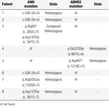
Table 2. AMH and AMHR2 gene sequencing results
Patient AMH
mutation State
AMHR2
mutation State
1 c.556-2A>g Homozygous nf -2 c.556-2A>g Homozygous nf -3 p.Arg95*
(c. 283c>T) p.Arg123Trp (c. 367c>T)
compound Heterozygous
nf
-4 nf - p.gly323Ser (c.967g>A)
Homozygous
5 nf - p.Arg407* (c.1219c>T)
Homozygous
6 c.556-2A>g Homozygous nf -7 p.Arg502Leu
(c.1505g>T)
Homozygous nf
-8 p.Arg123Trp (c. 367c>T)
Homozygous nf
-nf: not found.
Figure 2. Electropherograms showing the three novel mutations identiied in this study: A: c.556-2A>g; B: p.Arg502Leu (c.1505g>T); C:
p.gly323Ser (c.967g>A).
Normal sequences of the genes are shown above and mutated sequences are shown below. Arrows indicate nucleotide substitutions.
Cop
yright
© ABE&M t
odos os dir
eit
os r
eser
vados
.
post-pubertal age, AMH levels cannot be used to select which gene to study in PMDS patients (3).
Currently, the majority of AMH mutations de-scribed are located especially in exon 1, and in the 3’ portion of exon 5, which are important regions for AMH bioactivity (17). In the present study, the AMH
p.Arg95*, p.Arg123Trp and p.Arg502Leu mutations that were identiied were located in these regions, and
in silico analyses of p.Arg123Trp and p.Arg502Leu
mutations predicted that they were the genetic causes of PMDS in these patients. In addition, Arg502 amino acid is conserved in several organisms, showing its im-portance in this protein.
The p.Arg502Leu mutation was identiied in patient 7, who presented normal levels of AMH for his age, indicating that this mutation, because of its position, might affect hormone bioactivity, and not its secretion rate. Menabo and cols. also described an Italian patient with AMH mutations and normal AMH levels (10).
The previously described mutations in AMH, p.Arg95* and p.Arg123Trp, were found in compound heterozygous state in patient 3, demonstrated by exon 1 amplicon cloning. The p.Arg95* mutation leads to a premature stop codon, resulting in a truncated protein, lacking a large part of the normal protein sequence (19). Formerly, Imbeaud and cols. described a PMDS patient with undetectable AMH levels and p.Arg123Trp muta-tion (16). These indings were similar to those of pa-tient 8.
The novel homozygous c.556-2A>G mutation in patients 1, 2, and 6, located in the acceptor splicing site, abolished this site according to in silico analysis, indicating a possible genetic cause of the disease. Inter-estingly, these patients had the same three allelic vari-ants near this mutation, which might indicate a founder effect.
AMHR2 mutations were identiied in two patients.
The p.Gly323Ser mutation, present in patient 4, is lo-cated in exon 7, which is a component of the protein kinase domain in the cytoplasmic portion of AMHR2. Glycine is a polar and neutral amino acid, and serine is a nonpolar and hydrophobic amino acid; these distinct amino acid characteristics indicate that the amino acid change might affect protein conformation. In addition, Gly323 is highly conserved among mammals.
The p.Arg407* mutation leads to a premature stop codon. The resulting truncated protein lose a signii-cant portion of the serine/threonine kinase domain in the intracellular portion of the AMHR responsible for
the bioactivity of the protein (5). Abduljabbar and cols. also identiied this mutation in a family from Saudi Ara-bia that had consanguineous parents and four affected males. Molecular models of the intracellular domains of the wild type and mutated receptors showed that the mutated protein lost most of the αF helix and all of the four α-helices at the C terminus (5).
In conclusion, a likely molecular etiology was found in these eight patients with PMDS. Four mutations in the AMH gene and two in AMHR2 were identiied. Three of them are novel mutations, c.556-2A>G and p.Arg502Leu in AMH; and p.Gly323Ser in AMHR2. Further functional studies are necessary to conirm the
in silico analysis. The possibility of a founder effect of
the novel AMH mutation (c.556-2A>G) found in three Brazilian patients with the PMDS cannot be excluded.
Acknowledgements: this study was supported by grants from Conselho Nacional de Desenvolvimento Cientíico e Tecnológi-co (CNPq), n. 305743/2011-2 to B.B.M, n. 314392/2009-2 to G.G.J. and n. 302084/2009-6 to A.T.G.; and from Fun-dação de Amparo à Pesquisa do Estado de São Paulo (Fapesp) 06/50999-0 to A.Z.N.
Disclosure: no potential conlict of interest relevant to this article was reported.
REFERENCES
1. Jost A. Problems of fetal endocrinology: the gonadal and hy-pophyseal hormones. Recent Prog Horm Res. 1953;8:379-418. 2. Farikullah J, Ehtisham S, Nappo S, Patel L, Hennayake S.
Persis-tent Müllerian duct syndrome: lessons learned from managing a series of eight patients over a 10-year period and review of litera-ture regarding malignant risk from the Mullerian remnants. BJU Int. 2012; Epub 2012 Apr 30.
3. Rey R, Picard JY, Josso N. Síndrome de Persistência dos Ductos de Müller. In: Guerra ATM, Guerra Júnior G, editors. Menino ou Menina? Distúrbios da Diferenciação do Sexo. 2.ed. Rubio; 2010. p. 279-95.
4. Hutson JM, Davidson PM, Reece LA, Baker M, Zhou B. Failure of gubernacular development in the persistent Müllerian duct syndrome allows herniation of the testes. Pediatr Surg Int. 1994;9:544-6.
5. Abduljabbar M, Taheini K, Picard JY, Cate RL, Josso N. Mutations of the AMH Type II receptor in two extended families with persis-tent Müllerian duct syndrome: lack of phenotype/genotype cor-relation. Horm Res Paediatr. 2012;77(5):291-7.
6. Imbeaud S, Faure E, Lamarre I, Mattei MG, di Clemente N, Tizard R, et al. Insensitivity to anti-Müllerian hormone due to a muta-tion in the human anti-Müllerian hormone receptor. Nat Genet. 1995;11(4):382-8.
Cop
yright
© ABE&M t
odos os dir
eit
os r
eser
vados
.
8. Josso N, di Clemente N. TGF-beta family members and gonadal development. Trends Endocrinol Metab. 1999;10(6):216-22. 9. Josso N, Belville C, di Clemente N, Picard JY. AMH and AMH
re-ceptor defects in persistent Müllerian duct syndrome. Hum Re-prod Update. 2005;11(4):351-6.
10. Menabo S, Balsamo A, Nicoletti A, Gennari M, Pirazzoli P, Cicog-nani A, et al. Three novel AMH gene mutations in a patient with persistent Müllerian duct syndrome and normal AMH serum dos-age. Horm Res. 2008;70(2):124-8.
11. van der Zwan YG, Bruggenwirth HT, Drop SL, Wolffenbuttel KP, Madern GC, Looijenga LH, et al. A novel AMH missense mutation in a patient with persistent Müllerian duct syndrome. Sex Dev. 2012; Epub 2012 Jul 11.
12. Baarends WM, van Helmond MJ, Post M, van der Schoot PJ, Hoogerbrugge JW, de Winter JP, et al. A novel member of the transmembrane serine/threonine kinase receptor fam-ily is specifically expressed in the gonads and in mesen-chymal cells adjacent to the Müllerian duct. Development. 1994;120(1):189-97.
13. di Clemente N, Wilson C, Faure E, Boussin L, Carmillo P, Tizard R, et al. Cloning, expression, and alternative splicing of the receptor for anti-Müllerian hormone. Mol Endocrinol. 1994;8(8):1006-20.
14. Belville C, Marechal JD, Pennetier S, Carmillo P, Masgrau L, Mes-sika-Zeitoun L, et al. Natural mutations of the anti-Mülle rian hor-mone type II receptor found in persistent Müllerian duct syndrome affect ligand binding, signal transduction and cellular transport. Hum Mol Genet. 2009;18(16):3002-13.
15. Miller SA, Dykes DD, Polesky HF. A simple salting out procedure for extracting DNA from human nucleated cells. Nucleic Acids Res. 1988;16(3):1215.
16. Imbeaud S, Carre-Eusebe D, Rey R, Belville C, Josso N, Picard JY. Molecular genetics of the persistent Müllerian duct syndrome: a study of 19 families. Hum Mol Genet. 1994;3(1):125-31.
17. Josso N, Picard JY, Rey R, di Clemente N. Testicular anti-Müllerian hormone: history, genetics, regulation and clinical applications. Pediatr Endocrinol Rev. 2006;3(4):347-58.
18. Rey RA, Codner E, Iniguez G, Bedecarras P, Trigo R, Okuma C, et al. Low risk of impaired testicular Sertoli and Leydig cell func-tions in boys with isolated hypospadias. J Clin Endocrinol Metab. 2005;90(11):6035-40.