399
Communication/Comunicação
1. Programa Pós-graduação em Doenças Infecciosas, Núcleo de Doenças Infecciosas, Universidade Federal de Espírito Santo, Vitória, ES. 2. Departamento de Patologia, Universidade Federal do Espírito Santo, Vitória, ES.
Address to: Dra Cinthia Furst Leroy Gomes Bueloni. Av. Marechal Campos 1468, Maruípe, 29040-091 Vitória, ES, Brasil.
Phone/Fax: 55 27 2122-7295 e-mail: cinthiafurst@hotmail.com Received in 02/12/2010 Accepted in 01/02/2011
Revista da Sociedade Brasileira de Medicina Tropical 45(3):399-401, may-jun, 2012
Parasitological stool sample exam by spontaneous sedimentation
method using conical tubes: efectiveness, practice, and biosafety
Exame parasitológico de fezes pelo método de sedimentação espontânea utilizando tubos
cônicos: eicácia, praticidade e biossegurança
Steveen Rios Ribeiro
1and
Cinthia Furst
2ABSTACT
Introduction: Spontaneous sedimentation is an important procedure for stool examination. A modiication of this technique using conical tubes was performed and evaluated. Methods: Fity fecal samples were processed in sedimentation glass and in polypropylene conical tubes. Another 50 samples were used for quantitative evaluation of protozoan cysts. Results: Although no signiicant diferences occurred in the frequency of protozoa and helminths detected, signiicant diferences in protozoan cyst counts did occur. Conclusions: he use of tube predicts a shorter path in the sedimentation of the sample, increases concentration of parasites for microscopy analysis, minimizes the risks of contamination, reduces the odor, and optimizes the workspace.
Keywords: Spontaneous sedimentation. HPJ method. Intestinal parasites.
RESUMO
Introdução: A sedimentação espontânea é um procedimento importante para o exame parasitológico de fezes. Uma modificação desta técnica utilizando tubos cônicos foi realizada e avaliada. Métodos: Cinquenta amostras de fezes foram processadas em cálices de sedimentação de vidro e em tubos cônicos de polipropileno. Outras 50 amostras foram usadas para avaliação quantitativa de cistos de protozoários. Resultados: Embora não tenham sido encontradas diferenças signiicativas na frequência de helmintos e protozoários identiicados, houve diferença signiicativa na contagem de cistos de protozoários. Conclusões: O uso do tubo prevê um caminho mais curto na sedimentação da amostra, aumenta a concentração de parasitas para a análise microscópica, minimiza os riscos de contaminação, reduz o odor e otimiza o espaço de trabalho.
Palavras-chaves: Sedimentação espontânea. Método HPJ. Parasitas intestinais.
Intestinal parasite diseases are still considered a public health problem, especially in developing countries where sanitation conditions are inadequate1. Diagnosis of these parasites is based
on microscopic analysis of fecal samples aimed at detecting the evolutionary forms, whether as eggs, larvae, cysts, oocysts, or trophozoites; a successful outcome becomes more likely when using a technique based on the principle of concentrating these stages. Numerous laboratories use the sedimentation method Hofman, Pons and Janer (HPJ), because it is easy to perform, has low cost,
and shows good sensitivity for diagnosing both helminths and protozoa using fresh or preserved feces2. Since the description of
the use of sedimentation to diagnose parasites, reported by Lutz in 19193, several modiications of the technique have been proposed to
facilitate its implementation and increase its eiciency.
In this communication, our group proposes the use of 50-ml conical polypropylene tubes instead of traditional sedimentation glass to perform the HPJ method and evaluates the degree of agreement between the sedimentation methods.
Fity stool samples were used from the parasitology sector of the Department of Pathology of the Federal University of Espírito Santo. Feces were homogenized in the collection container, using a disposable spatula. Ater homogenization, they were separated into two aliquots of 10g: One aliquot was prepared by the HPJ method in sedimentation glass consisted of inverted cone of 200 ml capacity mounted on a base like a goblet, and the other was prepared for sedimentation in a 50-ml conical botom polypropylene tube (Falcon®-type tube). In both techniques, iltered water was used as homogenization solution, and the supernatant was discarded ater 1 hour of sedimentation. he sedimentation process was repeated twice to clarify the sediment. For each sediment sample, three slides (coverslip of 24×32mm) were examined under light microscopy on the same day by the same examiner using lugol for staining.
For quantitative evaluation of protozoan cysts, another 50 stool samples known to be positive for protozoa were used. Sedimentation of these samples was performed as described in the preceding paragraph. he supernatant of the inal sedimentation was discarded, and a 1-ml aliquot of sediment was transferred to a tube in which 4ml of 0.15M sodium chloride (NaCl) were added. Following homogenization, cyst counts were performed in a Neubauer Chamber, in duplicate.
Data analysis was performed using the Chi-square test to compare the frequency of parasites diagnosed with both methods and by the paired Student’s t-test to compare the results of the quantitative evaluation of protozoan cysts. Kappa statistics (k) were used to determine the strength of agreement between methods. he SPSS program, version 11.5 for Windows, was used, and p values less than 0.05 were considered statistically signiicant.
400
Ribeiro SR and Furst C - Parasitological stool sample exam by sedimentation method using conical tubes
TABLE 1 - Occurrence of helminths and protozoa detected by the sedimen-tation techniques in sedimensedimen-tation glass (HPJ) and in a 50-ml conical botom polypropylene tube (Tube) in 50 stool samples and the value of agreement (Kappa) between the two techniques.
Helminths Protozoa
k=0.82* k=0.95*
HPJ + HPJ - HPJ + HPJ
-Tube+ 15 3 15 1
Tube- 1 31 0 34
*Kappa value indicates almost perfect agreement. HPJ: Hofman, Pons and Janer.
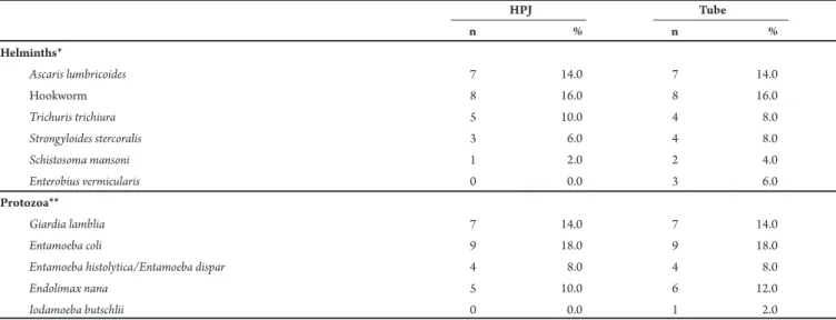
TABLE 2 - Comparison of the occurrence of helminths and protozoa observed in the parasitological exam of 50 stool samples, using sedimentation glass (HPJ) and a 50-ml conical botom polypropylene tube (Tube).
HPJ Tube
n % n %
Helminths*
Ascaris lumbricoides 7 14.0 7 14.0
Hookworm 8 16.0 8 16.0
Trichuris trichiura 5 10.0 4 8.0
Strongyloides stercoralis 3 6.0 4 8.0
Schistosoma mansoni 1 2.0 2 4.0
Enterobius vermicularis 0 0.0 3 6.0
Protozoa**
Giardia lamblia 7 14.0 7 14.0
Entamoeba coli 9 18.0 9 18.0
Entamoeba histolytica/Entamoeba dispar 4 8.0 4 8.0
Endolimax nana 5 10.0 6 12.0
Iodamoeba butschlii 0 0.0 1 2.0
*χ2: 3.30, p: 0.653; **χ2: 1.02, p: 0.907; HPJ: Hofman, Pons and Janer.
TABLE 3 - Mean number of cysts per ml of sediment, quantiied in a Neubauer Chamber, ater sedimentation in conventional sedimentation glass (HPJ) and conical botom polypropylene tube (Tube) of 50 stool samples positive for protozoa.
Mean ± SEM Mean ± SEM p
Protozoa N HPJ Tube value*
Entamoeba coli 16 17578.1±6707.6 24609.3±6390.8 0.040
Endolimax nana 17 86029.4±33663.8 106250.0±42497.8 0.296
Giardia lamblia 10 40000.0±14942.0 77500.0±25141.9 0.034
Isospora belli 5 1250.0±1250.0 3750.0±1530.9 0.374
Entamoeba histolytica/Entamoeba dispar 2 0.0±0.0 3125.0±3125.0 0.500
Total 50 43000.0±12673.0 60000.0±16159.4 0.022
SEM: standard error of the mean, HPJ: Hofman, Pons and Janer; N: number of stool samples. *paired Student’s t-test.
Of the 50 fecal samples examined, 16 were positive for helminths by the HPJ method and 18 using the Falcon®-type tube. Regarding protozoans, 15 were positive by HPJ and 16 by the Falcon®-type tube.
Almost perfect agreement was achieved in the diagnoses of helminths (κ=0.82) and protozoa (κ=0.95) (Table 1).
No statistically signiicant diferences in the data were veriied when the individual frequencies of species of nematodes and protozoa were compared between methods using the Chi-square test (Table 2).
Quantitative analysis of protozoan cysts showed a tendency to recover a greater number of cysts for the conical tube. Statistically signiicant diferences were veriied when using the paired Student’s t-test (Table 3).
Analysis of the results revealed that spontaneous sedimentation using the conical tube was consistent with that observed in the HPJ method (Table 1). When a nonsigniicant disagreement occurred between the two methods, in the majority of cases, it was favorable to the use of the Falcon®-type tube in relation to traditional sedimentation (Table 2). One example was Enterobius vermicularis, identiied in three cases using the conical tube method, but no cases were identiied using the traditional HPJ method. Considering that eggs of E. vermicularis can occur in reduced numbers in fecal samples, oten presenting false negative results in the HPJ method, the conical
tube showed the potential of increasing the chances of detecting the eggs due to improved sedimentation, aided by internal vertical walls, in contrast to the angled walls of the sedimentation glass.
401
Rev Soc Bras Med Trop 45(3):399-401, may-jun, 2012
ACKNOWLEDGMENTS
he authors declare that there is no conlict of interest.
CONFLICT OF INTEREST
REFERENCES
he space used to perform the method was smaller, as conical tubes can be maintained in compact supports, reducing the risk of accidents and improving safety during the laboratory routine.
Observation of the slides under an optical microscope veriied that the proposed change presented beter results in relation to the number of protozoan cysts visualized, a result that was conirmed by the quantitative evaluation (Student’s t-test).
In conclusion, the use of conical polypropylene tubes for spontaneous sedimentation presented practical advantages over the use of sedimentation glass, especially in relation to safety in the laboratory, the elimination of odors, and the smaller area used; it also permited greater concentration of protozoan cysts.
he authors would like to thank the staf of the Laboratory of Parasitology, particularly Mr. Fausto Edmundo Lima Pereira and Mrs. Adriana Oliveira Costa, and the Federal University of Espírito Santo.
1. Frei F, Juncansen C, Ribeiro-Paes JT. Epidemiological survey of intestinal parasite infections: analytical bias due to prophylactic treatment. Cad Saude Publica 2008; 24:2919-2925.
2. Hofman WA, Pons JA, Janer JL. he sedimentation-concentration method in Schistosomiasis mansoni. Puerto Rico J Publ Hlth 1934; 9:281-298.