1 Private Bilecik Orhangazi Dialysis Center, Bilecik, Türkiye
2 Department of Medical Biology, Faculty of Medicine, Turgut Özal University, Ankara, Türkiye 3 Department of Chemistry, Ankara Regional Office of Council of Forensic Medicine, Ankara, Türkiye
4 Division of Medical Biochemistry Laboratory, Gölbaşı Hasvak State Hospital, Ankara, Türkiye 5 Department of Periodontology, Faculty of Dentistry, Adnan Menderes University, Aydın, Türkiye
6 Department of Biology, Faculty of Education, Gazi University, Ankara, Türkiye Yazışma Adresi /Correspondence: Veli Uğurcu,
Private Bilecik Orhangazi Dialysis Center, Bilecik, Turkey Email:[email protected]
Geliş Tarihi / Received: 17.06.2014, Kabul Tarihi / Accepted: 15.07.2014 ORIGINAL ARTICLE / ÖZGÜN ARAŞTIRMA
The effects of insulin on the expression levels of ADAMTS6 & 19 in OUMS-27 cell
İnsülinin OUMS-27 hücrelerinde ADAMTS6 ve 19 ekspresyon düzeylerine etkileri
Veli Uğurcu1, Sümeyya Akyol2, Aynur Altuntaş3, Rıdvan Fırat4, S. Fatih Kurşunlu5, Özlem Çakmak6, Kadir Demircan2
ÖZET
Amaç: A Disintegrin-like Metalloproteinase with Throm-bospondin Motifs (ADAMTS) proteinleri ekstraselüler matrikste (ECM) bulunan bir çeşit matriks metalloprotei-naz enzimidir. İnsulin birçok dokuda etki gösteren önemli bir anabolik hormondur. Bu çalışmanın amacı OUMS-27 insan kondrosarkom hücre kültüründe insülinin, fonksi-yonları tam olarak bilinmeyen ADAMTS enzimlerinden ADAMTS6 ve 19 ekspresyonu üzerine zamana bağımlı olarak etkilerini değerlendirmek ve insülinin anabolik etki-lerinden dolayı ADAMTS ekspresyonunu azaltma hipote-zini test etmektir.
Yöntemler: OUMS-27 hücrelerinin, 10 μg/mL insulin içe-ren ve içermeyen Dulbecco’s modified Eagle besiyerinde (DMEM) kültürü yapıldı. 11. güne kadar iki günde bir be-siyeri ile birlikte insülin takviyesi yapıldı. Hücreler 1, 3, 7 ve 11. günlerde harvest edilerek uygun zamanlarda RNA izolasyonu yapıldı. ADAMTS6 ve 19 RNA ekspresyonları uygun primerler kullanılarak qRT-PCR yöntemi ile ölçüldü. Bulgular: qRT-PCR cihazının verilerine göre, insülin uy-gulanmasından 1 gün sonra başlamak üzere deneyin son günü olan 11. güne kadar ADAMTS6’nın mRNA düzeyinde ciddi bir azalma meydana geldi (p=0.008). İnsülin uygu-lanan gruplarda ADAMTS6 oranı kontrol grubuna kıyasla 1/2 ve 1/4 aralığında değişti. İnsülin verilen hücre grubun-da ADAMTS19 mRNA düzeyindeki değişiklikler ise kontrol grubuna kıyasla istatistiksel olarak anlamsızdı.
Sonuç: Sonuçlarımız, insülinin ADAMTS6 düzeylerini azaltarak ekstrasellüler matriks bileşenlerinin kaybının telafi edilmesinde potansiyel bir etkisinin olabileceğini gösterdi. Bu hipotez ve bulgunun anlaşılması için yetim ADAMTS proteinlerinin gerçek fonksiyonlarının incelene-ceği çok sayıda araştırma yapılmasına ihtiyaç vardır. Anahtar kelimeler: İnsulin, ADAMTS6, ADAMTS19, kondrosarkom, OUMS-27, RNA
ABSTRACT
Objective: A Disintegrin-like Metalloproteinase with Th-rombospondin Motifs (ADAMTS) proteins are kind of matrix metalloproteinase enzymes that primarily founds in the extracellular matrix (ECM). Insulin is an important anabolic hormone, which acts on many tissues. The aim of this study is to evaluate the time-dependent effects of insulin on the two functionally unknown enzyme expres-sions (ADAMTS6 & 19) in OUMS-27 human chondrosar-coma cell line.
Methods: OUMS-27 cells were cultured in Dulbecco’s modified Eagle’ medium (DMEM) alone and DMEM con-taining 10 μg/mL insulin. The medium was changed every other day up to 11th day. Cells were harvested at 1, 3, 7, and 11th days and RNA isolation was performed at ap-propriate times according to study setup. The levels of RNA expression of ADAMTS6 and 19 were estimated by qRT-PCR using appropriate primers.
INTRODUCTION
Articular cartilage is a specialized connective tissue found in many areas of the body such as joints, ear, nose, intervertebral disk etc. Cartilage matrix com
-prises of collagen type 2, 9, and 11, aggrecan, carti
-lage oligomeric matrix protein and other compounds [1]. Some members of matrix metalloproteinase (MMP) and A Disintegrin-like Metalloproteinase with Trombospondin Motifs (ADAMTS) have been implicated in cartilage matrix destruction [2].
ADAMTS proteins are a kind of matrix metal
-loproteinase and have 19 members [3]. They take part in many physiological mechanisms such as blood coagulation, ovulation, extracellular matrix turnover, and have many important roles at patho
-logical processes such as angiogenesis and can
-cer [4]. These proteins perform their functions by breakdown structural matrix proteins such as ag
-grecan, versican, and breavican. ADAMTS proteins are classiied according to their functions, while some ADAMTS proteins are classiied as “orphan ADAMTS” (including ADAMTS 6, 19, and etc.) because their functions are not known clearly [5].
Chondrosarcomas are heterogeneous tumors that produce cartilage [6], which are the third most common primary bone malignancy following my
-eloma and osteosarcoma [7]. These tumors grow slowly and have low potential of metastasis. They produce and secrete some types of ADAMTS pro
-teins. OUMS-27 cell line is one of the most known human chondrosarcoma cell lines that are used in cell culture experiments.
Insulin is an anabolic hormone that produced at β cells of Langerhans islets in response to high glucose levels. Many functions are described such as glucose to glycogen conversation, inhibiting glucose production at liver. Its metabolic effects begin with binding of insulin to a speciic receptor at plasma membrane [8]. Regardless of its mecha
-nism of action, a role of insulin in the metabolism of proteoglycans in the extracellular matrix of carti
-lage may have considerable biological implications [9]. The retarded wound healing and bone frac
-tures, long bones’ susceptibility to fractures [10], the unsuccessful attempts for the maxillary sinus graft procedure, also referred to as maxillary sinus loor elevation [11] operations are currently main problems in patients with uncontrolled diabetes
mellitus, especially in type 2 form. Therefore, this study was planned to evaluate the time-dependent effects of insulin on the chondrogenic ADAMTS 6 and ADAMTS 19 expressions in OUMS-27 chon
-drosarcoma cell line. Based on this, we also aimed to illuminate the regulatory role of insulin on these ADAMTS proteins and the function of ADAMTS 6 and 19 in OUMS-27 cells.
METHODS
OUMS-27 cell culture: OUMS-27 cell line is one
of the most known human chondrosarcoma cell lines that are frequently used in cell culture experiments. OUMS-27 chondrosarcoma cells were kindly provid
-ed by Dr. T. Kunisada (Okayama University Graduate School of Medicine and Dentistry, Okayama, Japan). Cells were cultured in Dulbecco’s modiied Eagle’s medium (DMEM) containing 10% fetal bovine serum and penicillin/streptomycin at 37°C in a humidiied atmosphere of 5% CO2 in air. The cells were sub-cultured at split ratios of 1:2-1:4 using trypsin plus EDTA every 7-10 days. Cells were used at passages 7-14 for all experiments. The medium was changed every other day with either control media or control media supplemented with 10 μg/mL insulin for a total of 11 days. Four groups of cells were subjected to in
-sulin: For 1st day experiment, 2x10⁵ cells, for 3th day
experiment 1x10⁵ cells, for 7th day experiment 5x10⁴
cells, and 3x10⁴ cells were plated in 20-mm dishes
and exposed to the same concentrations of insulin at the days indicated. After the experiment, cells were harvested and total RNA measurements were per
-formed.
Total RNA isolation: Total RNA was extracted
with TRIzol (Invitrogen, Carlsbad, CA, USA) ac
-cording to the manufacturer’s instructions. Two mi
-crograms RNA were reverse transcribed with Rever
-Tra Ace (Thermo Scientiic, Waltham, MA, USA) and random hexamers (Thermo Scientiic, Waltham, MA, USA) with random primers according to the manufacturer’s instruction (Table 1). Human glycer
-aldehyde-3-phosphate dehydrogenase (GAPDH) was ampliied as a control for the PCR reaction. Samples lacking reverse transcriptase were ampliied as a con
-trol for genomic DNA contamination. RNase-free water was used to elute total RNA from each sample. UV spectrophotometry was used to quantify and de
ADAMTS6 Forward GTGGCCCGCTTAATTGTTCTC 71bp product Reverse AGGGACTTGTCTGCATGGTG
ADAMTS19 Forward CAGTCTGAGTGTGCAGGTCA 137 bp product
Reverse TGCCAAATTCTTCATGTTGCCA
GAPDH Forward CCTGCACCACCAACTGCTTA 108 bp product
Reverse TCTTCTGGGTGGCAGTGATG Table 1. The forward and reverse
primers used in the real-time poly-merase chain reaction analyses for ADAMTS6, ADAMTS19, and GAPDH.
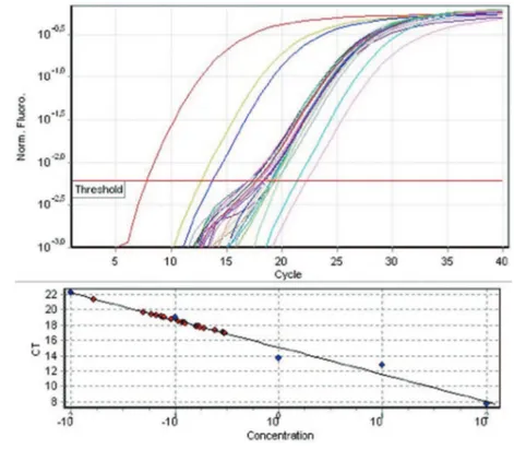
Real-time PCR: qRT-PCR was performed on
cDNA samples obtained (Qiagen Rotor-Gene Q RT-PCR, Limburg, Netherlands) as described in our previous report [1]. Total RNA RT-PCR sec
-tion uses the intercalating dye SYBR green (Ther
-mo Scientiic Maxima SYBR Green/ROX qPCR Master Mix) in the presence of primer pairs. The PCR mixture consisted of SYBR Green PCR Mas
-ter Mix, which includes DNA polymerase, SYBR Green I Dye, dNTPs including dUTP, PCR buffer, 10 pmol forward and reverse primers and cDNA of samples in a total volume of 20 μL. The ampliica
-tion of a housekeeping gene, GAPDH, was used for normalizing the eficiency of cDNA synthesis and the amount of RNA applied. PCR was performed with initial denaturation at 95°C for 5min, followed by ampliication for 40 cycles, each cycle consist
-ing of denaturation at 95°C for 10 s, anneal-ing at
57°C for 30 s, polymerization at 72°C for 30s and, the last stage, polymerization at 72°C for 5min. The results pertaining to ADAMTS6 and ADAMTS19 were represented as graphic charts. The bars and error bars represent mean and standard deviation, respectively.
Statistical Analyses
Statistical Package for Social Studies (SPSS) ver
-sion 16.0 was used for all statistical tests. Non
-parametric Kruskal Wallis Test was applied. The relationships between the variables were tested by Mann-Whitney U test. p<0.05 was accepted as sig
-niicant.
RESULTS
We irst examined whether the expressions of the ADAMTS6 and 19 genes are induced or suppressed
upon insulin application in OUMS-27 cells. As shown in Figure 2, according to qRT-PCR analy
-ses, ADAMTS6 mRNA expression signiicantly de
-creased as early as 1st day after insulin induction compared to control group (p=0.008). At day 3, AD
-AMTS6 level was the same as seen in day 1 when compared to control group (p=0.008). The decreas
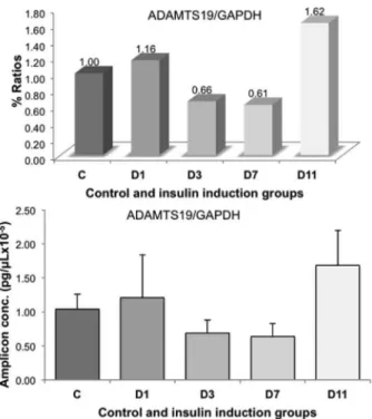
-es in ADAMTS6 mRNA levels were continued to decrease in day 7 up to 1/4 ratio compared to control group’s level (p=0.008). ADAMTS6 mRNA level was 1/2 of control values in day 11 again as seen in days 1 and 3 (p=0.027). There were statistically signiicant differences between the insulin-applied groups such as D1-D7 (p=0.047), D3-D7 (p=0.008), and D7-D11 (p=0.014). As shown in Figure 4, ac
-cording to qRT-PCR analyses, ADAMTS19 mRNA expression increased in day 1, decreased in days 3 and 7, and again increased at day 11 when compared to control group but the differences were not statis
-tically signiicant (p>0.05).
Figure 2. The results of ADAMTS6 qRT-PCR calculations of 5 different experiments. The values were standardized by division of ADAMTS6 to GAPDH. There is statistically signiicant differences between C-D1 (p=0.008), C-D3 (p=0.008), C-D7 (p=0.008), C-D11 (p=0.027), D1-D7 (p=0.047), D3-D7 (p=0.008), and D7-D11 (p=0.014)
Figure 4. The results of ADAMTS19 qRT-PCR calcula-tions of 5 different experiments. The values were stan-dardized by division of ADAMTS19 to GAPDH. There is no statistically signiicant difference neither between con-trol and insulin induction groups nor among insulin induc-tion groups
DISCUSSION
This study was undertaken to compare the mRNA levels of ADAMTS6 and 19 in OUMS-27 cells in response to insulin hormone application. We have shown that insulin had potential positive effects on matrix metabolism in isolated human chondrosarco
-ma cell line. Insulin suppressed ADAMTS6 mRNA, and probably as a result, ADAMTS6 enzyme activ
-ity, which is a potential factor for removal of sur
-rounding extracellular matrix. Therefore, we sug
-gested that insulin inhibited matrix breakdown and stimulated matrix synthesis in OUMS-27 cells at least by decreasing ADAMTS6 levels even if its function is not known exactly. On the other hand, another orphan ADAMTS member, ADAMTS19, was not affected by insulin induction.
ADAMTS family members of matrix metal
-loproteinases are zinc-dependent endopeptidases known for their ability to degrade one or more extracellular matrix constituents, as well as non-matrix proteins [12]. A broad spectrum of several pathological conditions are associated with the me
-talloproteinases such as Alzheimer’s disease, Par
-kinson’s disease, cerebral aneurysm, neurodegener
-ative diseases, cancer, human periodontal diseases, rheumatoid arthritis, atrial cardiomyopathy, cardiac ibrosis, congestive heart disease, atherosclerosis, chronic obstructive pulmonary diseases, asthma, acute respiratory distress syndrome, liver ibrosis, and chronic ulcers [12]. Orphan ADAMTSs are such an ADAMTS group that their function of sub
-strate cannot be shown to date. These are currently ADAMTS6, 7, 10, 12, 16, 17, 18, and 19. Although the exact mechanism is not known, ADAMTS16 has been shown to be dysregulated in osteoarthri
-tis [13]. The chromosomal location of ADAMTS6 gene is 5q12 and ADAMTS19 gene 5q31 [5]. These two ADAMTS are very closely located on chromo
-some 5 and most probably, they show similar func
-tioning pattern though it could not be enlightened to date.
Chondrocytes like OUMS-27 cells synthesize and secrete some macromolecules that constitute the extracellular matrix of cartilage. Glucose is a fundamental nutrient in cartilage tissues for both metabolic and structural needs. Because glucose also serves as a source for glucose-derived sugars such as glucosamine sulfate and vitamin C, which are of great importance in the maintenance, repair and remodeling of cartilage [14], insulin stands an important junction to moderate this anabolic/cata
-bolic events. The studies in this ield have dem
-onstrated that chondrosarcoma chondrocytes is uniquely responsive in vitro to physiological con
-centrations of pig insulin, producing cartilage-like proteoglycans [15, 16], type II collagen [17], hyal
-uronic acid [18] and some other secretory proteins [19]. Glade et al. showed the modulatory effects of insulin and thyroid hormones on chondrocyte me
-tabolism via multiple biosynthetic/receptor path
-ways [20]. It should be noted that our insulin ap
-plication procedure was limited to 11 days because of our previous experience that the cells could not be appropriately manipulated after that time. There
-fore, by adding supplementary materials to extend the viability time-course of cells, the experiments should be repeated in future to achieve more certain and clear results of long-term insulin induction ef
The effects of insulin to ADAMTS expression has not been studied much. Recently at our study we found that insulin increases the expression of ADAMTS13 but this increase was not statistically signiicant [21]. Insulin and other ADAMTS rela
-tionships should be investigated in detailed later. Our results demonstrated that insulin mitigated loss of extracellular matrix compounds and prob
-ably stimulated de novo synthesis of new molecules to replace lost ones. To test this hypothesis, future experiments investigating the extracellular matrix molecules such as versican, brevican, and aggre
-can as well as their breakdown products together with other orphan ADAMTS members should be planned. These studies will also play an important role in determining the functions of orphan AD
-AMTS members, which has not been explained up to now, and contributing to the related literature.
REFERENCES
1. Demircan K, Hirohata S, Nishida K, et al. ADAMTS-9 is synergistically induced by interleukin-1beta and tumor ne
-crosis factor alpha in OUMS-27 chondrosarcoma cells and in human chondrocytes. Arthritis Rheum 2005;52:1451-1460.
2. Koshy PJ, Lundy CJ, Rowan AD, et al. The modulation of matrix metalloproteinase and ADAM gene expression in human chondrocytes by interleukin-1 and oncostatin M: a time-course study using real-time quantitative reverse transcription-polymerase chain reaction. Arthritis Rheum 2002;46:961-967.
3. Apte SS. A disintegrin-like and metalloprotease (reprolysin type) with thrombospondin type 1 motifs: the ADAMTS family. Int J Biochem Cell Biol 2004;36:981-985.
4. Kumar S, Rao N, Ge R. Emerging Roles of ADAMTSs in Angiogenesis and Cancer. Cancers (Basel) 2012;4:1252-1299.
5. Porter S, Clark IM, Kevorkian L, Edwards DR. The AD
-AMTS metalloproteinases. Biochem J 2005;386:15-27. 6. WHO: Cartilage tumours. In: World health organization clas
-siication of tumours. Pathology and genetics - Tumours of soft tissue and bone, Fletcher CDM, Unni KK, Mertens F (Eds), 2002. p.234.
7. Dorfman HD, Czerniak B. Bone cancers. Cancer 1995; 75: 203-210.
8. Murray RK, Granner DK, Mayes PA, Rodwell VW, Harper’s Illustrated Biochemistry, 26th Edition. USA: McGraw-Hill Companies, 2003:466.
9. Schiller S, Dorfman A. The metabolism of mucopolysaccha
-rides in animals. IV. The inluence of insulin. J Biol Chem 1957;227:625-632.
10. Kayal RA, Alblowi J, McKenzie E, et al. Diabetes causes the accelerated loss of cartilage during fracture repair which is reversed by insulin treatment. Bone 2009;44:357-363. 11. Hou CJ, Liu JL, Li X, Bi LJ. Insulin promotes bone forma
-tion in augmented maxillary sinus in diabetic rabbits. Int J Oral Maxillofac Surg 2012;41:400-407.
12. Mandal M, Mandal A, Das S, et al. Clinical implica
-tions of matrix metalloproteinases. Mol Cell Biochem 2003;252:305-329.
13. Kevorkian L, Young DA, Darrah C, et al. Expression proil
-ing of metalloproteinases and their inhibitors in cartilage. Arthritis Rheum 2004;50:131-141.
14. Maor G, Vasiliver-Shamis G, Hazan-Brill R, et al. GLUT4 in murine bone growth: from uptake and translocation to proliferation and differentiation. Am J Physiol Endocrinol Metab 2011;300:E613-623.
15. Stevens RL, Nissley SP, Kimura JH, et al. Effects of insu
-lin and multiplication-stimulating activity on proteoglycan biosynthesis in chondrocytes from the Swarm rat chondro
-sarcoma. J Biol Chem 1981;256:2045-2052.
16. McCumbee WD, Lebovitz HE. Hormone responsiveness of a transplantable rat chondrosarcoma. I. In vitro effects of growth hormone-dependent serum factors and insulin. Endocrinology 1980;106:905-910.
17. Bembenek ME, Willis DH, Jr., Liberti JP. The effect of insulin on collagen production in isolated chondrosar
-coma chondrocytes. Biochem Biophys Res Commun 1982;106:338-345.
18. Mason RM, Kimura JH, Hascall VC. Biosynthesis of hyal
-uronic acid in cultures of chondrocytes from the Swarm rat chondrosarcoma. J Biol Chem 1982;257:2236-2245. 19. Stevens RL, Hascall VC. Characterization of proteoglycans
synthesized by rat chondrosarcoma chondrocytes treated with multiplication-stimulating activity and insulin. J Biol Chem 1981;256:2053-2058.
20. Glade MJ, Kanwar YS, Stern PH. Insulin and thyroid hor
-mones stimulate matrix metabolism in primary cultures of articular chondrocytes from young rabbits independently and in combination. Connect Tissue Res 1994;31:37-44. 21. Fırat R, Akyol S, Kurşunlu SF, et al. ADAMTS13 expres