Aline Ferreira de ALMEIDA(a)
Eliana do Nascimento TORRE(b)
Maicon dos Santos SELAYARAN(b)
Fábio Renato Manzolli LEITE(a)
Flávio Fernando DEMARCO(b)
Alessandro Dourado LOGUERCIO(c)
Adriana ETGES(a)
(a)Universidade Federal de Pelotas - UFPel, School of Dentistry, Department of Semiology and Clinics, Pelotas, RS, Brazil.
(b)Universidade Federal de Pelotas - UFPel, School of Dentistry, Department of Restorative Dentistry, School of Dentistry, Pelotas, RS, Brazil.
(c)Universidade Estadual de Ponta Grossa - UEPG, School of Dentistry, Department of Restorative Dentistry, Ponta Grossa, PR, Brazil.
Genotoxic potential of 10% and 16%
Carbamide Peroxide in dental bleaching
Abstract: Dental bleaching has become one of the most frequently
requested esthetic treatments in dental ofices. Despite the high clinical success observed with this procedure, some adverse effects have been reported, including a potential for developing premalignant lesions, root resorption and tooth sensitivity, especially when misused. The aim of this study was to evaluate the genotoxic response using a micronucleus (MN) assay, after the application of two concentrations of carbamide peroxide. Thirty-seven patients were divided into two groups and randomly received either a 10% carbamide peroxide (CP) (19) or a 16% carbamide peroxide (18) concentration for 21 days in individual dental trays. Gingival margin cells were collected immediately before the irst use (baseline), and then 15 and 45 days after baseline. The cells were placed on a histological slide, stained by the Feulgen technique, and evaluated by an experienced blinded examiner. One thousand cells per slide were counted, and the MN rate was determined. The two groups were analyzed by the Wilcoxon rank-sum test and the Kruskal-Wallis equality-of-populations rank test. A slight increase in MN was observed for both groups, in comparison with the baseline, at 15 days. However, no difference was observed between the two groups (10% and 16%), at either 15 or 45 days (p = 0.90). When bleaching is not prolonged or not performed
very frequently, bleaching agents containing carbamide peroxide alone will not cause mutagenic stress on gingival epithelial cells.
Keywords: Tooth Bleaching; Peroxides; Cytology; Micronucleus Tests.
Introduction
Tooth discoloration may be inluenced by a combination of intrinsic and extrinsic factors. Intrinsic stains are related to the properties of enamel and dentin, whereas extrinsic stains are associated with deposition of food and beverages onto the tooth surface.1
Tooth bleaching with custom trays for home use is considered the most common bleaching procedure recommended by dentists to patients.2
The procedure usually involves the use of 10% carbamide peroxide (CP) applied in a tray and worn by the patient overnight for at least 2 weeks. This bleaching agent is considered safe, has few side effects, and presents excellent esthetic results.3 CP at 10% is the only bleaching agent to receive
the seal of acceptance by the American Dental Association4 (ADA), assuring
its safety and eficacy for at-home tooth bleaching. Some authors have suggested that a higher concentration of bleaching agent could improve and
Declaration of Interests: The authors certify that they have no commercial or associative interest that represents a conflict of interest in connection with the manuscript.
Corresponding Author: Eliana do Nascimento Torre E-mail: [email protected]
DOI: 10.1590/1807-3107BOR-2015.vol29.0021
Submitted: Jun 13, 2014
accelerate the bleaching effect and make it last longer.5
However, long-term clinical trials have shown that both 10% and 16% CP produced a similar whitening effect.3 Moreover, a greater concentration of CP may
also increase the side effects.3 The most commonly
reported side effects of at-home bleaching treatments are tooth sensitivity and gingival irritation, which tend to disappear quickly after the bleaching treatment is stopped or when a remineralizing agent is applied.6
Concerns have also been raised about the potential of bleaching treatments to cause premalignant oral lesions.7 Hydrogen peroxide (H
202) has been found
to enhance the carcinogenic effects of potent DNA reactive carcinogens in experimental animals, but these experimental conditions are artiicial; they involve high levels of exposure and are of no relevance to potential human exposure to low quantities of H202 from tooth
whitening products.8 However, during the bleaching
process, CP breaks down into H202 and urea, which
are then dissociated into oxygen, water, ammonia, carbon dioxide,9 and reactive oxygen species (ROS)
that are considered potentially carcinogenic agents able to cause damage to proteins and changes in the cell nucleus.10 Thus, there is a gap in the literature on the
real genotoxicity of dental whitening agents in humans. A modern phenomenon has emerged called “bleachorexics” or “whitening junkies,” i.e., individuals
who are constantly bleaching their teeth.1 CP or H 202 may cause genotoxic effects when associated with
other well-known carcinogenic products (e.g., alcohol
and tobacco), or when whitening agents are used frequently at high concentrations.11
The DNA of cells exposed to chemical or physical agents may become damaged; chromosomal fragments, called micronucleus (MN), are observed as a result of atypical mitoses. Depending on the extent of cellular damage, the consequences may include impairment of the cell cycle, cell death, and even formation of a neoplasm.11,12 However, there are few scientiic reports available on in vivo studies regarding the genotoxic
effects of CP agents; most reports have evaluated
in vitro studies.13 Thus, the aim of this study was
to evaluate the in vivo genotoxic effect of two CP
concentrations (10% and 16%) in gingival epithelial cells of patients undergoing tooth whitening using the tray technique.
Methodology
Tooth bleaching
This study was approved by the Local Ethics Committee (protocol number 51/04). Each volunteer
received written information about treatment risks
and beneits, and signed an informed consent form before enrolling in the study. The patients were selected from a previously conducted double-blind clinical trial aimed at evaluating the eficacy and safety of two CP concentrations (10% and 16%; Whiteness Perfect, FGM Dental Products, Joinville, Brazil) for a home bleaching treatment. Patients with active caries, periodontal disease, previous hypersensitivity, smokers, alcohol drinkers, and pregnant or lactating women were excluded. The examiners and participants
were blinded to the concentration of the agent that
was being delivered. A more detailed description of
the methodology of the earlier clinical trial is given in Meireles et al.3
An alginate impression (Jeltrate regular set, Dentsply International Inc., Milford, USA) was taken, and a stone
mold was prepared. The buccal surfaces of the anterior teeth in each mold received ive coats of nail polish, starting approximately 1.0 mm above the gingival margin. The custom trays were fabricated using a 0.9 mm thick soft vinyl material (FGM Dental Products, Joinville, Brazil) and a vacuum process (Bio-art, Sao Paulo, Brazil). The trays were trimmed on the buccal and lingual surfaces 1.0 mm above the gingival margin.
Before starting treatment, the participants were given the trays and three tubes of bleaching gel. They were instructed to dispense the same amount
of gel into the trays each day and insert them into their mouth to cover at least the anterior teeth for
2 h daily for 3 weeks. Participants bleached both
their maxillary and mandibular arches at the same
time. The use of bleaching agents was standardized according to the manufacturer’s instructions. All patients received a hands-on practical demonstration and written instructions regarding both the proper
use of the bleaching agents and the dietary restrictions
during treatment. The participants also received
toothbrushes and dentifrice without whitening agents
to standardize their oral hygiene regimen.3
The participants included 30 women and 7 men. The average age of the volunteers was 28.14 ± 7.94 years for men and 27.5 ± 6.82 years for women.The researchers and participants were blinded to the CP concentration used by each patient. For the genotoxicity study, gingival margin cells were collected from each patient on three occasions: immediately before the bleaching treatment (baseline), at 15 days and at 45 days after starting the bleaching treatment. Cells were collected from marginal gingiva, from premolar to premolar of both jaws.
Cytology and MN assay
Cells from the gingival margin were collected with cytological brushes (Vagispec, Adlin Plásticos Ltda., Jaraguá do Sul, Brazil) and transferred to a
centrifuge tube containing phosphate-buffered saline, pH 6.8 (Gibco, Invitrogen, Carlsbad, USA). They were then centrifuged for 10 min before being ixed with methanol (Vetec, Xerém, Brazil) /acetic acid (3:1) (Synth, Diadema, Brazil). Hydrolysis was performed using 1 N HCl (Synth, Diadema, Brazil) at 60°C for 10 min, and the slides were stained using Schiff Fast Green (Sigma Aldrich, St Louis, USA), according to the method described by Roth et al.14
An experienced examiner (AE), pre-calibrated and blinded to the experimental conditions, evaluated the presence of MN in an optical microscopic (Olympus CX 21, São Paulo, Brazil) under 400× magniication. When doubt arose in counting the cells under 400× magniication, the magniication was increased to 1000×. To determine the MN rate, 1000 cells were counted per slide (for each volunteer), for each period of time, and the number of MN in these cells was recorded.
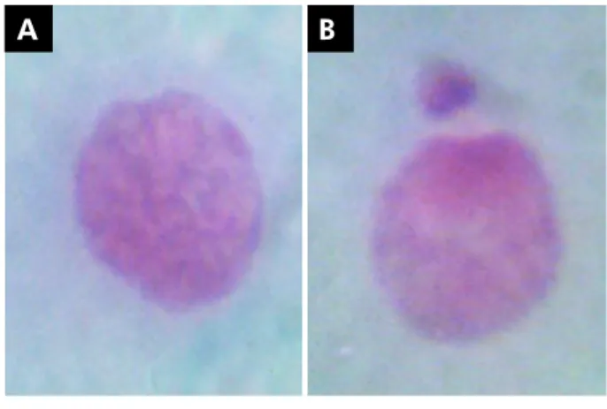
The MN were characterized according to the criteria specified in a previous report:15 (a)
regular contour, round or elliptical, and inside the cell cytoplasm; (b) similar color to the principal nucleus; (c) less than one-third of the diameter of the nucleus; (d) completely separated from the nucleus, allowing clear identification between the nucleus and MN limits (Figure 1).
The data were subjected to non-parametric tests, the Wilcoxon rank-sum test and the Kruskal-Wallis equality-of-populations rank test, using SPSS software (SPSS, Inc., Chicago, USA).
Results
Thirty-seven patients enrolled in the original clinical trial were selected for the genotoxicity study.3
Nineteen volunteers were assigned to the 10% CP group and 18, to the 16% CP group. There was no loss to follow-up during the study.
Figure 2 shows the descriptive analysis of MN presence in the different groups. Comparing the median of both groups, there was no difference between the rates, at the three time periods.
Table 1 presents the evolution of the median MN rate over the follow-up period. The interquartile range (IQR) at the 15-day follow-up is larger for the 10% CP group, compared with the 16% CP group. At the 45-day
R
ate of micronucleus presence
0
Baseline 15 days 45 days
10% CP concentration 2
4 6 8 10
16% CP concentration
Figure 2. MN rate at baseline, 15 days and 45 days, in 10% and 16% CP concentrations. No differences were found between the two groups at either point of time.
A B
follow-up, the IQR is wider for the16% CP group than for the 10% CP group. Despite these differences, we found no statistically signiicant difference between the IQR for the two follow-up periods. Although there was no signiicant difference between the groups,
the median MN rate was slightly higher among the
patients of the 10% CP concentration group. In the 15 days after beginning the bleaching treatment, there was an increase in the MN rates for both groups,
whereas the median MN rate was equal for both
groups at day 15. After this point, we observed a decrease in MN rates, reaching a median count of 2 MN per 1000 cells for both groups.
Analyzing the two groups as independent samples, we carried out the Kruskal-Wallis equality-of-populations rank test, which showed that there was no difference between the two groups, comparing the three follow-up points (baseline, 15 days and 45 days) (p = 0.90) (Table 1).
Discussion
Exfoliative cytology was used to analyze MN formation in the oral mucosa. This is a simple, painless and cheap method, and has been used as an adjunct in molecular epidemiology. The buccal micronucleus cytome (BMNcyt) assay is a non-invasive method for studying DNA damage, chromosomal instability, cell death and the regenerative potential of human buccal mucosal tissue. The MN assay detects damage at the chromosomal level, leading to more severe genome instability correlated with a health risk. Another advantage of this minimally invasive approach is that it can be used without establishing cell cultures.16
The spontaneous frequency of MN in human buccal exfoliated cells is between 0.3% and 1.7%. These numbers were previously determined in a multicenter study with 30 laboratories worldwide, which analyzed 5000 individuals for formation of
MN and other nuclear anomalies.16 Our MN levels at baseline were close to the lower value of the mean
worldwide frequency found by Bonassi et al.16 In the
present study, 1000 cells/individual were counted,
although the literature suggests using a minimum
of 2000 cells/individual.17,18 The study evaluated the
genotoxicity of a local agent, as is the case of the effect of bleaching gels on the cells of the gingival epithelium (stratiied parakeratinized squamous epithelium). International recommendations (HUMN-XL)18 suggest
oral mucosa cells (non-keratinized stratiied squamous tissue) for these assessments. Cell exfoliation is easier to perform in the oral mucosa than in gingival tissue,
where the number of exfoliated cells is much smaller
than in the buccal mucosa; moreover, these tissues have different histological features. Four to ive slides were prepared for each patient at each stage of the analysis, and 1000 cells/individual could be counted. To count 2000 cells would require increased local friction, causing trauma in some cases, and painful symptoms in the volunteers. Our protocol was an adaptation of the original protocol of genotoxicity in exfoliated buccal mucosa cells.19
The counting of micronuclei is done to monitor cell changes in areas exposed to genotoxic agents,19
as was done in this study. These DNA fragments appear only in cells that have completed at least one cell division after being affected by a genotoxic agent. Non-incorporation of the fragments is usually due to a lack of centromeres, which prevents the fragments from migrating toward the spindle poles, late in anaphase. The DNA fragments that are left behind are incorporated into a secondary nucleus and kept in the cell cytoplasm.20
In vivo studies cannot be standardized, because
the oral cavity is a multifactorial environment, and each patient has his/her own specific biological variation. This may be an advantage, because it allows
Table 1. Number of micronucleated cells at different points of time for both CP concentrations.
CP Concentration Baseline 15 days 45 days p-value*
N Median IQR N Median IQR N Median IQR
10% 19 1 1 19 3 4 19 2 2 0.9093
16% 18 0 2 18 3 2 18 2 3
evaluation of the effects of dental materials in their
natural setting. Thus, the use of gingival epithelial cells can be beneficial, because these cells are in
direct contact with these materials when using the
tray technique for dental bleaching. Patient progress
was tracked regularly over time in order to eliminate
individual variations.21
Products used in dental treatments may be toxic and genotoxic to oral tissues.22 Reactive oxygen and
free radicals released by bleaching products in contact with cells have been shown to interact with DNA, causing some oxidative damage, such as strands and chromosomal breakage, and may alter DNA repairability.23 This study evaluated two groups of
patients treated with two different CP concentrations. According to our knowledge, this is the only in vivo
study evaluating the genotoxic effects of dental
bleaching directly on patients.
We observed a moderate level of damage when evaluating the MN (frequency of micronucleated
cells) after exposure for 15 and 45 days. These results were expected, because the damage that leads to MN formation takes place in the basal layer of the epithelial tissue, where cells undergo mitosis. The rapid turnover of epithelial tissues brings the cells to the surface, where they exfoliate. The turnover time of the epithelium is the time needed for a cell to divide and pass through the entire epithelium. It ranges from 10 to 12 days.21
According to the literature, the spontaneous
frequency of MN in buccal cells in men versus women
is not signiicantly different. In contrast, the age factor
is well characterized by a steady increase in MN
cells according to advancing age.16 Since the average
age of the volunteers was 27.62 ± 7.05 years, their age did not inluence the outcome. The participants in this study were recruited from a well-controlled, randomized clinical trial.3 The volunteers had to
complete a questionnaire before being submitted to the oral examination, and only those who met the inclusion
criteria3 could participate in the present study. Thus,
confounding factors, such as smoking, alcohol and occupational exposure to potentially genotoxic agents, led to exclusion, making this sample homogeneous.
In vitro studies using bleaching agents showed
DNA damage in immortalized cell lines.10,13 The
mutagenicity of substances in in vitro systems should
be analyzed carefully; these assays do not have the
in vivo enzyme levels responsible for detoxiication
of the bleaching agents.8 Animal models can also be used to characterize the toxic effects of bleaching
substances.24 These studies show the genotoxic
effect caused by oxidative stress to the DNA, due to the action of peroxide, but the effects are smaller
than those found in in vitro studies. This is probably
related to the quick detoxiication of H202 and the elimination of free radicals before any interaction
with the DNA.8 Moreover, the concentration of CP
decreases rapidly when evaluated clinically, probably as a result of wash-out by saliva.25
Tooth sensitivity and gingival irritation are the most common side effects reported in the literature6
for clinical studies, and there has been agreement regarding the direct relationship between the
amount of damage and the concentration of the
bleaching substance.3 However, the literature has not yet reached a consensus on genetic alterations of the oral mucosa due to the action of dental
bleaching products in clinical studies,11 and, to our
knowledge, studies on genotoxicity and the use of these substances have not yet been published. The possible genetic damage caused by indiscriminate
use of bleaching substances without a dentist’s
supervision1 should be taken into consideration. MN assays on oral mucosa cells have shown that these biomarkers tend to increase when individuals are
exposed temporarily to different substances, such as dental composite materials21 and mouthwash.26
The conditions caused by this temporary exposure corroborate our indings, insofar as the greatest increase in the number of MN cells occurred up to 15 days after exposure to CP, regardless of the concentration. Thus, our results suggest that the reduction in the MN production rate from day 15 to 45 indicates that the primary lesions in DNA induced by CP were repaired. In addition, micronucleated cells with irreparable damage detected at day 15 were no longer seen after 45 days.
Conclusions
peroxide alone do not cause mutagenic stress on gingival epithelial cells. However, repetitive exposure to bleaching agents should be avoided, at least in the short term. Future studies should explore whether exposure to these products, in association with other factors, such as tobacco, alcohol and hot beverages, has the potential to cause genetic damage.
Acknowledgments
The authors are grateful to the Conselho Nacional
de Desenvolvimento Científico e Tecnológico - CNPq
for scholarships 113010/2005-2, 134831/2009-8, and 119431/2010-6. We gratefully acknowledge the assistance of Maria da Graça Roth and Giovanny França – a PhD student of epidemiology, for their technical advice and help with the statistics.
1. Demarco FF, Meireles SS, Masotti AS. Over-the-coun-ter whitening agents: a concise review. Braz Oral Res. 2009 Jun;23 Suppl 1:64-70.
2. Hasson H, Ismail AI, Neiva G. Home-based chemically-in-duced whitening of teeth in adults. Cochrane Database Syst Rev. 2006 Oct 18;(4):CD006202.
3. Meireles SS, Heckmann SS, Leida FL, Santos IS, Della Bona A, Demarco FF. Efficacy and safety of 10% and 16% carbamide peroxide tooth-whitening gels: a randomized clinical trial. Oper Dent. 2008 Nov-Dec;33(6):606-12.
4. American Dental Association (ADA). Consumer products with the ADA Seal of Acceptance [Internet]. [cited 2010 Feb 4]. Available from: http://www.ada.org/ada/seal/adaseal_con-sumer_shopping.pdf 2010.
5. Kihn PW, Barnes DM, Romberg E, Peterson K. A clinical eval-uation of 10 percent vs. 15 percent carbamide peroxide tooth-whitening agents. J Am Dent Assoc. 2000 Oct;131(10):1478-84. 6. Armênio RV, Fitarelli F, Armênio MF, Demarco FF, Reis A,
Loguercio AD. The effect of fluoride gel use on bleaching sensitivity: a double-blind randomized controlled clinical trial. J Am Dent Assoc. 2008 May;139(5):592-7.
7. Tredwin CJ, Naik S, Lewis NJ, Scully C. Hydrogen peroxide tooth-whitening (bleaching) products: review of adverse ef-fects and safety issues. Br Dent J. 2006 Apr;200(7):371-6. 8. Munro IC, Williams GM, Heymann HO, Kroes R. Use of
hydro-gen peroxide-based tooth whitening products and its relation-ship to oral cancer. J Esthet Restor Dent. 2006 Jun;18(3):119-25. 9. Li Y. Biological properties of peroxide-containing tooth
whit-eners. Food Chem Toxicol. 1996 Sep;34(9):887-904.
10. Ribeiro DA, Marques MEA, Salvadori DMF. Study of DNA damage induced by dental bleaching agents in vitro. Braz Oral Res. 2006 Jan-Mar;20(1):47-51.
11. Goldberg M, Grootveld M, Lynch E. Undesirable and adverse effects of tooth-whitening products: a review. Clin Oral In-vestig. 2010 Feb;14(1):1-10.
12. Igarashi M, Nagata M, Itoh S, Yamoto T, Tsuda S. Relationship between DNA damage and micronucleous in mouse liver. J Toxicol Sci. 2010 Dec;35(6):881-9.
13. Fernández MR, Carvalho RV, Ogliari FA, Beira FA, Etges A, Bueno M. Cytotoxicity and genotoxicity of sodium
percarbon-ate: a comparison with bleaching agents commonly used in discoloured pulpless teeth. Int Endod J. 2010 Feb;43(2):102-8. 14. Roth JM, Restani RG, Gonçalves TT, Sphor SL, Ness AB,
Marti-no-Roth MG, et al. Genotoxicity evaluation in chronic renal pa-tients undergoing hemodialysis and peritoneal dialysis, using the micronucleus test. Genet Mol Res. 2008 May 20;7(2):433-43. 15. Countryman PI, Heddle JA. The production of micronuclei
from chromosome aberrations in irradiated cultures of hu-man lymphocytes. Mutat Res. 1976 Dec;41(2-3):321-32. 16. Bonassi S, Coskun E, Ceppi M, Lando C, Bolognesi C, Burgaz
S, et al. The Human Micro Nucleus project on exfoliated buccal cells (HUMN(XL)): the role of life-style, host factors, occupational exposures, health status, and assay protocol. Mutat Res. 2011 Nov-Dec;728(3):88-97.
17. Bolognesi C, Knasmueller S, Nersesyan A, Thomas P, Fenech M. The HUMNxl scoring criteria for different cell types and nuclear anomalies in the buccal micronucleus cytome assay - an update and expanded photogallery. Mutat Res. 2013 Oct-Dec;753(2):100-13.
18. Fenech M, Holland N, Zeiger E, Chang WP, Burgaz S, Thomas P, et al. The HUMN and HUMNXL international collaboration proj-ects on human micronucleus assays in lymphocytes and buccal cells-past, present and future. Mutagenesis. 2011 Jan;26(1):239-45. 19. Thomas P, Holland N, Bolognesi C, Kirsch-Volders M, Bonas-si S, Zeiger E, et al. Buccal micronucleus cytome assay. Nat Protoc. 2009 May 7;4(6):825-37.
20. Fenech M. The in vitro micronucleus technique. Mutat Res. 2000 Nov;455(1-2):81-95.
21. Tadin A, Galic N, Mladinic M, Marovic D, Kovacic I, Zeljezic D. Genotoxicity in gingival cells of patients undergoing tooth restoration with two different dental composite materials. Clin Oral Investig. 2014 Jan;18(1):87-96.
22. Kirsch-Volders M, Sofuni T, Aardema M, Albertini S, East-mond D, Fenech M, et al. Report from the in vitro micronucle-us assay working group. Mutat Res. 2003 Oct 7;540(2):153-63. 23. Kawamoto K, Tsujimoto Y. Effects of the hydroxyl radical and hy-drogen peroxide on tooth bleaching. J Endod. 2004 Jan;30(1):45-50. 24. Cherry DV, Bowers DE Jr, Thomas L, Redmond AF. Acute
toxicological effects of ingested tooth whiteners in female rats. J Dent Res. 1993 Sep;72(9):1298-303.
25. Ladeira C, Viegas S, Carolino E, Prista J, Gomes MC, Brito M. Genotoxicity biomarkers in occupational exposure to formaldehyde the case of histopathology laboratories. Mutat Res. 2011 Mar 18;721(1):15-20.