Arq Neuropsiquiatr 2001;59(2-B):362-364
TERATOGENIC EFFECTS OF LAMOTRIGINE
ON RAT FETAL BRAIN
A morphometric study
Nely Silvia Aragão de Marchi
1, Reinaldo Azoubel
2, Waldir Antonio Tognola
3ABSTRACT - A study of the teratogenic activity of an antiepileptic drug - lamotrigine – was carried out in the brain of fetuses of rats who had received the drug. The dosage levels studied corresponded to four times the median effective dose (ED50) in rats. The drug was administered during the organogenesis period. Rats were sacrificed one day prior to term and fetuses were macroscopically examined, weighted and cephalic segments sectioned (Wilson technique), for histological study by stereological analysis, using Merz’s grid for drawing and point counts. Cortex, subcortex, ependyma and lateral ventricles were analyzed. The same methodology was applied to the control group; data were compared with by the non-parametric Mann-Whitney statistical analysis test. Results showed that fetuses of the experimental group had reduced body weight at birth, increased volume and diameter of the cerebral structure, increased density of the subcortical layer, and ventricle dilatation .Possible mechanisms of this teratogenicity were discussed.
KEY WORDS: teratogenesis, lamotrigine.
Efeitos teratogênicos da lamotrigina em cérebro de fetos de ratos: estudos morfométrico
RESUMO – Foi realizado estudo da atividade teratogênica de uma droga antiepiléptica - lamotrigina - em cérebro de fetos de ratas que receberam a droga. Estudamos dose que corresponde a 4 vezes a dose efetiva mediana (ED50) em ratos. A droga foi administrada durante o período de organogênese, as ratas foram sacrificadas 1 dia antes do termo e os fetos foram examinados macroscópicamente, pesados e foram realizados cortes no segmento cefálico (técnica de Wilson) e feito preparo histológico para análise estereológica (utilizado grade de Merz para desenho e contagem dos pontos). Foram analisados: córtex, subcórtex, epêndima e ventrículos laterais. A mesma metodologia foi aplicada ao grupo controle e os dados foram submetidos a análise estatística por teste não paramétrico de Mann-Whitney. Os resultados mostraram nos fetos do grupo tratado: redução do peso ao nascimento, aumento do diâmetro e volume da estrutura cerebral, aumento da densidade da camada subcortical e dilatação ventricular. Possíveis mecanismos de teratogenicidade foram discutidos.
PALAVRAS-CHAVES: teratogênese, lamotrigina.
Faculdade de Medicina de São José do Rio Preto, São José do Rio Preto SP, Brazil: 1Master in science; 2Professor; 3Associated Professor.
Received 1 September 2000, received in final form 12 January 2001. Accepted 22 January 2001.
Dr. Reinaldo Azoubel – Avenida Brigadeiro Faria Lima 5416 – 15090-000 São José do Rio Preto SP – Brasil.
There is no completely satisfactory definition for epilepsy, generally a chronic condition comprising a group of illnesses having in common, recurrent epi-leptic seizures, occurring in the absence of toxic, metabolic, or febrile situations. According to the World Health Organization, epilepsy has a prevalence of around 5% of the total population1. Therapeutic
and surgical practices allow 80% of the patients to have a normal life, with little interference of epilep-tic seizures in it2. Although in the past, reproduction
was discouraged in epileptic women, over 90% of pregnancies in such patients currently have an un-eventful outcome when appropriately managed3. In
approximately one third of pregnant epileptic women an increase of seizure frequency relative to the period prior to pregnancy occurs and among other factors, has been attributed to increased es-trogen and reduced AED (antiepileptic drug) serum levels2.
The first systematic study of AEDs teratogenicity, conducted by Jans and Fuchs in 19644, demonstrated
an average of 2.2% malformations in 262 children exposed intra-uterus to AED, a value not significantly greater than that of the general population. How-ever other authors - Lindhout and Omtizigt5 in 1992
Arq Neuropsiquiatr 2001;59(2-B) 363
5% higher than that in the general population. The number and doses of AEDs utilized can also inter-fere with the final result , high doses and polytherapy increasing this risk6,7.
Anomalies and malformations commonly found in association with the use of AED are: a) neural tube defects – “spina bifida”, mostly related to the use of sodium valproate8 and carbamazepine9; b) facial cleft
- labial or palatal10 and, c) congenital cardiac defects
(atrial septal defect, Fallot’s tetralogy, ventricular septal defect, aortic coarctation, pulmonary steno-sis and persteno-sistence of the arterial channel)11. The
majority of studies on fetal malformations and AEDs, have been made in patients treated with the five leading AEDs - phenytoin (DPH), carbamazepine (CBZ), phenobarbital (PB), primidone (PR), and so-dium valproate (VPA)4-11. In general, investigators
agree that the risk of fetal malformations due to AEDs is about twofold above normal, and is enhan-ced by polytherapy and high AED serum levels.
Lamotrigine (LTG) is a recent drug considered to be effective against partial tonico-clonic seizures, secondarily generalized. The drug’s mechanism of action is related to blockade of voltage dependent sodium channels which stabilize pre-synaptic mem-branes and inhibit excitatory neurotransmitter re-lease, in special of glutamate and aspartate12.
Analy-sis of 42 pregnancies in 1993, did not reveal clear-cut evidence of a relationship between LTG and ter-atogenesis13; however, information on LTG’s
terato-genicity remains insufficient.
METHOD
In this study, a dose of 1.5 mg of LTG, corresponding to four times the effective mean dose (ED50), was admin-istered to white rats by gastric intubation on days 9, 10
and 11 of pregnancy (corresponding to the organogen-esis period). Animals were sacrificed one day prior to term (treated rats = 04; litters = 51 fetuses / control rats = 04; litters = 39 fetuses). Fetuses were examined macroscopi-cally, weighted, and portions of their cephalic segments dissected by Wilson’s technique as follows: palate surface down, 3 coronal sections made immediately frontal to the eyes, through the eyes and retro-occularly passing the lat-eral ventricles, the last section was utilized in the present study14. Histological preparations from the cortex,
subcor-tex, ependyma, and lateral ventricles from aleatorily cho-sen 30% of the offspring were prepared by inclusion in paraffin, sectiong at intervals of 2mm with a 6 microme-ter thickness, and dyeing with Hematoxilin – Eosin. Nine histological sections from each fetus were prepared for stereological analysis using Merz’s grid for drawing and point counts, maintaining a space of 56 microns per cut. The same methodology was applied to the control group. Data were statistically analyzed by the non-parametric Mann-Whitney test15.
RESULTS
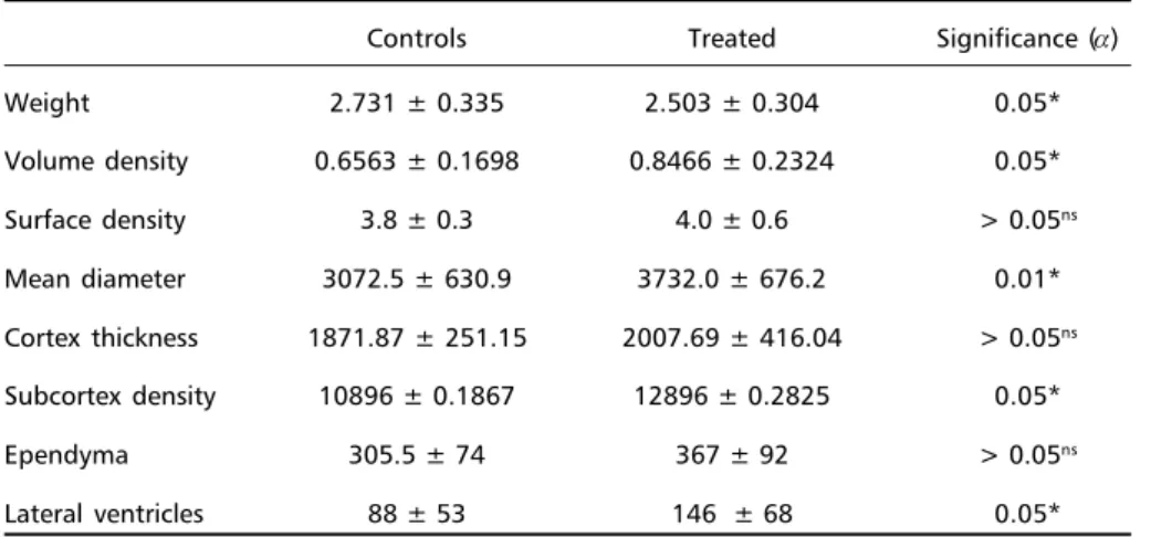
As demonstrated in the Table 1, in the treated group we found lowered body weight at birth, in-creased structural diameter, inin-creased volume den-sity, increased lateral ventricles and increased sub-cortical density. Other items analyzed did not show significant alterations.
DISCUSSION
Teratogenicity can be expressed by interference in proliferation, migration or differentiation at the cellular level. The basis for recognition of teratoge-nicity is a reproducible repetition and association of a given agent with a recognizable pattern of mal-formation, growth delay, mutagenesis and embryo or fetal death. Due to the fact that certain agents
Table 1. Results.
Controls Treated Significance (α)
Weight 2.731 ± 0.335 2.503 ± 0.304 0.05*
Volume density 0.6563 ± 0.1698 0.8466 ± 0.2324 0.05*
Surface density 3.8 ± 0.3 4.0 ± 0.6 > 0.05ns
Mean diameter 3072.5 ± 630.9 3732.0 ± 676.2 0.01* Cortex thickness 1871.87 ± 251.15 2007.69 ± 416.04 > 0.05ns
Subcortex density 10896 ± 0.1867 12896 ± 0.2825 0.05*
Ependyma 305.5 ± 74 367 ± 92 > 0.05ns
Lateral ventricles 88 ± 53 146 ± 68 0.05*
Results expressed as averages ± standard deviation: ns, not statistically significant; *, statistically significant
364 Arq Neuropsiquiatr 2001;59(2-B)
have the same metabolic pattern, they are also as-sociated with similar patterns of malformation, re-sulting in a recognizable syndrome. Some good ex-amples of these effects are the AEDs16.
In this research we observed that alterations like low birth weight, ventricle dilatation, subcortical density enhancement with consequent increase in cerebral volume and diameter, were associated with the use of lamotrigine during the organogenesis pe-riod. Genetic-molecular susceptibly to teratogenesis is probably heterogeneous. The equilibrium between metabolic activation and detoxification determines the levels of reactive intermediates; besides this, not only inhibition of detoxification by drug interaction, but also genetically determined deficiencies of en-zymes for detoxification are potential factors for ter-atogenesis5.
Nau in 199517, showed that AED therapy can have
a significant effect on endogenous retinoid metabo-lism; due to the importance of retinoids in signaliz-ing crucial biological events dursignaliz-ing embryonic de-velopment, alterations in their metabolism can be important factors for AED teratogenesis.
Wells et al. in 199718, argued that bioactivation
of cytochrome P450, prostaglandin H synthetase, lipooxygenation, and/or free radical reactivation, contribute to the oxidation of macromolecules like DNA, proteins and lipids which can determine intra-uterine death or teratogenesis.
Animal studies are limited by inter-species vari-ability and by the fact that in many studies doses used were much higher than those utilized in hu-mans. These characteristics decrease the reliability of the results of studies on animals, on human ter-atogenesis evaluation, although these studies may aid in the localization of events related to biological plausibility19. The occurrence of embryopathy
asso-ciated with talidomide, preceded the erroneous be-lief that human teratogenicity cannot be predicted on the base of animal studies. Drugs that have been
found to be teratogenic in man have caused similar effects in animals20.
Our research concludes that lamotrigine has ter-atogenic effects on the brain of rats. Further research must be carried out to corroborate these findings and establish their applicability to humans. One of the major purposes of teratology is to anticipate risks before they materialize16.
REFERENCES
1. Shorvon SD. Epidemiologia, classificação, história natural e genética da epilepsia. In Costa JC. Epilepsy: a lancet review. London: Biogalenica, 1990:5-13.
2. Yerby MS. Pregnancy and epilepsy. Epilepsia 1991;32(supl 6):51-59. 3. Shuster EA. Epilepsy in women. Mayo clin proc 1996;71:991-999. 4. Jans D, Fuchs V. Are antiepileptic drugs harmful when given during
pregnancy? Ger Med Mon 1964; 9:20-22. [In Yerby MS. Pregnancy and epilepsy. Epilepsia 1991;32(Suppl 6): 51-59].
5. Lindhout D, Omtzigt JGC. Pregnancy and the risk of teratogenicity. Epilepsia 1992; 33(Suppl 4):41-48.
6. Nakane Y, Okuma T, Takahashi R, et al. Multiinstitutional study on the teratogenicity and fetal toxicity of antiepileptic drugs: a report of a collaborative study group in Japan. Epilepsia 1980;21:663-680. 7. Samrén EB, Van Duijn CM, Koch S, et al. Maternal Use of major
con-genital malformations: a joint european prospective study of human teratogenesis associated with maternal epilepsy. Epilepsia 1997; 38(Suppl 9):981-990.
8. Yerby MS, Leavi HA, Ericson DM, McCormick KB, Loewensoon RB, Sells CJ, et al. Antiepileptics and the development of congenital anoma-lies. Neurology 1992;42(Suppl 5):132-140.
9. Rosa F. Spina bifida in infants of women treated with carbamazepine during pregnancy. N Engl J Med 1991;324:674-677.
10. Friis ML, Holm NV, Sindrup EH, Andersen PF, Hauge M. Facial clefts in sibs and children of epileptic patients. Neurology 1986;36:346-350. 11. Annergers JF, Hauser WA, Elveback LR, Anderson VE, Kurland LT.
Congenital malformations and seizure disorders in the offspring of parents with epilepsy. Intl J Epidemiol 1978;7:241-247.
12. Messenheimer JA. Lamotrigine. Epilepsia 1995;36(Suppl 2):87-94. 13. Richens A. Safety of lamotrigine. Epilepsia 1994;35(Suppl 5):37-40. 14. Wilson JG. Embryological considerations in teratology. In Wilson JG,
Warkany J. Teratology principles and techniques. Chicago: Univer Chicago Press, 1965.
15. Siegel S. Estatística não paramétrica para as ciências do comportamento. São Paulo: McGraw Hill, 1995.
16. Wilson JG. Current status of teratology. In Wilson JG, Fraser FC. The handbook of teratology. 2.Ed. New York: Plenum Press 1979;47-74. 17. Nau H. Chemical struture: teratogenicity relationships, toxicokinetics
and metabolism in risk assessment of retinoids. Toxicology Letters 1995;82/83:975-979.
18. Wells PG, Kim PM, Laposa RR, Nicol CJ, Parman T, Winn LM. Oxida-tive damage in chemical teratogenesis. Mutation Res 1997;396:65-78. 19. Sharony R, Graham JM. Identification of fetal problems associated with
anticonvulsant usage and maternal epilepsy. Obst Gynecol Clin N Am 1991;18:933-951.