Immunolocalization of HLA-DR and
Metallothionein on Amalgam Tattoos
Camila M.A. LEITE1
Amanda S. BOTELHO1
Jamila R. OLIVEIRA2
Sérgio V. CARDOSO2
Adriano M. LOYOLA3
Ricardo S. GOMEZ2
Ricardo R. VAZ2
1Dental Surgeon, Private Practice, Belo Horizonte, MG, Brazil
2Department of Oral Surgery and Pathology, Faculty of Dentistry, Federal University of Minas Gerais
Belo Horizonte, MG, Brazil
3Department of Pathology, Faculty of Dentistry,Federal University of Uberlândia, Uberlândia, MG, Brazil
Despite studies concerning toxic reactions related to amalgam components in the literature, few studies have been devoted to evaluate local noxious effects of amalgam tattoos (AT) on biological tissues. In addition, little is known about activation of inflammatory cells by mucosa-implanted amalgam debris. Tissue reaction to AT depends on the particle size. Human leukocyte antigen DR (HLA-DR) is an activation marker of inflammatory cells associated with antigen presentation. Metallothioneins (MT) are proteins involved with metal detoxication, including mercury and silver. The purpose of the present study was to investigate the immunolocalization of HLA-DR and MT in AT with large or powdered particles. Paraffin-embedded AT tissue blocks were sectioned and subjected to immunohistochemistry for HLA-DR and MT localization. The results demonstrated a dense mononuclear inflammatory infiltrate associated with large and powdered debris and positivity for HLA-DR and MT in inflammatory cells. While blood vessel walls and connective fibers impregnated with powdered particles were negative for HLA-DR, they were positive for MT. In addition, wherever epithelial basement membrane impregnation by powdered amalgam particles was observed, a strong positivity for MT was detected. These findings demonstrate that residual elements of AT still have noxious local effects over tissues.
Key Words: amalgam tattoo, dental amalgam, HLA-DR, metallothionein, toxicity.
Correspondence: Prof. Dr. Ricardo Santiago Gomez, Laboratório de Patologia Experimental 3 – Sala 3203, Faculdade de Odontologia, Universidade Federal de Minas Gerais, Avenida Antônio Carlos 6627, 31270-901 Belo Horizonte, MG, Brasil. [email protected]
INTRODUCTION
Amalgam tattoo is a common finding in dental practice (1,2). It is characterized by the deposit of restorative debris composed of a mixture of silver (Ag), mercury (Hg), tin (Sn), zinc (Zn), and copper (Cu) in subepithelial connective tissue (3-5). It has been con-sidered that such deposits are often due to mucosa abrasion by dental drilling or flossing during restor-ative procedures, implanting of residual fragment of amalgam following tooth extraction, deposit of amal-gam into wounds during endodontic treatment, or
components, amalgam tattoos have been considered innocuous lesions (2,6,9,10), and surgical removal of the lesions has been indicated for aesthetic purposes or for differential diagnosis with other pigmented lesions, notably melanoma (6).
The tissue reaction to amalgam tattoo depends on the particle size. Powdered amalgam undergoes intracellular degradation by macrophages and giant cells in the soft tissue, with continuing mercury release. Residual particles containing mainly silver become diffusely distributed throughout the tissues. Larger frag-ments are surrounded initially by macrophages but are then progressively enclosed by a fibrous capsule, asso-ciated with a very slow breakdown process. Energy dispersive x-ray microanalysis has shown that Cu and Zn are rapidly lost from the lesion, while Hg and Sn are lost slowly and only Ag remains in older lesions (3,4). Despite studies concerning tissue reactions related to amalgam components in the literature, few studies have evaluated local activation of inflammatory cells by amalgam tattoos (10-14). In addition, little is known about local noxious effects of amalgam tattoos on biological tissues (14,15).
Human leukocyte antigen DR (HLA-DR) is con-stituted by transmembranous glycoproteins. It is one of the major histocompatibility complex class II antigens (16). HLA-DR molecules are abundantly expressed in human monocytes and macrophages, and are respon-sible for antigen presentation to CD4+ T lymphocytes. Irritants are able to induce HLA-DR internalization by antigen-presenting cells (17). Metallothioneins (MT) are evolutionary conserved proteins associated with detoxication of heavy metals, among other likely cellu-lar functions, and have been widely used as environ-mental markers of intoxication (18,19). The only study accessing MT in amalgam tattoos observed higher immunoexpression in histiocytes adjacent to large globu-lar particles, but very weak or even absent staining when related to fine amalgam particulate (15). The authors associated MT induction with the presence of Hg, and considered the findings compatible with the temporal loss of this metal by the particles. No differ-ence in the immunostaining of epithelial cells associ-ated with amalgam dust was observed when compared with normal tissue (15). These findings are astonishing, since Ag, the last component of amalgam tattoos, is able to induce MT expression, as well as other compo-nents of metallic restorations (20).
Considering that tissue reaction to amalgam tat-too depends on the particle size and HLA-DR antigens are activation markers of inflammatory cells associated with antigen presentation (16), together with the evi-dence that metallothioneins are proteins involved with metal detoxication, including Hg and Ag (19), the purpose of the present study was to investigate the immunolocalization of HLA-DR and MT in amalgam tattoos with large or powdered particles.
MATERIAL AND METHODS
RESULTS
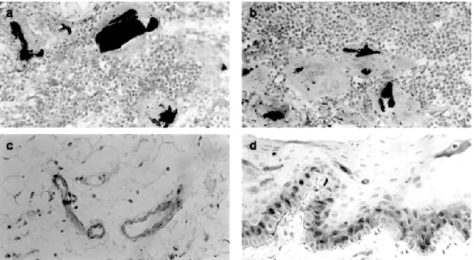
Larger amalgam particles were generally associ-ated with a dense inflammatory infiltrate of mono-nuclear cells with some foreign body giant cell forma-tion. Mononuclear and giant cells were usually positive for HLA-DR (Figure 1a), with less conspicuous reac-tion for MT (Figure 1b). Powdered amalgam particles were associated with a decreased number of inflamma-tory cells presenting HLA-DR or MT immunopositivity. Pigmented dust was also found associated with blood vessel walls and connective tissue fibers. In these cases, endothelial cells were negative for HLA-DR, but some-times positive for MT immunostaining (Figure 1c). The basal cell layer of the mucosa epithelium demonstrated positive immunostaining for MT in all samples. Im-pregnation of the epithelial basement membrane with powdered amalgam particles was associated with strong MT immunopositivity (Figure 1d).
DISCUSSION
Amalgam tattoos have been considered innocu-ous lesions (1,2,6,9). The toxicity of this compound has raised questions about its use in routine dental practice (9,10,12,14). Local effects of amalgam debris over tissues have been sparsely studied (13,15). The present results show that amalgam remnants are able to activate immunologic adaptive reaction and are also precipi-tants of the expression of an important detoxication protein.
HLA-DR is abundantly expressed on human monocytes and macrophages. It is one of the major histocompatibility complex class II-antigens and is re-sponsible for antigen presentation to CD4-positive T-lymphocytes (16). It is reported that, albeit using dis-tinct patterns of endocytosis, irritants are able to induce HLA-DR internalization by antigen-presenting cells as well as allergens (17). MT is a protein ubiquitously
distributed from single-cell organisms to human tis-sues, and has been used as a marker of intoxication in a variety of environmental casualties (19). It is a small protein with a high cysteine content that allows up to seven metal-binding sites, each one formed by four residues of thiolate ligands. Detoxication occurs when each of the residues binds with bivalent metals, free-radicals and other toxic compounds (18). MT immuno-expression in amalgam tattoos has been observed and interpreted as a protective reaction against local or systemic toxicity of amalgam components (15). Con-sidering that different tissue reactions are found on amalgam tattoo according to its size and composition, in the present study we investigated the immunolocali-zation of HLA-DR and MT antigens in amalgam tattoo looking for insights to its pathogenesis and biological effects.
As mentioned, tissue reaction to amalgam tattoo depends on its particle size and composition (2,3). While copper and zinc are rapidly lost from the area of the tattoo, mercury and tin are lost more slowly and finally silver remains permanent in the tissues. Accord-ing to Eley (2), the process of intracellular digestion of amalgam in biological tissues results in the release of Hg and the formation of two kinds of secondary fine particles, those containing Sn and those containing Ag. Ionic Hg associates with biological ligands and be-comes bound to protein. In this way, it would be prob-ably transferred from the tissue fluid to the blood. The secondary particles containing Sn are progressively lost from the local lesion and are excreted by the kidneys and liver. Finally, the secondary particles con-taining Ag are degraded into silver cations or small molecules inside the phagocyting cells. Afterwards these cations gain access to the extracellular environ-ment where they are re-aggregated into fine particles in the extracellular tissues. The association of these par-ticles with blood vessel walls, basal lamina and connec-tive tissue seemed to be responsible for the develop-ment and permanence of the tattoo. Forsell et al. (11) reported that the inflammatory reaction to amalgam tattoo becomes more severe as Hg content in the tissue increases, and that the latter was also correlated with Ag presence. Nadarajah et al. (13) also suggested that Hg accumulation may lead to altered expression of MHC class II determinants. In the present study it was found that inflammatory infiltrate associated with large and powdered debris were HLA-DR positive.
There-fore, Hg may induce HLA-DR on macrophages during its degradation. While the blood vessel walls and con-nective fibers impregnated by the powdered particles were negative for HLA-DR, they were positive for MT. In addition, wherever epithelial basement membrane impregnation by powdered amalgam particles was ob-served, a strong positivity for MT was detected. These findings suggest that residual elements of amalgam tattoos, mainly containing Ag, still have noxious ef-fects over tissues. MT immunoexpression may be a local tissue detoxication reaction against amalgam resi-dues.
Lau et al. (15) reported higher MT immunoex-pression in mononuclear cells adjacent to large globu-lar particles compared to the very weak or even absent staining associated with fine amalgam particulate. It was associated by these authors with the presence and progressive loss of Hg. They did not observe differ-ences in the immunostaining of epithelial cells associ-ated with amalgam dust when compared with normal tissue. Blood vessels and connective fibers containing debris were also negative for MT immunolocalization. Our results present a somewhat different picture. A weak but distinct reaction was observed in inflamma-tory cells, while basal and parabasal epithelial cells, blood vessels and connective fibers were associated with immunostaining when amalgam residues were present. A possible explanation for these results lays in the use of different antigen retrieval solution.
In conclusion, the present data demonstrate that residual elements of amalgam tattoo develop a noxious effect in biological tissues. However, we did not look for clinical implications of this finding for the manage-ment of the lesion, and further studies are necessary to elucidate this topic.
RESUMO
tamanhos. Cortes histológicos de lesões fixadas em formol e embebidas em parafina foram submetidos a técnica imunoistoquímica para a detecção dos antígenos mencionados. Os resultados demonstraram denso infiltrado inflamatório associado com partículas grandes ou pulverizadas, observando-se preobservando-sença de células HLA-DR e MT positivas. Paredes de vasos sangüíneos e fibras de tecido conjuntivo impregnadas por restos de amálgama foram negativas para HLA-DR, mas positivas para MT. Impregnação da membrana basal por partículas de amálgama correspondia a forte positividade para MT no epitélio. Esses resultados demonstram a existência de efeitos nocivos locais das TA sobre os tecidos.
ACKNOWLEDGEMENTS
This study was supported in part by grants from PRONEX, PADCT, FAPEMIG and CNPq, Brazil. Drs. R.S. Gomez and A.M. Loyola are research fellows of CNPq.
REFERENCES
1. Buchner A, Hansen LS. Amalgam pigmentation (amalgam tattoo) of the oral mucosa. A clinicopathologic study of 268 cases. Oral Surg Oral Med Oral Pathol 1980;49:139-147.
2. Owens BM, Johnson WW, Shuman NJ. Oral amalgam pigmenta-tions (Tattoos): A retrospective study. Quintessence Int 1992;23:805-10.
3. Eley BM. Tissue reactions to implanted dental amalgam, includ-ing assessment by energy dispersive x-ray micro-analysis. J Pathol 1982;138:251-72.
4. Eley BM, Garrett JR. Tissue reactions to the separate implanta-tion of individual constituent of dental amalgam, including as-sessment by energy dispersive x-ray microanalysis. Biomaterials 1983;4:73-80.
5. Daley TD, Gibson D. Practical applications of energy dispersive x-ray microanalysis in diagnostic oral pathology. Oral Surg Oral Med Oral Pathol 1990;69:339-44.
6. Neville BW, Damm DD, Allen CM, Bouquot JE. Oral and maxil-lofacial pathology. 2nd ed. EUA: WB Saunders, 2001.
7. Peters E, Gardner DG. A method for distinguishing between amalgam and graphite in tissue. Oral Surg Oral Med Oral Pathol 1986;62:73-76.
8. Mohr W, Görz E. Die assoziation von Silbergranula mit elastischen Fasern bei der Amalgamose der Mundschleimhaut. HNO 2001;49:454-457.
9. Holmstrup P. Reactions of the oral mucosa related to silver amalgam: a review. J Oral Pathol Med, 1991;20:1-7.
10. Wahl MJ. Amalgam: resurrection and redemption. Quintessence Int 2001;32:696-710.
11. Forsell M, Larsson B, Lungqvist A, Carlmark B, Johansson O. Mercury content in amalgam tattoos of human oral mucosa and its relation to local tissue reactions. Eur J Oral Sci 1998;106:582-7.
12. Skoner JR, Wallace JA, Fochtman F, Moore PA, Zullo T, Hoffman D. Blood mercury levels with amalgam retroseals: a longitudinal study. J Endod 1996;22:140-141.
13. Nadarajah V, Neiders ME, Aguirre A, Cohen RE. Localized cellular inflammatory responses to subcutaneously implanted den-tal mercury. Toxicol Environ Health 1996;49:113-125. 14. Zhu Q, Safavi KE, Spangberg LS. Cytotoxic evaluation of
root-end filling materials in cultures of human osteoblast-like cells and periodontal ligament cells. J Endod 1999;25:410-412. 15. Lau JC, Jackson-Boeters L, Daley TD, Wysocki GP, Cherian
MG. Metallothionein in human gingival amalgam tattoos. Arch Oral Biol 2001;46:1015-1020.
16. Triantafilou K, Triantafilou M, Wilson KM, Fernandez N. Hu-man major histocompatibility molecules have the intrinsic ability to form homotypic associations. Hum Immunol 2000;61:585-598.
17. Rizova H, Carayon P, Barbier A, Lacheretz F, Dubertret L, Michel L. Contact allergens, but not irritants, alter receptor-mediated endocytosis by human epidermal Langerhans cells. Br J Dermatol 1999;140:200-9.
18. Palmiter RD. The elusive function of metallothionein. Proc Natl Acad Sci USA 1998;95:8428-30.
19. Coyle P, Philcox JC, Carey LC, Rofe AM. Metallothionein: the multipurpose protein. Cell Mol Life Sci 2002;59:627-647. 20. Murata M, Gong P, Suzuki K, Koizumi S. Differential metal
response and regulation of human heavy metal-inducible genes. J Cell Physiol 1999;180:105-13.