Cross-contamination in the Dental Laboratory
Through the Polishing Procedure of
Complete Dentures
Alessandra Marçal AGOSTINHO1 Paula Regina MIYOSHI1
Nelson GNOATTO1
Helena de Freitas Oliveira PARANHOS1 Luciene Cristina de FIGUEIREDO2
Sérgio Luiz SALVADOR3
1Department of Dental Materials and Prosthodontics, Faculty of Dentistry at Ribeirão Preto, University of São Paulo,
Ribeirão Preto, SP, Brazil
2Department of Research, School of Dentistry, University of Guarulhos, Guarulhos, SP, Brazil
3Department of Clinical Analysis, Faculty of Pharmaceutical Sciences at Ribeirão Preto, University of São Paulo,
Ribeirão Preto, SP, Brazil
Polishing of dental prostheses can cause a dangerous cycle of cross-contamination involving dentists, laboratory technicians, patients and auxiliary personnel. The aim of this study was to show the microbial contamination in the dental laboratory during the polishing procedure of complete dentures. For this purpose, 4 experiments were conducted. Experiment I - Determination of the total colony-forming units (CFU) counts contaminating complete maxillary dentures. During the polishing procedure, determination of the CFU counts transferred to the operator (Experiment II) and of the total CFU counts transferred to previously sterilized complete dentures (Experiment III). Experiment IV - The total counts of remaining CFU in the lathe spindle after Experiments II and III. Complete dentures were highly contaminated (mean = 1.4 x 10⁷ CFU/mL). There was a elevated level of contamination by splatter and aerosols.
There was high microbial transfer from the contaminated lathe spindle to the sterile prostheses (mean = 1.7 x 10⁷ CFU/mL). The
spindles were highly contaminated after polishing procedures (mean = 3.5 x 10⁸ CFU/mL). The polishing of dental prostheses is a
possible source of transmission of communicable diseases in the laboratory and requires improved techniques for infection control.
Key Words: cross infection, dental laboratory, dental prostheses.
Correspondence: Dr. Sergio L. Salvador, Departamento de Análise Clínica (Microbiologia), Faculdade de Ciências Farmaceuticas, Universidade de São Paulo, Avenida do Café s/nº, 14040-904 Ribeirão Preto, SP, Brasil. Tel: +55-16-602-4167. e-mail: sldssalv@usp.br
INTRODUCTION
Cross contamination is a severe problem that involves health professionals, especially in dentistry. The transmission of diseases during treatment between patients and dentists, auxiliary personnel and dental laboratory technicians can occur if preventive mea-sures are not taken. The risk of cross-contamination in dental clinics as well as transmission of microorgan-isms in prosthetic laboratories has been reported in various studies (1-3). More than 60% of the prostheses delivered to clinics from laboratories are contaminated with pathogenic microorganisms, i.e., streptococci,
lac-tobacilli, diphtheroids originating in the oral cavity of other patients (1-3). In prosthetic laboratories, lathes and pumice, usually used for polishing procedures and finishing of prostheses have been described as the greatest sources of contamination with levels of con-tamination reported of 1.4-8.0 x 10⁵ colony forming units (CFU) in pumice pans (4).
long periods of time when using lathes for the polishing of prostheses (4,5). In spite of the fact that it is not possible to eliminate all sources of contamination in the laboratory, a series of prevention measures to decrease these levels should be adopted. The use of sterile pum-ice and rag wheels or the association of disinfectants with pumice for polishing is a viable alternative to significantly reduce cross-contamination in the labora-tory (6-8).
Potentially pathogenic microorganisms, such as Gram-negative bacilli of the genus Acinetobacter, as well as Micrococcus, Pseudomonas, Moraxella and
Alcaligenes, have been detected contaminating pumice in commercial laboratories (6,9). These bacteria, which are not part of normal oral flora, can cause serious diseases if passed to patients whose dentures are pol-ished with contaminated material and to the technician by exposure to contaminated aerosol. Williams et al. (10) reported, in 1985, the increase of cases of pneumo-nia in individuals exposed to lathe aerosol. Sande et al.(11) reported 10 cases of infection by Mycoplasma pneumoniae involving persons working in dental pros-thetic laboratories, suspecting that these infections de-rived from manipulation of prostheses contaminated by these microorganisms. Therefore, the use of aprons, gloves and protective glasses by the professionals should be a routine (12).
Dental prostheses should be disinfected before they are sent to the laboratory and upon return to the dental clinic (13) but, despite rigorous control of steril-ization and disinfection of instruments in dental clinics, prosthetic appliances do not receive adequate infection control.
Jagger et al. (8), in 1995, published a study about attitudes to cross-infection control of dental laboratories in the U.K. They found that only 49% of the respondents had a cross-infection policy and of these, 61% used no disinfectant in the pumice and 93% did not disinfect the polishing instruments. The need of changing this pan-orama has led to publications on the possibility of transmission of microorganisms between patients and professionals who, directly or indirectly, handle dental material in both the clinic and the laboratory.
The objective of this research is to show, by reproducing the routine conditions of polishing com-plete dentures, the transmission of potentially patho-genic microorganisms to the operator, polishing cones and new prostheses.
MATERIAL AND METHODS
Selection of Patients
A total of 40 edentulous patients from the Com-plete Dentures Clinic of the School of Dentistry at Ribeirão Preto of the University of São Paulo partici-pated of this study. They were of both sexes, ranging in age from 35 to 79 years and had complete maxillary dentures made of thermopolymerized resin and acrylic resin teeth. Patients who fulfilled 3 requirements were selected: had not taken antibiotics during the previous 6 months, had used a maxillary denture for at least 2 months which had not been polished during this time and used only toothbrushes and tooth paste or soaps for cleaning.
Microbial Processing
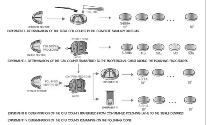
To verify the transfer of microorganisms from the polishing of complete dentures, four experiments were conducted (Figure 1).
Experiment I: Determination of CFU/mL of complete maxillary dentures demonstrating the contamination level of prostheses
Ten maxillary dentures were placed on sterile Petri plates and transferred to the Microbiology Labo-ratory. Under laminar flow, the dentures were washed with 10 mL PBS (phosphate buffered saline) using sterile toothbrushes for removal of microorganisms. The resulting suspensions were serial diluted (100-106) in PBS, pH = 7.2, seeded on Petri plates containing BHI agar, supplemented with 5% defibrinated sheep blood (S-BHIA), and incubated at 37oC for 48 h under anaero-bic conditions (GasPack Anaerobic System, BBL, Cockeysville, MD, USA).
The counts of anaerobe colony-forming units per milliliter of sterile PBS used to wash the dentures were determined.
Experiment II: Determination of CFU transferred to the professional during the polishing process of used complete dentures
Prosthesis Laboratory for routine polishing with a lathe (style 15.2014, SS White, Dayton, OH, USA), disin-fected with 2% iodophor and a frontal protection stand (VH Equipments, Araraquara, São Paulo, Brazil).
The polishing cones and pumice were submitted to sterilization in an ethylene oxide gas chamber. Fol-lowing sterilization, random samples were tested to determine sterility. All tested materials were negative for culture growth.
The technician used sterile gloves, mask, protec-tive glasses and apron. Four open Petri plates with the following culture media were attached to the techni-cian: BHI agar, supplemented with 5% S-BHIA, Mitis Salivarius agar (MS) selective for Streptococci, MacConkey agar (MC) selective for Gram-negative microorganisms, Sabouraud dextrose agar (SDA) se-lective for yeast (all media were from Difco, Detroit, MI, USA) and Sucrose-Bacitracine agar (SB20) selec-tive for mutans streptococci (14). Each denture was polished for 4 min at 2,600 rpm, with the culture plates exposed on the thorax and abdomen of the technician for 2 min, respectively. The plates were then closed and incubated at 37oC for 48 h. MC and SDA cultures were
maintained under aerobic conditions; SB20, MS and S-BHIA used the anaerobic GasPacksystem.
Experiment III: Determination of CFU transferred from contaminated polishing lathe to sterile dentures
Complete maxillary prostheses were made from a standard model to obtain samples with similar anatomy, size and rugosity to standardize conditions of microor-ganism transmission. Each denture received individual “blister” packaging and was submitted to sterilization in an ethylene oxide gas chamber, considered inert for prosthetic materials (7). A random sample of the den-tures was tested for sterility. There was no culture growth.
After each polishing procedure of patient’s den-ture in Experiment II, the technician, wearing new sterile gloves, began polishing of the sterile denture using the same cone and pumice used in Experiment II. After polishing, each denture was placed immediately on a sterile Petri plate and taken to the Microbiology Laboratory for processing with washing with PBS and brushing similar to the method of Experiment I.
Experiment IV: Determination of CFU remaining on the polishing cone after experiments II and III
After polishing the sterile denture in Experiment III, the 30 cones were removed aseptically, placed in a sterile container and taken to the Microbiology Labora-tory for processing. Glass beads and 20 ml PBS were added to the container under laminar flow and were closed and spun for 1 min. Ten milliliters of the result-ing suspension were diluted and seeded in S-BHIA.
RESULTS
The results are reported in Tables 1 and 2. Ex-periment I, determining the CFU in used complete dentures, showed that these dentures were highly colo-nized (mean 1.4 x 10⁷ CFU per milliliter of fluid used to wash the dentures). In Experiment II, there was a high level of contamination by splatter and aerosols in the different selective culture media used to show the trans-mission of potentially pathogenic microorganisms to the operator (Table 2). Experiment III showed a mean transfer of 1.7 x 10⁷ CFU/mL from patient´s prostheses to sterile prostheses. The mean number of remaining microorganisms on the cone after experiments II and III was 3.5 x 10⁸ CFU/mL (Experiment IV).
DISCUSSION
Dental laboratory technicians are particularly vulnerable to microbial cross-contamination from the elastomeric impressions and from the dental prostheses they receive from dental offices (15,16). Casts poured from impressions can also harbor infectious microor-ganisms that can be distributed throughout the labora-tory when the casts or dies are trimed (16).
The results of the four experiments conducted in
this investigation revealed massive cross contamina-tion in prosthesis laboratory routines and a strongly contaminating source in complete dentures of patients. The process of polishing using high-speed lathes can transmit disease between the dental clinic and the labo-ratory technician.
Polishing lathes are considered to be a source of contamination in prostheses laboratories. However, in-fection control measures are not being effectively ap-plied. This research studied the transmission of micro-organisms in the dental laboratory by means of lathes, using a method which reproduced laboratory polishing procedures.
Experiment I, determining the CFU in used com-plete dentures, showed that these dentures were densely colonized indicating a high level of contamination, especially considering that the patients in this study did not present any debilitating disease. Dentures of pa-tients who are diseased, debilitated and/or immuno-compromised have been reported to have even higher levels of contamination (12). In a similar study, Powell et al. (3)reported a high level of contamination of complete dentures, with the presence of α-hemolytic streptococci, β-hemolytic streptococci, Klebsiella oxytoca and Pseudomonas sp.
Bacterial contamination of scrub jackets during dental hygiene procedures was studied by Huntley and Campbell (17). They demonstrated that aerosols are produced during examination and scaling when hand instruments alone are used. The number of
microorgan-Table 1. Number of colony forming units per milliliter found in experiments I, III and IV.
Experiment N CFU/mL (mean)
I 10 1.4 x 10⁷ ± 0.8 x 10⁷
III 30 1.7 x 10⁷ ± 1.5 x 10⁷
IV 30 3.5 x 10⁸ ± 9.0 x 10⁸
CFU/mL = colony-forming units per milliliter of sterile PBS used to wash denture/cone.
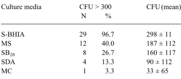
Table 2. Mean and frequency of colony forming units (CFU) grown in different culture media transferred to the technician after polishing 30 dentures (Experiment II).
Culture media CFU > 300 CFU (mean)
N %
S-BHIA 29 96.7 298 ± 11
MS 12 40.0 187 ± 112
SB20 8 26.7 160 ± 117
SDA 4 13.3 90 ± 112
MC 1 3.3 33 ± 65
S-BHIA: Brain heart infusion agar, supplemented with 5% defibrinated sheep blood
MS: Mitis Salivarius agar SB20: Sucrose-Bacitracine agar
isms is higher on sleeves than on the chest of scrub jackets, and is higher when ultrasonic or sonic scalers or air polishers are used.
Experiment II verified the transmission of mi-croorganisms to the professional by aerosol contamina-tion produced during the polishing process. Oral micro-organisms such as Streptococcus mutans and non-oral potentially pathogenic microorganisms such as yeast and Gram-negative bacteria, which can cause eye and respiratory infections, were found in aerosol and splat-ter. Williams et al. (10) found Gram-negative Acineto-bacter in cultures of pumice from laboratories. This non-oral bacteria has been associated with infections such as pneumonia, meningitis, septicemia and eye infections (9).
Experiment III showed a transfer of microorgan-isms from patient prostheses to sterile prostheses. These results can have serious implications because this ex-periment reproduced the conduct of most prosthesis laboratories where pumice and polishing cones are not changed or disinfected regularly between procedures on different prostheses. Because the typical users of dentures are the elderly who can have lowered immu-nological resistance, the transfer of microorganisms confirmed in Experiment III places these patients at risk for the development of infection caused by cross-con-tamination. In a similar study, Kahn et al. (1) reported a mean transfer of 5.0 x 10⁵ CFU/mL of patient dentures to sterile dentures, noting the presence of pathogenic microorganisms such as Staphylococcus aureus, Es-cherichia coli, Candida albicans and α-hemolytic strep-tococci.
In Experiment IV, the remaining microorgan-isms on the cone after experiments II and III showed that the cone was highly contaminated even after the transfer of a large quantity of microorganisms to the sterile prosthesis, remaining as a source of infection ready to contaminate the environment, professional and future dentures continuing the cycle of cross infection. According to Molinari et al. (7), the cone should be changed after each polishing and then sterilized.
The 4 experiments showed that the handling of dentures between the dentist and the laboratory pre-sents a dangerous source of cross-contamination that will continue placing the dentist, technician, patient and auxiliary personnel at risk until efficient measures of infection control are instituted.
The use of aprons, gloves and protective glasses (12) by professionals, the use of lathes with efficient shields (5), the association of disinfectants with pumice (7,8), the sterilization or disposal of the cone after each use (7), and the disinfection of dentures before sending them to the laboratory and upon return to the dental clinic (13) are means which can reduce the risk of cross-contamination.
Infection control measures such as the use of barriers during polishing, the disinfection of dentures before being sent to the laboratory and upon return to the dental clinic, the disposal or sterilization of the cone after each use, as well as the addition of disinfectants to pumice, and unit doses of pumice should be adopted with the objective of reducing the risk of cross infection.
In conclusion, complete dentures are massively contaminated with microorganisms and can serve as the primary source in the cycle of cross infection within dental laboratories. The polishing of dentures without previous disinfection leads to a high level of transfer of microorganisms to the professional, the polishing cone and the new dentures.
RESUMO
O polimento de próteses dentais pode causar um ciclo de contaminação cruzada envolvendo cirurgões-dentistas, técnicos de laboratório, pacientes e pessoal auxiliar. O objetivo deste estudo foi demonstrar a contaminação microbiana em laboratório dental durante os procedimentos de polimento de próteses totais. Com esse propósito, 4 experimentos foram idealizados: Experimento I - Determinação da contagem total de unidades formadoras de colônias (UFC) presentes em próteses totais superiores. Durante o procedimento de polimento, determinação da contagem de UFC transferidas para o operador (Experimento II) e contagem total transferida para próteses totais previamente esterilizadas (Experimento III). Experimento IV - Contagem total de UFC remanescentes no cone da politriz após a realização dos experimentos II e III. As próteses totais estavam altamente contaminadas (média = 1,4 x 10⁷ UFC/mL). Observou-se um
elevado nível de contaminação pelo aerosol. Houve transferência de microrganismos da politriz contaminada para as próteses esterilizadas (média = 1,7 x 10⁷ UFC/mL). Os cones estavam
altamente contaminados depois dos procedimentos de polimento (média = 3,5 x 10⁸ UFC/mL). O polimento de próteses dentais é um
possível veículo de transmissão de doenças no ambiente do laboratório e requer técnicas adequadas para o controle de infecção.
ACKNOWLEDGMENTS
REFERENCES
1. Kahn RC, Lancaster MV, Kate Jr W. The microbiologic cross-contamination of dental prostheses. J Prosthet Dent 1982;47:556-559.
2. Wakefield CW. Laboratory contamination of dental prostheses. J Prosthet Dent 1980;44:143-146.
3. Powel GL, Runnells RD, Saxon BA, Whisenant BS. The presence and identification of organisms transmitted to dental laboratories. J Prosthet Dent 1990;64:235-237.
4. Miller RL, Micik RE. Air pollution and its control in the dental office.Dent Clin North Am 1978;22:453-476.
5. Fisher WT, Chandler HT, Brudvik JS. Reducing laboratory con-tamination. J Prosthet Dent 1972;27:221-225.
6. Katberg Jr JW. Cross-contamination via the prosthodontic labo-ratory. J Prosthet Dent 1974;32:412-418.
7. Molinari JA, Merchant VA, Gleason MJ. Controversies in infec-tion control.Dent Clin North Am 1990;34:55-69.
8. Jagger DC, Huggett R, Harrison A. Cross-infection control in dental laboratories. Br Dent J 1995;179:93-96.
9. Williams HN, Falkler Jr WA, Hasler JF. Acinetobacter contami-nation of laboratory dental pumice. J Dent Res 1983;62:1073-1075.
10. Williams HN, Falkler Jr WA, Hasler JF, Libonati JP. The
recov-ery and significance of nonoral opportunistic pathogenic bacteria in dental laboratory pumice. J Prosthet Dent 1985;54:725-730. 11. Sande MA, Gadot F, Wenzel RP. Point source epidemic of
Myco-plasma pneumoniae infection in a prosthodontics laboratory.Am Rev Respir Dis 1975;112:213-217.
12. Henderson CW, Schwartz RS, Herbold ET, Mayhew RB. Evalua-tion of the barrier system, an infecEvalua-tion control system for the dental laboratory. J Prosthet Dent 1987;58:517-521.
13. The Council on Dental Therapeutics, The Council on Prosthetic Services and Dental Laboratory Relations. Guidelines for infec-tion control in the dental office and the commercial dental labora-tory. J Am Dent Assoc 1985;110:969-972.
14. Davey AL, Rogers AH. Multiple types of the bacterium Strepto-coccus mutans in the human mouth and their intra-family trans-mission. Archs Oral Biol 1984; 29:453-460.
15. Nagamatsu Y, Tajima K, Kakigawa H, Kozono Y. Application of electrolyzed acid water to sterilization of denture base. Part 1. Examination of sterilization effects on resin plate. Dent Mater J 2001;20:148-155.
16. Kugel G, Perry RD, Ferrari M, Lalicata P. Disinfection and communication practices: a survey of U.S. dental laboratories. J Am Dent Assoc 2000;131:786-792.
17. Huntley DE, Campbell J. Bacterial contamination of scrub jack-ets during dental hygiene procedures. J Dent Hygiene 1998;72:19-23.