berghei
Sporozoite Antigens by Hepatocytes
Saidou Balam1., Jackeline F. Romero1.¤a
, Silayuv E. Bongfen1¤b, Philippe Guillaume2,
Giampietro Corradin1*
1Department of Biochemistry, University of Lausanne, Epalinges, Switzerland,2Ludwig Institute for Cancer Research, University of Lausanne, Epalinges, Switzerland
Abstract
One target of protective immunity against the Plasmodium liver stage in BALB/c mice is represented by the circumsporozoite protein (CSP), and mainly involves its recognition by IFN-cproducing specific CD8+T-cells. In a previous
in vitrostudy we showed that primary hepatocytes from BALB/c mice processPlasmodium berghei (Pb) CSP (PbCSP) and present CSP-derived peptides to specific H-2kdrestricted CD8+T-cells with subsequent killing of the presenting cells. We now extend these observations to an in vivoinfection model in which infected hepatocytes and antigen specific T-cell clones are transferred into recipient mice inducing protection from sporozoite (SPZ) challenge. In addition, using a similar protocol, we suggest the capacity of hepatocytes in priming of naı¨ve T-cells to provide protection, as further confirmed by induction of protection after depletion of cross-presenting dendritic cells (DCs) by cytochrome c (cyt c) treatment or using traversal deficient parasites. Our results clearly show that hepatocytes presentPlasmodiumCSP to specific-primed CD8+ T-cells, and could also prime naı¨ve T-T-cells, leading to protection from infection. These results could contribute to a better understanding of liver stage immune response and design of malaria vaccines.
Citation:Balam S, Romero JF, Bongfen SE, Guillaume P, Corradin G (2012) CSP—A Model for In Vivo Presentation ofPlasmodium bergheiSporozoite Antigens by Hepatocytes. PLoS ONE 7(12): e51875. doi:10.1371/journal.pone.0051875
Editor:Anne Charlotte Gruner, Museum National d’Histoire Naturelle, France
ReceivedMay 30, 2012;AcceptedNovember 8, 2012;PublishedDecember 18, 2012
Copyright:ß2012 Balam et al. This is an open-access article distributed under the terms of the Creative Commons Attribution License, which permits unrestricted use, distribution, and reproduction in any medium, provided the original author and source are credited.
Funding:The authors have no support or funding to report.
Competing Interests:The authors have declared that no competing interests exist. * E-mail: [email protected]
¤a Current address: EPFL SB IPSB LIFMET Station 6, Lausanne, Switzerland
¤b Current address: Department of Biochemistry, McGill University, Montreal, Quebec, Canada
.These authors contributed equally to this work.
Introduction
Immunization of rodents and humans with radiation- or genetically-attenuated sporozoites (SPZ) (RAS, GAS) confers pre-erythrocytic stage specific protective immunity to an infectious challenge [1,2]. This protective immunity is mediated in part by CD8+ T-cells specific for the CSP and other not yet identified proteins [2–5]. Recently, it was demonstrated that both infected and sporozoite-traversed mouse primary hepatocytes can process thePbCSP and present CSP-derived peptides to a specific H-2Kd -restricted CD8+ T-cell clone in vitro but recognition of infected hepatocytes was the only relevant step in the elimination of infection [6]. Using bone marrow cell transfer into totally irradiated mice, it was also concluded that activation of protective CD8+ T-cell clones was due to antigen presentation by nonhematopoietic parenchymal cells [7]. Thus, the role of hepatocytes as antigen presenting cells (APCs) in the activation of primed T-cells to provide sterile protection seems to be accepted. Nevertheless, the role of liver cells and, in particular, hepatocytes in the activation ofPlasmodium-specific CD8+T-cells is not clearly elucidated. Chakravarty et al. [7] presented data supporting the role of lymph node dentritic cells (DCs) in presentation to parasite-specific T-cells while Leiriao et al. [8] supported the notion that apoptotic Plasmodium-infected hepato-cytes provide antigen to liver DCs. While both pathways of antigen presentation co-exist, their role in providing a protective CD8+
T-cell response has not been established. In both studies, critical experiments such as the elimination of DCs and its consequence on sterile protection are missing. On the other hand, several publications point to the role of liver cells and hepatocytes in the mechanism of protection [2,5,9–11]. In addition, intravenous (iv) injection in mice of RAS gives rise to a more robust immune response and to sterile immunity compared to intradermal (id) immunization [12]. On the other hand, this difference in protection between iv and id injection is overcome by a higher dose of sporozoites [13]. Furthermore, immunization of mice with GAS is associated with a better immune response, probably due to the development of GAS liver stages and presentation of antigens other than CSP to specific T-cells [14].
hepatocytes present CSP that is secreted directly into the cytosol, unlike DCs that cross-presentPlasmodiumantigens via endosomes [16]. These findings suggested that both in DCs and hepatocytes, the presentation of antigens requires the transporter associated with antigen processing (TAP) proteins [16–18] and that the cross-presenting DCs could be abrogated in mice after treatment with cytochrome c (cyt c) [16]. Determining if hepatocytes in vivo process and presentPlasmodiumantigens to naı¨ve and primed T-cells may help in the rational identification of pre-erythrocytic vaccine candidates. To this purpose, we have established anin vivo protocol to address two questions: are hepatocytes capable of 1) in vivostimulation of primed CD8+cells and 2) priming naı¨ve T-cells to protect mice against parasite challenge? We used different methods to address these two questions. First, we used intrasplenic (IS) transfer of PbSPZ-infected hepatocytes from BALB/c mice into naı¨ve,TAP2/2 deficient mice (H-2Kb)and BALB/c mice (H-2Kd) in the presence or absence of H-2Kd-specific CD8+ T-cells (C7 clone) in order to determine the ability of primary hepatocytes to present antigen to primed CD8+T-cells. Secondly, to show that hepatocytes could present antigen in the absence of dendritic cells, mice treated with cytcwere injected with irradiated SPZ (iSPZ) and challenged with live SPZ. Thirdly, mice were immunized with iSPZs deficient for the sporozoite microneme protein essential for cell traversal (spect (2)iSPZs), known for being incapable of cell traversal, but capable of infection and normal development [6,19,20] in order to show the role of infected hepatocytes in inducing a protective immune response. In all cases, mice were partially or totally protected from an infectious SPZ challenge as assessed by their level of blood parasitemia up to 14 days post infection.
Materials and Methods
Peptides
Peptides PbCS245–253 (YIPSAEKI) and PbCS253–260 modified
with iodo-azidosalicylic acid (IASA) and azidobenzoic acid (ABA) groups, (IASA)-YIPSAEK(ABA)I representing the epitope for C7 clone (H-2Kd-restricted andPbCSP specific CTL) and S14 (H-2Kd-restricted and an irrelevant CTL), respectively [3,21], were synthesized by solid-phase F-moc chemistry. Peptide stock solutions (2 mg/ml) were prepared in PBS and stored at220uC.
Parasites
Plasmodium bergheiANKA wild-type (wt) andspect (2)SPZ were obtained after salivary gland dissection of infected femaleAnopheles stephensimosquitos raised in the mosquito facility at the Depart-ment of Biochemistry, University of Lausanne, Switzerland as described previously [6,20]. After dissection, salivary glands were
homogenized in a glass grinder and released SPZ were counted and then diluted in sterile Dulbecco’s Modified Eagle Medium, DMEM (GibcoH, Life TechnologiesTM, New York, NY).
Animals
Six-to-12-week-old BALB/c (H-2Kd) or TAP2/2 (H-2Kb) mice were obtained from Harlan Laboratories B.V. (Venray, Netherlands) or bred at the animal facility at the Department of Biochemistry (University of Lausanne, Switzerland). All mice were housed under pathogen-free conditions and handled according to the guidelines of the authorization Nu805.7 of the Service de la consommation et des affaires ve´te´rinaires (Lausanne, Switzerland).
Hepatocyte Isolation
SPZ-infected and naı¨ve hepatocytes from BALB/c mice were obtained after collagenase perfusion of the liver as previously described [6,20,22]. Briefly, mice were sacrificed by CO2
inhalation, dissected, and the biggest lobule of liver was cut out. The lobule was perfused for 10 min with Ca2+
-free HEPES (4-(2-hydroxyethyl)-1-piperazineethanesulfonic acid) buffer, pH 7.6 (GibcoH InvitrogenTM, New York, NY) at 37uC at a rate of 5 ml/min. The lobule was then perfused with type IV collagenase (Sigma-AldrichH, Steinheim, Germany) (HEPES buffer containing 0.04% type IV collagenase and 0.075% CaCl2-2H2O) for 5 min at
37uC. The perfused lobule was incubated for 10 min at 37uC in the collagenase solution. Using sterile pipettes, the tissue was gently teased apart to release cells and washed once with Ca2+
-free HEPES buffer at 800 rpm for 30 s at 4uC. The pellet was gently re-suspended in DMEM, layered on 60% Percoll (GE Healthcare Bio-Sciences AB, Uppsala, Sweden) and centrifuged at 2000 rpm for 2 min at 4uC. The resulting pellet was re-suspended in complete culture medium (DMEM supplemented with 10% FCS, 1% penicillin streptomycin, 1% HEPES, and 0.05 mM of b -mecarptoethanol (b-ME, Sigma-AldrichH), and centrifuged again at 800 rpm for 30 s at 4uC. The pellet was finally re-suspended again in 5 ml of DMEM for counting. Viability of isolated hepatocytes was assessed by light microscopy and trypan blue dye exclusion. As reported previously [6] hepatocyte contamination with Kupffer/dendritic cell markers is less than 1% as determined by FACS analysis [6,10,23,24]. Then, mice were injected iv with 76105live hepatocytes re-suspended in 200ml of sterile DMEM. To obtain infected hepatocytes, 16106 of live or irradiated wt PbSPZwere injected iv through the tail vein of BALB/c mice. Two hours later, the liver was removed from the infected mouse and perfused as described above.
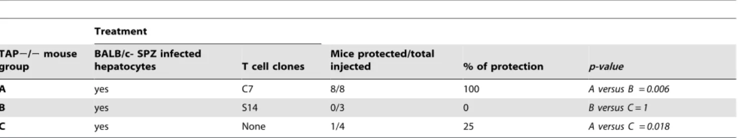
Table 1.Infected hepatocytes present aPbCSP-specific epitope to primed CD8+T-cells and protect mice against SPZ challenge.
Treatment
TAP2/2mouse group
BALB/c- SPZ infected
hepatocytes T cell clones
Mice protected/total
injected % of protection p-value
A yes C7 8/8 100 A versus B = 0.006
B yes S14 0/3 0 B versus C = 1
C yes None 1/4 25 A versus C = 0.018
Each recipientTAP2/2mouse (H-2Kb) received 7
Intrasplenic (IS) Transfer
Hepatocellular transplantation was carried out by direct injection of 76105 hepatocytes in 200ml of sterile DMEM with a syringe into the splenic parenchyma of recipient mice that were anaesthetized with Isoflurane (Provet AG, Berne, Switzerland). Briefly, under aseptic and anaesthesia conditions, cautiously, with a chisel and a clip, the abdomen of the mouse was opened on the left flank. Using a clamp fitted, the spleen was gently pulled out and placed on a sterile piece of paper. The injection of cell suspension in the spleen parenchyma was carefully carried out at a rate of 10ml per 10 respiratory cycles of the mouse. Suturing and clamping of the skin minimized leakage from the site of operation. Four hours later, 20 millionPbCSP-restricted CD8+T-cells (C7-clone) or irrelevant CD8+ T-cells (clone S14) were injected iv through the tail vein in a volume of 500ml of DMEM. Control mice received only hepatocytes.
T-cell Clone Re-stimulation
The C7 and irrelevant S14 clones [3,21] were re-stimulated weekly, maintained at 37uC and used as effector cells. Briefly, P815 cells (mastocytoma cells as antigen presenting cells, APC) were re-suspended (16106cells/ml) in complete culture medium (DMEM supplemented with 10% FCS +1% of pyrimethamine-streptomycin+0.1% ofb-ME and 1% of HEPES) and pulsed for 1 hour withPbCSP-epitope peptides (1mg/ml) specific for C7-and S14 clones. C7 and S14 were washed and re-suspended (26106/ ml/well) in CTL culture medium (complete culture medium
supplemented by 30 U/ml of mouse IL-2) in a 6-well plate. For re-stimulation, the respective clones were added to the 16106pulsed and irradiated P815 cells (10000 rads/20 minutes) in the presence of 156106/well of irradiated-BALB/c spleen cells (5000 rads/10 minutes) in 6-well flat bottom plates. In the intrasplenic experiment, clones were re-stimulated and kept for two weeks in culture to make sure they were resting at the time of injection.
Cytochrome c Treatment and Induction of CD8+T-cells BALB/c mice were depleted of cross-presenting DCs by iv treatment for 3 days with 15 mg/ml (5 mg/ml per day) of horse cyt c (Sigma-AldrichH, St Louis, LA) in 100ml of PBS (GibcoH
InvitrogenTM). Control mice received 100ml of PBS alone. On the last day of treatment, mice were immunized iv with 16105 wt PbiSPZ). Seven-to-ten days later, the frequency of both PE-conjugated SYIPSAEKI-tetramer and FITC-PE-conjugated anti-mouse CD8 antibody (BD Biosciences, Allschwil, Switzerland) specific CD8+ T-cells was measured in different mouse tissues (blood = PBL, spleen, liver and lymph nodes (LN)) by flow cytometry.
Real Time PCR (RT-PCR)
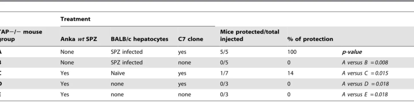
In vivoassessment of parasite loads was performed by RT-PCR as described previously [25]. Briefly, BALB/c mice were injected iv with 16105PbiSPZ (wtorspect (2)). Two hours later, total livers and spleens were isolated, perfused with PBS alone and total RNA was extracted. Then, cDNA was synthesized using specific primers Table 2.Specificity of activation of C7 clone and protection.
Treatment
TAP2/2mouse
group AnkawtSPZ BALB/c hepatocytes C7 clone
Mice protected/total
injected % of protection
A None SPZ infected yes 5/5 100 p-value
B None SPZ infected none 0/5 0 A versus B = 0.008
C Yes Naı¨ve yes 1/7 14 A versus C = 0.015
D Yes none yes 0/3 0 A versus D = 0.018
E Yes none none 0/3 0 A versus E = 0.018
Mice were first injected (iv) or not with 103live wild-type (wt) ANKAPbSPZ 10 h before they received or not 7
6105naı¨ve-BALB/c hepatocytes and C7 clone as indicated above. Infected hepatocytes were isolated from BALB/c mice injected with 106ANKAwt PbSPZ 2 h earlier; and naı¨ve BALB/c hepatocytes were isolated from naı¨ve BALB/c mice. Mice were considered protected if they remained parasite negative 2 weeks after infection.
doi:10.1371/journal.pone.0051875.t002
Table 3.BALB/c mice injected with iSPZ-loaded hepatocytes are protected against SPZ challenge.
2 weeks after challenge
Mouse group (n of SPZ for challenge) Treatment Mice protected/total challenged % of protection p-value: IS versus naive
A (2000) IS 5/5 100 0.167
Naive 2/5 40
B (5000) IS 2/6 33 0.125
Naive 0/10 0
Total IS 7/11 64 0.014
Naive 2/15 13
BALB/c mice were injected (IS transfer) with 76105PbiSPZ-infected or naı¨ve BALB/c hepatocytes. Infected hepatocytes were obtained from BALB/c mice immunized with 106ANKAwtiSPZ 2 h earlier. Recipient mice were then challenged with two different doses (2
6103and 56103, respectively, A and B) of livePbSPZ one week later. Mice were considered protected if they remained parasite negative 2 weeks after challenge.
for the P. berghei 18S rRNA (forward: 59
AAGCATTAAA-TAAAGCGAATACATCCTTAC-39 and reverse: 59
GGA-GATTGGTTTTGAC GTTTATGTG-39) as described previous-ly [25]. The DNA was thus amplified in the LightCycler 2.0 Instrument (Roche Diagnostics, Basel, Switzerland) using the program Roche LightCycler Run 5.32, and the relative parasite
DNA load was thus determined in liver and spleen for each type of SPZ.
Parasitemia Assessment
At different time points after the IS transfer or challenge with livePbSPZs (iv), parasitemia was assessed by 10% Giemsa (FlukaH, Sigma-AldrichH)-stained blood smears. Blood smear slides were air-dried and read by light microscopy (Olympus CH-2, Micro-scope Company, Hicksville, NY) to determine infected red blood cells (iRBC). Animals were protected against malaria if they remained negative in Giemsa-stained blood smears 2 weeks after receiving infected hepatocytes or live SPZ. Control animals were included to verify infectivity of SPZ or infected hepatocytes. In each control animal, parasitemia was detectable 7 days after IS transfer or challenge.
Statistical Analysis
Different statistic tests were performed using GraphPad Prism software (version 6). Fisher’s exact test compares the proportion of mouse protection among various groups. The Mann-Whitney test was performed to compare parasite DNA load and frequency of PbCSP-specific CD8+ T-cells in PBL in two independent experiments or in different organs in the same experiment in which mice underwent different treatments (wild-type and Spect (2) iSPZ or PBS and cytcgroups). All p-values equal to or lower than 0.05 were considered significant.
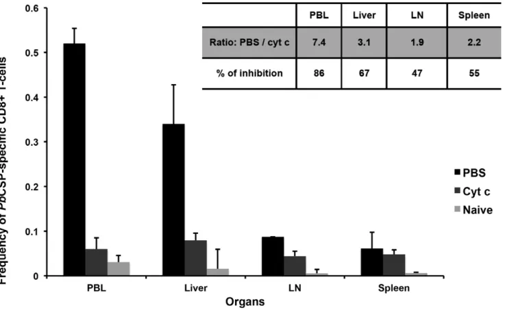
Figure 1. Level ofPbCSP-specific CD8+T-cells in different organs of BALB/C mice after treatment with either cyt c or PBS and
immunization withPbiSPZ.Two groups of BALB/c mouse were pre-treated (iv through the tail vein) with 5 mg/mouse of cyt c per day for 3 days in 100ml of PBS or with 100ml of PBS alone. Immediately after the last treatment, mice were immunized with 16105PbiSPZ in 100ml of RPMI.
One-week later,PbCSP epitope-specific CD8+T-cell frequency (2 mice per group of treatment) was evaluated in peripheral blood lymphocytes (PBL), liver, lymph nodes (LN) and spleen. Naı¨ve (receiving neither cyt c, PBS nor iSPZ) mice were used as negative control. Inserted panel shows the fold change in frequency and the percentage of inhibition ofPbCSP-specific CD8+T-cells in cyt c-treated compared to PBS alone-treated groups.
doi:10.1371/journal.pone.0051875.g001
Table 4.Protection from infection in cyt c- and PBS- treated mice.
Treatment
Mice protected/total
challenged % of protection
Cyt c 10/10 100
PBS 10/10 100
Naive 1/10 10
Protection of mice after cyt c or PBS treatment from live SPZ challenge of two independent experiments (5 mice per group in each experiment). Mice were either pre-treated iv for 3 days with 5 mg/mouse of horse cyt c per day in 100ml of PBS or with 100ml of PBS alone. On the last day of treatment, mice were immunized iv with 16105PbiSPZ in 100ml of RPMI. One week later, mice
were challenged with 56103livePbSPZ. Parasitemia was checked at 1 and 2 weeks after challenge. Mice were considered protected if they remained parasite negative 2 weeks after challenge.
Results
Infected Hepatocytes Present thePbCSP-specific Epitope to Cloned CD8+ T-cells with Subsequent Protection against Malaria
To show that infected hepatocytes process and presentPbCSP, TAP2/2mice received BALB/cPbSPZ-infected hepatocytes by IS transfer followed by iv injection ofPbCSP specific (clone C7) or irrelevant (S14 clone) CD8+ T-cells 4 h later. TAP 2/2 mice were selected to bypass the possibility that presentation could be performed by professional APCs through processing of apoptotic, infected hepatocytes or live SPZs possibly externally associated with BALB/c hepatocytes. In addition, the host vs graft immune response is minimized. All mice (8/8; 100%) that received infected hepatocytes and the specific C7 clone were protected from an infective sporozoite challenge(Table 1, group A). In contrast, all mice receiving the irrelevant S14 clone were infected(Table 1, group B). In addition, 75% of naiveTAP2/2mice treated with infected BALB/c hepatocytes were infected(Table 1, group C).
In order to show that the protective immune response is specific and not due to a bystander effect or a continuous secretion of cytokine by the C7 clone,TAP2/2mice were first infected with live PbSPZ (1000/mouse in iv) or not. Ten hours later, mice received infected or naı¨ve BALB/c hepatocytes together or without C7 clone as indicated in Table 2. Parasitemia determi-nation of group C showed that only 1/7 (14%) of mice was protected (Table 2, group C). Other controls showed that protection occurred only in the group that received infected BALB/c hepatocyte and C7 clone (Table 2, group A).
Infected Hepatocytes PrimePbCSP-specific CD8+ T-cells and Protect Mice against SPZ Challenge
Since the infected hepatocytes can reactivate resting CSP-specific CD8+T-cells and induce protective immunity(Table 1 and 2), the next step was to determine if they could also prime naı¨ve T-cells to protect mice against live SPZ challenge. To this effect, we injected iSPZ-infected BALB/c hepatocytes into naive BALB/c mice before challenging with live SPZ. Considering that the immunization procedure may give rise to a sub-optimal immunity (about 700 infected hepatocytes injected IS if the overall infection efficacy is estimated to be 10%), mice were challenged with a sub-optimal or optimal dose of live SPZ (26103and 56103) that led to 60% and 100% infection in naı¨ve mice, respectively, with an overall protection of 13%(Table 3). In contrast, mice receiving iSPZ-infected hepatocytes were protected at 64% (7/11; p = 0.014) (Table 3), suggesting that the infected hepatocytes could contribute to the priming of naı¨ve T-cells and protection of mice against infection.
To further corroborate that hepatocytes could prime naı¨ve T-cells and induce protection, BALB/c mice were treated with cyt c to delete cross-presenting DCs before and during iSPZ immuni-zation. This protocol was established according to previous studies which showed that cross-presenting DCs can be largely depleted afterin vivocyt c treatment [16,26]. The first experiment (as pilot) showed a significant reduction of 60% ofPbCSP245–253 specific
CD8+T-cells in PBL after cyt c treatment (data not shown). In the second experiment, the analysis was extended to other organs (LN, spleen and liver) (Fig. 1). Thus, FACS analysis showed that induction ofPbCSP-specific CD8+T-cells was highly reduced in the cyt c- compared to the PBS -treated mice in PBL and liver (86% and 67%, respectively), while this reduction was about 50%
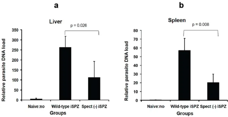
Figure 2. Relative comparativewtandspect (2)parasite DNA load in spleen and liver of immunized mice.To compare relative load of wtandspect (2)parasite DNA in liver and spleen, BALB/c (3 mice per group) were immunized iv on tail with 16105iSPZ (wtorspect (2)) in 500ml of
RPMI. Two hours later, a real-time PCR was performed following extraction of respective parasite RNA. Every sample was done in duplicate. To avoid any contamination by eventual parasite from blood, the liver and spleen were perfused with PBS before performing the RT-PCR. Figuresaandb representwtandspect (2)parasite DNA load, respectively, in the liver and spleen 2 h after iSPZ injection (iv). Naı¨ve mice (receiving only 500ml iv of
in LN and spleen(Fig. 1,inserted panel). Taking the PBL data from the two experiments or from all organs in the second experiment, normalizing and combining them, we obtained p values of 0.003 (PBL) and 0.001 (organs) for the cyt c-treated compared to PBS groups. In spite of these differences, both groups were protected after SPZ challenge(Table 4).
Protective Immune Response Induced in BALB/c Mice Immunized withspect (2)iSPZ
Wild-type SPZs are known to be able to cross several cells (leaving a trail of the CSP behind) before infecting a single hepatocyte, unlike spect (2) SPZ that infect hepatocytes without cell traversal. It has been shown that both infected and traversed hepatocytes are able to process and presentPbSPZ CSP to primed CD8+ T-cells to induce IFN-c secretion invitro but only the infected hepatocytes were responsible for their own elimination
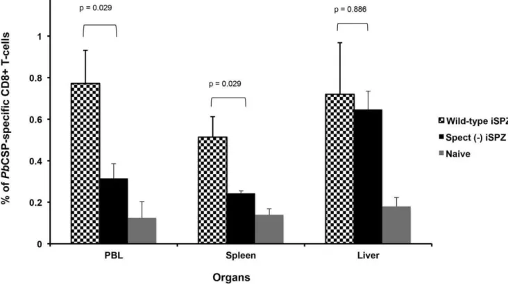
[6,20]. Similar experiments were then performedin vivo. Thus, it was expected thatspect (2)SPZ would activate a lower number of thePbCSP-specific CD8+ T-cells in the periphery and present a lower parasite load in the liver. RT-PCR clearly showed that the relative parasite DNA load in liver and spleen was significantly higher in mice receivingwtiSPZ compared to thespect (2)iSPZ parasite group (p = 0.026 and 0.008, respectively) (Fig. 2a and b). In addition, FACS analysis showed that the level of thePb CSP-specific CD8+ T-cell response was significantly higher in the wt compared to thespect (2) group in PBL and spleen (p = 0.029) suggesting a role of cross-presenting DCs in these compartments
(Fig. 3). In contrast, in the liver, the frequency of CD8+T-cells was similar (p = 0.886) for the two kinds of iSPZ(Fig. 3)in spite of a lower parasite load forspect (2). Both groups of mice were protected against live SPZ challenge(Table 5). Together, these data indicate that T-cell traversal by SPZ induces a higher level of immune response in PBL and spleen, but not in the liver, further
Figure 3. Frequency ofPbCSP-specific CD8+T-cells in different organs of BALB/c mice immunized withwtorspect (2)iSPZ.BALB/c mice (4 mice per group) were immunized (iv) withwtorspect (2)ANKAPbiSPZ (one dose of 16105iSPZ). Seven (7) days later, PBL, liver and spleen cells of mice were isolated to determine frequency ofPbCSP-specific CD8+T-cells by flow cytometry (FACScan). The CD8+T-cells were double stained
with PE-conjugatedPbCSP epitope tetramer and FITC-conjugated anti-mouse CD8b antibody. The naive group received neitherwtnorspect (2)iSPZ. p-value compares statistically significant mean of CD8+T-cell frequency betweenwtandspect (2)iSPZ-immunized groups.
doi:10.1371/journal.pone.0051875.g003
Table 5.Bothwtandspect (2) PbiSPZ immunization protect mice against SPZ challenge.
Immunization (number ofPbISPZ) Challenge withPbSPZ Mice protected/total challenged % of protection
Wild-type (16105) 16104 5/5 100
Spect (2) (16105) 16104 5/5 100
Naive (0) 16104 0/5 0
One week after 16105PbiSPZ (wtorspect (2)) immunization, BALB/c mice were challenged with 16104live ANKAwt PbSPZ. Parasitemia was checked at 1 week and 2 weeks post challenge and mice were considered protected when they remained parasite negative 2 weeks after challenge.
supporting the key role of infected hepatocytes in antigen presentation and induction of protective immunity.
Discussion
Data presented in this manuscript provide further evidence for the role of Plasmodiuminfected hepatocytes in the stimulation of protective secondary CD8+T-cells leading to the elimination ofP. berghei pre-erythrocytic stages. In addition, they indicate that hepatocytes can indeed prime sporozoite-specific, protective naı¨ve T-cells. While the antigen-presenting role of infected hepatocytes in the secondary CD8+T-cell response seems to be accepted, the mode of priming naı¨ve CD8+T-cells is still, in our opinion, not yet established. With regard to the first point, the in vivo data presented here are fully consistent with our previousin vitroresults [6,20] and within vivodata published by Zavala and collaborators [7] via bone marrow cell transfer experiments. In our case, we transferred either BALB/c infected hepatocytes or a CS specific T-cell clone or both to TAP-deficient H-2kb mice. Tap2/2mice were chosen to bypass the possibility that antigen presentation is mediated by professional antigen presenting cells which might have ingested apoptotic infected hepatocytes [8] or live sporozoites possibly externally associated with BALB/c hepatocytes, and minimize host vs graft immune responses. In addition, we have determined protection as lack of infection in mice 14 days post-challenge, which represents a stringent, but the only significant standard for protection for pre-erythrocytic vaccines. Our results show that protection from infection is antigen-specific since mice are not protected if an irrelevant CD8+ T-cell clone is used. In addition, as observedin vitro[6], our results indicate that infected hepatocytes are directly killed by the antigen- specific T cells and not by a bystander effect through continuous secretion of IFN-cor other lymphokines by the injected T-cell clones or host vs graft immune response since concomitant infection of TAP-deficient mice does not lead to protection after treatment with the CS specific T-cell clone and/or naı¨ve BALB/c hepatocytes. These and the previous data [6,7] clearly establish the central role of infected hepatocytes in the total clearance ofPlasmodiuminfection in vivo once an immune response has been induced (secondary response). However, in our opinion, the role of hepatocytes in priming naı¨ve, T-cells is not yet elucidated. Zavala and collaborators [7,16] claim that peripheral dendritic cells prime CD8+ T-cells, while Leiriao et al. [8] suggest that apoptotic infected hepatocytes provide antigens to liver dendritic cells. In addition, these claims seem to be supported by the notion that hepatocytes act as tolerizing cells [27,28]. On the other hand,
given the large number of hepatocyte genes affected by sporozoites and salivary gland components, including some related to antigen processing and presentation and chemokine production it is not far fetched to hypothesize that infected hepatocytes become full-fledged antigen presenting cells upon infection [29,30]. This would allow the activation of T-cells specific to sporozoite and late liver stage antigens (14). This would optimize the balance between infection and immune responses that parasites and hosts have developed through co-evolution. Evidence that the liver is central to obtaining an optimal immune response was provided early on by Reniaet al[10] in which similar results as obtained here were presented, where immunization with non-parenchymal cells did not result in protection. Recent data by Epstein etal. [12] in which a better immune response was obtained by immunization of mice with irradiated sporozoites via iv than id or sc injection also point to the antigen-presenting role of the liver. In this study, we specifically target the role of infected hepatocytes in the immune response by immunizing (iv) mice withspect (2) iSPZ that infect hepatocytes without prior traversal. We show that, in spite of a significant decrease in parasite DNA load, the liver CSP-specific CD8+ T-cell response was similar to that inwtiSPZ-immunized groups. This and cyt c treatment of mice prior to immunization with iSPZs further support the key role of infected hepatocytes in T-cell priming to provide protective immune responses.
In conclusion, the data provided show that hepatocytes can indeed presentPlasmodium bergheiCSP epitopes to primed CD8+ T-cells and strongly suggest that they could also prime parasite-specific naı¨ve T-cells to fully protect mice against a live parasite challenge. But in our opinion, formal proof of the role of hepatocytes in antigen presentation can only be obtained by isolating malaria parasite infected hepatocytes for in vivo and invitroexperiments.
Acknowledgments
We thank HR McDonald for the gift of TAP2/2mice, I Lu¨scher for help in preparing the tetramers, R. Menard for providing thespect (2)parasites and the University of Lausanne for in-house support.
Author Contributions
Conceived and designed the experiments: S. Balam JR S. Bongfen GC. Performed the experiments: S. Balam JR S. Bongfen. Analyzed the data: S. Balam JR S. Bongfen GC. Contributed reagents/materials/analysis tools: PG. Wrote the paper: S. Balam GC. Review and revision: JR S. Bongfen PG.
References
1. Jobe O, Lumsden J, Mueller AK, Williams J, Silva-Rivera H, et al. (2007)
Genetically Attenuated Plasmodium berghei Liver Stages Induce Sterile Protracted Protection That Is Mediated by Major Histocompatibility Complex Class I–Dependent Interferon-g– Producing CD8+T-Cells.J Infect Dis 196: 599–607.
2. Scheller LF, Azad AF (1995)Maintenance of protective immunity against malaria by persistent hepatic parasites derived from irradiated sporozoites.Proc Natl Acad Sci U S A 92: 4066–4068.
3. Romero P, Maryanski JL, Corradin G, Nussenzweig RS, Nussenzweig V, et al. (1989)Cloned cytotoxic T-cells recognize an epitope in the circumsporozoite protein and protect against malaria.Nature 341: 323–326.
4. Schofield L, Villaquiran J, Ferreira A, Schellekens H, Nussenzweig R, et al. (1987)Gamma interferon, CD8+T-cells and antibodies required for immunity to malaria sporozoites.Nature 330: 664–666.
5. Gru¨ner AC, Mauduit M, Tewari R, Romero JF, Depinay N, et al. (2007)Sterile Protection against Malaria Is Independent of Immune Responses to the Circumsporozoite Protein.PLoSOne 2: e1371.
6. Bongfen SE, Torgler R, Romero JF, Renia L, Corradin G (2007)Plasmodium berghei-infected primary hepatocytes process and present the circumsporozoite protein to specific CD8+T-cells in vitro.J Immunol 178: 7054–7063.
7. Chakravarty S, Cockburn IA, Kuk S, Overstreet MG, Sacci JB, et al. (2007)
CD8+T lymphocytes protective against malaria liver stages are primed in skin-draining lymph nodes.Nat Med 13: 1035–1041.
8. Leiriao P, Mota MM, Rodriguez A (2005)Apoptotic Plasmodium-Infected Hepatocytes Provide Antigens to Liver Dendritic Cells.J Infect Dis 191: 1576–81.
9. Renia L, Maranon C, Hosmalin A, Gruner AC, Silvie O, et al. (2006)Do apoptotic Plasmodium-infected hepatocytes initiate protective immune responses?J Infect Dis 193: 163–4.
10. Renia L, Rodrigues MM, Nussenzweig V (1994)Intrasplenic immunization with infected hepatocytes: a mouse model for studying protective immunity against malaria pre-erythrocytic stage.Immunology 82: 1164–168.
11. Mota MM, Pradel G, Vanderberg JP, Hafalla JC, Frevert U, et al. (2001)
Migration of Plasmodium sporozoites through cells before infection.Science 291: 141–144. 12. Epstein JE, Tewari K, Lyke KE, Sim BK, Billingsley PF, et al. (2011)Live attenuated malaria vaccine designed to protect through hepatic CD8+T-cell immunity.
Science 334: 475–80.
13. Voza T, Kebaier C, Vanderberg JP (2010)Intradermal immunization of mice with radiation-attenuated sporozoites of Plasmodium yoelii induces effective protective immunity.
Malar J 9: 362.
14. Douradinha B, van Dijk M, van Gemert GJ, Khan SM, Janse CJ (2011)
induces a long-lasting effector memory CD8+T-cell response in the liver.J Immune Based Ther Vaccines 9: 6.
15. Frevert U, Engelmann S, Zougbe´de´ S, Stange J, Ng B, et al. (2005)Intravital observation of Plasmodium berghei sporozoite infection of the liver.PLoS Biol 3: e192. 16. Cockburn IA, Tse SW, Radtke AJ, Srinivasan P, Chen YC, et al. (2011)Dendritic
Cells and Hepatocytes Use Distinct Pathways to Process Protective Antigen from Plasmodium in vivo.PLoS Pathog 7: e1001318.
17. Van Kaer L, Ashton-Rickardt PG, Ploegh HL, Tonegawa S (1992)TAP1 Mutant are deficient in Antigen presentation, Surface Class I Molecules, and CD4–8+T cells.Cell press 71: 1205–1214.
18. Chefalo PJ, Grandea AG 3rd, Van Kaer L, Harding CV (2003)Tapasin2/2
and TAP12/2Macrophages Are Deficient in Vacuolar Alternate Class I MHC (MHC-I) Processing due to Decreased MHC-I Stability at Phagolysosomal pH1.J Immunol 170: 5825–5833.
19. Ishino T, Yano K, Chinzei Y, Yuda M (2004)Cell-passage activity is required for the malarial parasite to cross the liver sinusoidal cell layer.PloS Biol 2: E4.
20. Bongfen SE, Balam S, Torgler R, Romero JF, Corradin G (2008)Processing of the circumsporozoite protein in infected hepatocytes is not dependent on aspartic proteases.Parasite Immunol 30: 375–378.
21. Luescher IF, Anjue`re F, Peitsch MC, Jongeneel CV, Cerottini JC, et al. (1995)
Structural analysis of TCR-ligand interactions studied on H-2Kd-restricted cloned CTL specific for a photoreactive peptide derivative.Immunity 3: 51–63.
22. Seglen PO (1976)Preparation of isolated rat liver cells.Methods Cell Biol 13: 29–83.
23. Meis JF, Verhave JP, Jap PH, Meuwissen JH (1983)An ultrastructural study on the role of Kupffer cells in the process of infection by Plasmodium berghei sporozoites in rats.
Parasitology 86: 231–242.
24. Seguin MC, Ballou WR, Nacy CA (1989)Interaction of Plasmodium berghei sporozoites and murine kupffur cells.J Immunol 143: 1716.
25. Torgler R, Bongfen SE, Romero JC, Tardivel A, Thome M, et al. (2008)
Sporozoite-mediated hepatocyte wounding limits plasmodium parasite development via MyD88-mediated NF-kB cctivation and inducible NO synthase expression.J Immunol 180: 3990–
3999.
26. Lin ML, Zhan Y, Proietto AI, Prato S, Wu L, et al. (2008)Selective suicide of cross-presenting CD8+dendritic cells by cytochrome c injection shows functional heterogeneity within this subset.Proc Natl Acad Sci U S A 105: 3029–34.
27. Mehal WZ, Azzaroli F, Crispe IN (2001)Antigen presentation by liver cells controls intrahepatic t cell trapping, whereas bone marrow-derived cells preferentially promote intrahepatic T cell apoptosis.J Immunol 167: 667–673.
28. Crispe IN, Giannandrea M, Klein I, John B, Sampson B, et al. (2006)Cellular and molecular mechanisms of liver tolerance.Immunol Rev 213: 101–118.
29. Chattopadhyay R, de la Vega P, Paik SH, Murata Y, Ferguson EW, et al. (2011)
Early transcriptional responses of HepG2-A16 liver cells to infection by Plasmodium falciparum sporozoites.J Biol Chem 286: 26396–405.