Arq Neuropsiquiatr 2006;64(3-B):862-864
Pontíficia Universidade Católica do Paraná, Hospital Universitário Cajuru, Curitiba PR, Brazil (HUC-PUCPR): 1Neurocirurgião do
Serviço de Neurocirurgia; 2Médico Residente de Neurocirurgia; 3Médica Residente de Radiologia; 4Chefe do Serviço de Neurocirurgia.
Received 16 January 2006, received in final form 8 June 2006. Accepted 28 June 2006.
Dr. Carlos A.F. Lobão - Hospital Universitário Cajuru - Avenida São José 300 - 80050-350 Curitiba PR - Brasil. E-mail: menegottom@ yahoo.com.br
VEIN OF GALEN ANEURYSM IN AN ADULT
Case report
Robinson M. Marques
1, Carlos A.F. Lobão
2, Viviane S. Sassaki
3, Luiz R. Aguiar
4ABSTRACT - Vein of Galen aneurysm is a rare pathology, representing less than 1% of intracranial vascu-lar malformations. We report on a 65 years-old man who experienced a generalized tonic-clonic seizure. Brain imaging showed a large calcified expanding mass in the pineal region, confirming the diagnosis of a vein of Galen aneurysm. Because of the spontaneous thrombosis of the malformation, there was no need for microsurgical or endovascular treatment and he is been regularly followed since that.
KEY WORDS: vein of Galen aneurysm, vein of Galen malformation, cerebral vein, congenital vascular mal-formation.
Aneurisma da veia de Galeno em adulto: relato de caso
RESUMO - Aneurisma da veia de Galeno é patologia rara, representando menos de 1% das malformações vasculares intracranianas. Apresentamos o caso de um homem de 65 anos que teve episódio de crise con-vulsiva tônico-clônica generalizada. Exames de imagem evidenciaram grande processo expansivo calcifi-cado na região pineal, confirmando o diagnóstico de aneurisma trombosado de veia de Galeno. Devido à trombose espontânea da malformação, foi excluída a possibilidade de tratamento endovascular bem como microcirúrgico, mantendo-se o acompanhamento clínico.
PALAVRAS-CHAVE: aneurisma da veia de Galeno, formação da veia de Galeno, veias cerebrais, mal-formação vascular congênita.
Vein of Galen aneurysm (VGA) is a rare vascular malformation representing less than 1% of vascular intracranial abnormalities. It is a congenital process frequently detected between the 6thand 11thmonths
of gestational age1, during early childhood or
neona-tal period2. This finding in adult age is very rare,
pre-senting or not symptoms throughout childhood3.
Cli-nical manifestations can present at any age in the form of heart failure, delayed neuropsychomotor de-velopment, hydrocephalus and seizures4.
We describe a case of VGA reporting clinical symp-toms, radiological findings and management relat-ing to current published literature.
CASE
A 65 year-old man was brought to the emergency room at the University Hospital of the Pontifical Catholic Univer-sity of Paraná, Brazil because of generalized tonic-clonic seizures. On admission, he was treated with anticonvulsant therapy for status epilepticus. Past medical history was
sig-nificant for treated high blood pressure and heart failure diagnosed late in life. Also significant for an episode of ischemic cerebrovascular disease two years before, result-ing in left hemiparesis. Durresult-ing childhood and adult life the-re was no neuropsycomotor development delay, no signs of cardiopathy, no neurological signs or symptoms as well as no history of epilepsy.
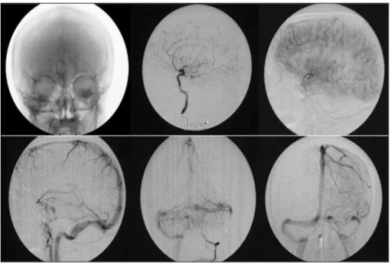
CT scan showed a calcified expanding process in the pineal region (Fig 1). MRI showed a heterogenic round-shaped mass localized in the supra-vermian cistern with dimensions of 35x32x30 mm showing high signal in T2-weight imaging (Fig 2). The MRI images were suggestive of a thrombosed VGA. Cerebral angiography showed a thrombosed VGA also (Fig 3).
Considering the fact that the aneurysm was already thrombosed and that there were no signs or symptoms of intracranial hypertension, nor hydrocephalus, the patient was not referred for surgery or endovascular treatment. Improvement in the level of consciousness and absence of seizures episodes were noted during the follow-up period while maintaining the clinical therapy for seizures.
Arq Neuropsiquiatr 2006;64(3-B) 863
Fig 3. Angiography evidencing the thrombosed vein of Galen aneurysm. Fig 2. MRI showing thrombosed vein of Galen aneurysm.
Fig 1. CT scan showing calcified expanding process at the pineal region.
DISCUSSION
VGA is a vascular abnormality typically found in children5. Raybaud et al.1believe it is a reminiscent
of fetal anatomy produced by frequent occlusions of posterior fossa dural sinus, especially at the sigmoid sinus. Even though the VGA can be asymptomatic in adults6,7, it is typically diagnosed during the
neona-tal period or in childhood with heart failure signs, macrocephaly and/or cranial murmurs8. Children with
slow flow fistulas have a better extra uterine adap-tation5. Adults usually present slow flow fistulas,
hea-daches, seizures, hydrocephalus and rounded calci-fied masses in the pineal region8. Subarachnoid and
intracerebral hemorrhages can occur because of blood flow reorganization to pial veins9.
propos-864 Arq Neuropsiquiatr 2006;64(3-B)
ed by Yasargil10and Lasjaunias et al.4. Yasargil´s lesions
types 1, 2 and 3 are direct fistulas between the mal-formations and the vein of Galen. Lesion type 4 are parenchymal arteriovenous malformations, which drain directly into the vein of Galen. According to Lasjaunias et al.4, these malformations can be
divid-ed into mural and coroidal types depending on the fistula localization. The coroidal type is characterized by multiple fistulas at the anterior and terminal seg-ment of the median prosencephalic vein. This type usually presents at the neonatal period causing seri-ous heart failure unfolding to multiple organ failure and death. The mural type has the fistula at the vas-cular wall usually at the lateral-inferior wall of the median prosencephalic vein. They are commonly slow flow and asymptomatic9. VGA differ from Galen
di-latation, which results from an obstruction of the normal vein of Galen. This alteration is mentioned as a dilatation of the persistent median prosencephal-ic vein, also known as the Markowski vein11.
Cerebral angiography is the gold standard for the diagnosis of VGA. The exam shows the dynamic as-pect of the cerebral venous system and vascular re-lationships to the fistula12,13.
In adults, this pathology has been described by some authors3,6,7,14-20. Because of its rarity in adults,
and despite what is known from these studies, there is still insufficient information about this disease dur-ing adult age13. Use of oral contraceptives21,
postpar-tum status22, sickle cell anemia23, and aseptic
menin-gitis24were risk factors related to thrombosis of the
vein of Galen.
Even considering recent micro-neurosurgery ad-vances, the lesion resection usually is not possible. Therefore, advances in interventional neuroradiolo-gy have assured good results. The indications of these treatments are based on case-specific clinical mani-festations9. Sasidharan et al.20, suggested
conserva-tive conduct and patient monitoring for those with severe heart failure. The absence of blood flow at the deep cerebral venous system due to malforma-tion thrombosis, absence of signs or symptoms of in-tracranial hypertension, and pharmacological con-trol of seizures led us to conservative treatment of the patient presented here.
Treating a VGA must be analyzed case-by-case. There is no standard treatment that applies univer-sally. Nowadays, endovascular techniques are the pro-cedures of choice considering interventional
treat-ment. However, conservative treatment must be con-sidered, especially for the elderly with co-morbidi-ties, and neurological stable patients without severe neurological signs or symptoms.
REFERENCES
1. Raybaud CA, Strother CM, Hald JK. Aneurysms of the vein of Galen: embryonic considerations and anatomical features relating to the patho-genesis of the malformation. Neuroradiology 1989;31:109-128. 2. Jones BV, Bal WS, Tomsick TA, Millard J, Crone KR. Vein of Galen
aneu-rysmal malformation: diagnosis and treatment of 13 children with ex-tended clinical follow-up. AJNR 2002;23:1717-1724.
3. Abe T, Matsumoto K, Kiyota K, Tanaka H. Vein of Galen aneurysmal malformation in an adult: a case report. Surg Neurol 1996;45:39-43. 4. Lasjaunias P, Ter Brugge K, Ibor LL, et al. The role of dural anomalies
in vein of Galen aneurysms: report of six cases and review of the liter-ature. AJNR 1987;8:185-192.
5. Paumier A, Winer N, Joubert M, et al. Galen vein aneurysm: review of the literature and report of two cases. J Gynecol Obstet Biol Reprod (Paris) 1998;27:814-820.
6. Rosenfeld JV, Fabinyi GC. Acute hydrocephalus in an elderly woman with an aneurysm of the vein of Galen. Neurosurgery 1984;15:852-854. 7. Mylonas C, Booth AE. Vein of Galen aneurysm presenting in middle
age. Br J Neurosurg 1992;6:491-494.
8. Sung KB, Chang DI, Kim JH, Kim MH, Hahm CK, Jeon SC. A case of aneurysm of the vein of Galen. J Trauma 1994;36:565-567.
9. Gupta AK, Varma DR. Vein of Galen malformations: review. Neurology (India) 2004;52:43-53.
10. Yasargil MG. Microneurosurgery IIIB. New York: Thieme Medical Publishers, 1988: 323-357.
11. Levrier O, Gailloud PH, Souei M, Manera L, Brunel H, Raybaud C. Normal galenic drainage of the deep cerebral venous system in two cases of vein of Galen aneurysmal malformation. Childs Nerv Syst 2004;20:91-97.
12. Seidenwurm D, Berenstein A, Hyman A, Kowalski H. Vein of Galen malformation: correlation of clinical presentation, arteriography, and MR imaging. AJNR 1991;12:347-354.
13. Hassan T, Timofeev EV, Ezura M, et al. Hemodynamic analysis of an adult vein of Galen aneurysm malformation by use of 3D image-based computational fluid dynamics. AJNR 2003;24:1075-1082.
14. Shin M, Kurita H, Tago M, Kirino T. Stereotactic radiosurgery for ten-torial dural arteriovenous fistulae draining into the vein of Galen: report of two cases. Neurosurgery 2000;46:730-733.
15. Cordonnier C, Lucas C, Leclerc X, Gauvrit JY, Leys D. Giant arteriove-nous malformation of the vein of Galen in a 50 year old man. Rev Neurol (Paris) 2002;158:731-733.
16. Haffajee MR, Naidoo S. Giant intracavernous internal carotid artery aneurysm with fatal epistaxis. Clin Anat 2003;16:277-281.
17. Porzionato A, Macchi V, Parenti A, De Caro R. Vein of Galen aneurysm: anatomical study of an adult autopsy case. Clin Anat 2004;17:458-462. 18. Xavier J, Alves V, Vasconcelos C, Leite A, Cruz R. Aneurysmal malfor-mation of the vein of Galen in adults. Acta Med Port 2003;16:203-206. 19. Ribeiro VT, Botelho LF, Lopes AC, et al. Choroidal type aneurysmal malformation of the vein of Galen associated with Dandy-Walker mal-formation in an adult. Acta Med Port 2003;16:217-220.
20. Sasidharan CK, Anoop P, Vijayakumar M, Jayakrishnan MP, Reetha G, Sindhu TG. Spectrum of clinical presentations of vein of Galen aneu-rysm. Indian J Pediatr 2004;71:459-463.
21. Marques MCP, Pires LA, Damasceno CA, Felício AC, Atala A, Franco GM. Galen vein thrombosis: case report. Arq Neuropsiquiatr 2003;61: 285-287.
22. Krenz I, Power KJ. Postpartum thrombosis of the great vein of Galen. Anaesthesia 1990;45:643-645.
23. Ildan F, Cetinalp E, Bagdatoglu H, Boyar B. Evolution of thrombosis of the vein of Galen in sickle cell disease. J Child Neurol 1993;8:189-191.