Abstract
Androtrichum
has only one species,
A. trigynum
(Spr.) Pfeiffer, occurring in coastal regions of the southwestern
Atlantic coast. It presents an underground system consisting of rhizomes and adventitious roots. The rhizome
is thickened, plagiotropic, sympodial, and floral scapes and roots arise from it. From the stem promeristem,
the protoderm, procambium and ground meristem are differentiated. At the apex region, the intercalary
me-ristem and primary thickening meme-ristem (PTM) are observed. The adventitious roots originate from the PTM,
and present root apex with closed organization. The young epidermis has papillose cells, and meristematic
endoderm activity is observed. In a mature root, the outer cortex, or hypodermis, and the internal cortex can
be identified. The endoderm presents radially elongated cells with thin walls and the pericycle is biseriate.
The anatomical features observed in the present study are found in other species of Cyperaceae and some
xeromorphic characters can be identified.
Key-words
: xeromorphic features, adventitious roots, rhizomes.
Resumo
Androtrichum
apresenta uma única espécie,
A. trigynum
(Spr.) Pfeiffer, ocorrendo em regiões litorâneas da costa
sudoeste atlântica. Apresenta o sistema subterrâneo constituído por rizomas e raízes adventícias. O rizoma é
espessado, plagiotrópico e simpodial, do qual partem os escapos florais e as raízes. A partir do promeristema
caulinar diferenciam-se a protoderme, o procâmbio e o meristema fundamental. Na região apical
observam--se os meristemas intercalar e de espessamento primário (MEP). As raízes adventícias se originam do MEP
e apresentam o ápice radicular do tipo fechado. A epiderme jovem apresenta células papilosas. Observa-se
também a endoderme meristemática em atividade. Na maturidade, identifica-se o córtex externo ou
hipoder-me e córtex interno diferenciado em aerênquima. A endoderhipoder-me tem células alongadas no sentido radial com
paredes finas e o periciclo é bisseriado. As características anatômicas observadas no presente estudo condizem
com outras espécies de Cyperaceae, e caracteres xeromorfos podem ser identificados.
Palavras-chave
: caracteres xeromorfos, raízes adventícias, rizoma.
Morphoanatomy of the underground system of
Androtrichum trigynum
(Cyperaceae)
Morfoanatomia do sistema subterrâneo de Androtrichum trigynum (Cyperaceae)
Roberta Andressa Pereira
1&
Ana Claudia Rodrigues
2,3 http://rodriguesia.jbrj.gov.br1 Part of the first author’s Master’s thesis
2 Universidade Federal de Santa Catarina, Depto. Botânica, 88040-900, Florianópolis, SC, Brasil 3 Corresponding author: [email protected]
Introduction
Androtrichum trigynum (Spr.) Pfeiffer is the only species of the genus Androtrichum, Cyperaceae (Alves et al. 2009). Popularly known as junco-da-praia (Cordazzo et al. 2006), its geographic distribution is restricted to the southwest Atlantic coast, occurring from Argentina to southern Brazil (Costa et al. 1988). It is considered to be a heath, halophyte plant, abundant on the dunes settled in
dry and humid areas. According to Cordazzo et al. (2006) swamps and depressions, periodically flooded, constitute habitats of great complexity and biological diversity of the coastal dunes, and
A. trigynum has great importance in ecological structuring of these habitats due to its dominance in these areas.
anatomical, physiological and reproductive adaptations (Cordazzo et al. 2006). Thus, this study aims to describe the underground system of
Androtrichum trigynum (Spr.) Pfeiffer, focusing on the principal phases of differentiation of the tissues and highlighting possible morphoanatomical adaptations to the environment.
Materials and Methods
Specimens of Androtrichum trigynum (Spr.) Pfeiffer (Cyperaceae) were collected from the restinga of Parque Municipal das Dunas da Lagoa da Conceição, Florianópolis, located on the eastern coast of Santa Catarina Island (Florianópolis, SC), between the coordinates 27º38’20,7’’S; 48º27’69,1’’W.
According to Strahler ’s classification, the city of Florianópolis (SC) has a subtropical humid climate, categorized as sub-hot (CECCA 1996). According to Beltrame et al. (2006), the temperatures of the studied area vary between 15ºC and 18ºC in winter, and between 24ºC and 30ºC in summer. Precipitation is well distributed during the whole year (average rate of 1,521mm) (CECCA 1996), and relative humidity of the air has an annual average rate of 82% (Herrmann 1989).
Part of the collected material was deposited in the “FLOR” herbarium under the number 37,276.
S a m p l e s o f r o o t s a n d r h i z o m e s , a t different stages of development, were fixed in glutaraldehyde 2.5% in 0.1M, pH 7.2 sodium phosphate buffer (Feder & O’Brien 1968) for 24 hours. Posteriorly, they were washed in the same buffer and preserved in ethanol 70%. Permanent slides were made with material embedded in paraffin and hydroxyethylmethacrylate resin. For paraffin, apical parts of the material were dehydrated in increasing tertiary butanol series (Johansen 1940), pre-infiltrated in paraffin oil and butanol, infiltrated in pure paraffin in three stages in an incubator at 60ºC. For resin, Wills & Briscoe (1970) describe the development
of the underground stems of C. rotundus L., and Rodrigues & Estelita (2002, 2004, 2009) studied the activity of the primary and secondary thickening meristems in the stem system of many species of the family, and also the development of
C. giganteus Vahl. roots.
According to Rudall (1991), monocots species show, near the stem apex, the primary thickening meristem (PTM), located at a pericyclic region. The PTM has been mentioned mostly for the families Cyperaceae, Araceae, Eriocaulaceae, and Juncaceae (Rudall 1991); however, there is still some controversy regarding the definition or origin of the PTM. For some authors, the PTM is the pericycle itself, or, in some species, the pericycle and the endoderm with meristematic activity (Menezes et al. 2005), or the PTM has a pericyclic origin (Martins & Appezzato-da-Glória 2006). In the Cyperaceae species studied by Rodrigues & Estelita (2002, 2009), the PTM is a sole meristem originated near the stem apex, as also observed in other monocot species by DeManson (1979a, b), Rudall (1991) and Gifford & Bayer (1995).
Regarding root ontogeny in general, studies related to meristematic activity of the endoderm (Williams 1947; Melo-de-Pinna & Menezes 2002, 2003; Rodrigues & Estelita 2004; Alonso & Moraes-Dallaqua 2004 and Menezes et al. 2005); and to the development of the aerenchyma in the cortical region are especially noteworthy in the literature, by authors such as Beckel (1956); Heimsch (1960); Clarck & Harris (1981); Seago & Marsh (1989); Drew et al. (2000) and Seago
the manufacturer’s instructions were followed. The material immersed using both techniques was sectioned with a steel razor using a Leica RM2125 rotary microtome, 7 to 10 µm thick. The sections in hystoresin were stained with toluidine blue (O’Brien et al. 1965), and the sections in paraffin were stained with astra blue and safranin (Bukatsch 1972).
Semi-permanent slides were made from handmade sections of the mature material, with the aid of a proper razor; stained with safranin and astra blue (Bukatsch 1972) and mounted with glycerinated gelatin (Kraus & Arduin 1997). Histochemical tests were carried out on fixed material, using Lugol for starch (Johansen 1940), Sudan III for lipophilic substances (Sass 1951), ferric chloride for phenolic substances, acidified phloroglucinol for lignin and ruthenium red for mucilage (Johansen 1940). The photomicrographs were made using a Leica MPS 30 DMLS microscope with a built-in digital camera.
Results Morphology
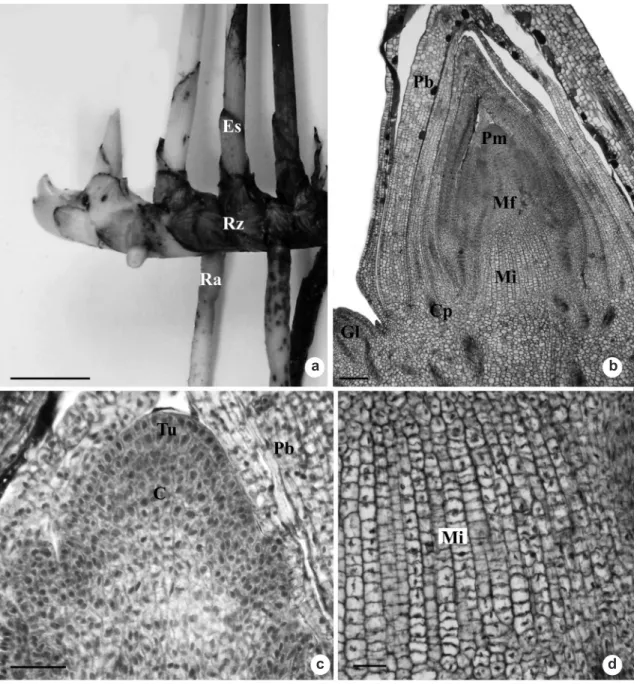
Androtrichum trigynum is aphyllous, with a perennial and photosynthetic floral scape. The underground system consists of rhizomes and adventitious roots (Fig. 1a). The rhizome is thickened, plagiotropic and sympodial, with small internodes, and covered by reddish-brown cataphylls; from the axils, side buds emerge. From the rhizome, arise the floral scapes whose base is also covered by reddish-brown cataphylls. Roots arise from the rhizome internodes. When young, they have a light coloration; when mature, a dark-brown coloration.
Anatomy - rhizome
In a longitudinal section from the stem apex, the promeristem can be observed surrounded by early cataphylls (Fig. 1b). The apical meristem has a tunic-body formation, the tunic consisting of two to three cell layers (Fig. 1c), and the body, of many cell layers. From the promeristem, the protoderm, the procambium and the fundamental meristem are distinguished (Fig. 1b). The intercalary meristem can also be seen at the apical region (Fig. 1b) consisting of, approximately, 15 cell layers with thin walls and arranged in a stratified way (Fig. 1d) and the formation of side buds of the same composition as the apical bud (Fig. 2a).
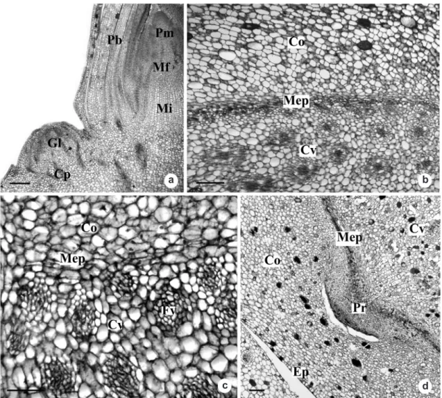
From approximately 720 μm from the apex, the primary thickening meristem (PTM) starts its activity. It has a circular shape, delimiting the cortical region and the vascular cylinder (Fig. 2b), producing centripetally, through periclinal divisions, vascular strands and parenchymatous cells, and only parenchymatous cells centrifugally (Fig. 2c). The PTM also originates adventitious roots (Fig. 2d). The epidermis is uniseriate, and the cortex, initially, is homogeneous (Fig. 2d).
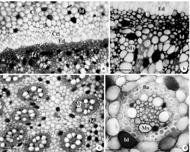
Following the development of the organ, the activity of the PTM gradually diminishes, and its endoderm and pericycle are distinguished (Fig. 3a-b). The endoderm presents radially elongated cells with thin walls (Fig. 3b). The pericycle is uniseriate, with isodiametric cells and thickened walls (Fig. 3b). At the vascular cylinder, there are isodiametric, conspicuous parenchymatous cells, with thin walls and few intercellular spaces (Fig. 3c). All of the vascular bundles are amphyvasal and present a sheath of cells with thickened walls (Fig. 3c-d). The outer bundles are formed by the PTM (Fig. 2c, 3a-b), and the inner bundles are formed by the procambium (Fig. 3c-d).
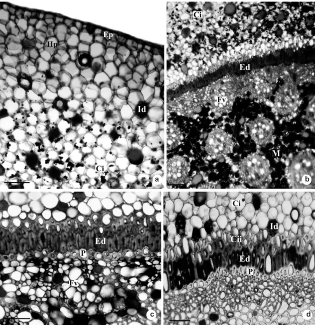
At maturity, the inner cortex and the outer cortex are distinguished (Fig. 4a). The outer cortex, or hypodermis, consists of compact hexagonal parenchymatous cells with thin walls (Fig. 4a). The inner cortex is formed by smaller cells in relation to the external cortical cells. They are isodiametric and with small intercellular spaces (Fig. 4a). It can also be observed that the innermost cells of this cortex, including the endoderm cells, have thickened walls (Fig. 4b-c). Many idioblasts having phenolic compounds are found throughout the whole rhizome (Fig. 2d, 3c, 4b-d), and in a lesser amount at the outer cortex (Fig. 4a). Phenolic compounds can also be observed on the walls of the endoderm cells and of the innermost cortical cells (Fig. 4c-d). Starch grains occur at the inner cortex (Fig. 4a), as well as at the vascular cylinder (Fig. 4b).
Anatomy – roots
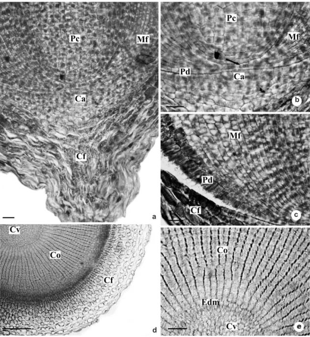
Figure 1 –
Underground system of
Androtrichum trigynum
(Spr.) Pfeiffer. a. General aspect. b-d. Rhizome longitudinal
sections. b. Stem apex.
c
. Detail of the previous figure showing promeristem organization. d. Detail of the intercalary
meristem. C = corpus; Cp = procambial strand; Es = scape; Gl = lateral bud; Mf = ground meristem; Mi = intercalary
meristem; Pb = bract primordium; Pm = promeristem; Ra = adventitious root; Rz = rhizome; Tu = tunica. Bars: 5
cm (a), 100
μm (b), 25
μm (c-d).
a b
c d
From the promeristem, the initial cell common to the protoderm and the meristem is observed (Fig. 5b). The cylinder of procambium is easily observed, the promeristem region being very small (Fig. 5a-b). The protoderm cells gradually elongate radially as they drift away from the initial cell (Fig. 5c).
Figure 2
– Rhizome of
Androtrichum trigynum
(Spr.) Pfeiffer. Longitudinal section (a) and transverse sections (b-d).
a. Shoot apex showing lateral bud. b. PTM arranged in circular shape between the cortex and vascular cylinder. c.
Detail of the PTM. d. Origin of adventitious roots from the PTM. Co = cortex; Cp = procambial strand; Cv = vascular
cylinder; Ep = epidermis; Fv = vascular strand; Gl =lateral bud; Mep = primary thickening meristem; Mf = ground
meristem; Mi = intercalary meristem; Pb = bract primordium; Pm = promeristem; Pr = root primordium. Bars: 100
μm.
On the sections 280 μm from the root apex (Fig. 6a), we can observe 3 of 4 layers of root cap cells and the epidermis differentiated in papillose cells. Those have dense content and secrete a thick layer of substance, which is deposited between those cells and the root cap cells (Fig. 6b). The cortical region, at this stage of development, consists of outer cortex or hypodermis and inner cortex. The hypodermis (Fig. 6a, 6c) originates from the fundamental meristem and presents approximately six layers of isodiametric cells, with no intercellular spaces. The inner cortex
(Fig. 6a, 6d) results from periclinal divisions of the meristematic endoderm, showing about 22 cell layers radially disposed, with thin walls and conspicuous intercellular spaces (Fig. 6d). At the vascular cylinder, the vascular elements are in differentiation (Fig. 6a, 6d).
From 1610 μm from the apex (Fig. 6e-g), the root cap cells disappear, and a considerable layer of substance secreted by the epidermal cells can still be observed, which present dense content as well as the outermost cells of the hypodermis (Fig. 6e-f). The meristematic endoderm ceases
a b
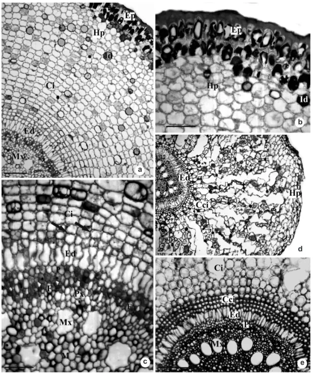
Figure 3
– Rhizome transverse sections of
Androtrichum trigynum
(Spr.) Pfeiffer. a. Endodermis and pericycle
differentiated from PTM. b. Detail of the previous figure, showing the endodermis, pericycle and peripheral vascular
bundles. c. Vascular bundles originated from the procambium. d. Detail of amphivasal vascular bundle. Ba = bundle
sheath of thick-walled cells; Ci = inner cortex; Ed = endodermis; F = phloem; Cci = innermost cells of the inner
cortex; Fv = vascular bundle; Id = idioblasts; Mx = metaxylem; P = pericycle. Bars: 100
μm (c), 50
μm (a-b, d).
its activity and begins to differentiate in radially elongated cells (Fig. 6g). At the vascular cylinder, the biserial pericycle and the elements of xylem and phloem still in differentiation are observed (Fig. 6e, 6g). Idioblasts with phenolic compounds begin to appear at the inner cortex and at the pith (Fig. 6e).
On the transverse sections 2870 µm from the apex (Fig. 7a), the thick layer of substance secreted by the epidermis becomes thin (Fig. 7a-b). The intercellular spaces of the inner cortex become more conspicuous (Fig. 7c). The endoderm is now completely differentiated, which can be observed by the high level of vacuolation of the cells, as well as the pericycle and vascular elements (Fig. 7c).
At maturity, part of the inner cortex transforms into schizo-lysigenous aerenchyma (Fig. 7d), starting with the dissolution of the middle lamella increasing the intercellular spaces, which then become more evident with the lysis of the cells. The endoderm cells remain with thin walls, and the innermost cell layers of the cortex start to have their walls thickened (Fig. 7e). The root is polyarc, with a great number of metaxylem elements surrounding the pith region (Fig. 7e).
Discussion
The underground stem of Androtrichum t r i g y n u m i s a r h i z o m e , a s i t s h o w s t h e morphological characteristics described by many authors to define this organ (Font Quer 1982;
a b
Figure 4
– Rhizome transverse sections of
Androtrichum trigynum
(Spr.) Pfeiffer. a. Hypodermis with no starch grains
is observed. b. Parenchyma cells of the inner cortex and vascular cylinder with starch grains. c. Note the thickening
of the innermost cortical cells and the endodermis and pericycle cells. d. Detail of the innermost cortical cells and
the endodermis cells with phenolic compounds on their walls. Cci = innermost cells of the inner cortex; Ci = inner
cortex; Ed = endodermis; Ep = epidermis; Fv = vascular bundle; Hp = hypodermis; Id = idioblasts; M = pith; P =
pericycle. Bars: 50
μm (c-d), 100
μm (a-b).
a b
c d
Bell 1991; Appezzato-da-Glória 2003). The aerial stem, is spite of being perennial, is classified as reproductive, as it is the axis of the inflorescence or floral scape, as occurs in other Cyperaceae species described by Estelita & Rodrigues (2007). According to these authors, the main anatomical characteristic that distinguishes the stem from the
scape in Cyperaceae is the presence of the PTM in the former, as occurs in A. trigynum.
Figure 5
– Root apex of
Androtrichum trigynum
(Spr.) Pfeiffer. a-c. Longitudinal sections of root apical meristem.
a. Organization of the apical meristem. b. Detail showing the initial cell (arrow) common to the ground meristem and
protoderm. c. Detail showing the radial elongation of the protoderm cells. d-e. Transverse sections at 120 µm from
the root apex. d. Note the vascular cylinder and cortical region in differentiation. e. Detail of the previous figure,
showing the meristematic activity of the endodermis forming the cortical cells. Ca – calyptrogen; Cf – root cap;
Co – cortex; Cv – vascular cylinder; Edm – meristematic endodermis; Mf – ground meristem; Pc – procambium;
Pd – protoderm. Bars: 25
μm (a-c), 100
μm (d), 50
μm (e).
a c
e d
b
epidermal cells of the root of A. trigynum, which do not form root hairs, but secrete mucilage, detected with an specific test. For Mauseth (1988), the mucilaginous substance secreted by the apical region of the roots is known as mucigel, which
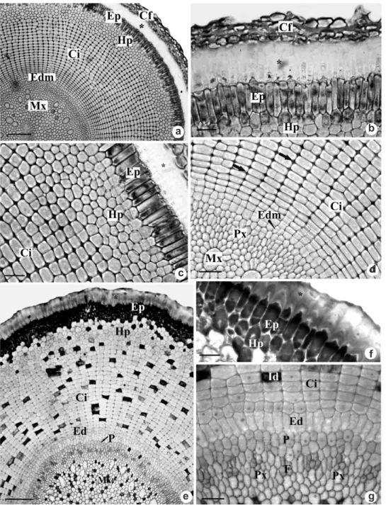
Figure 6
– Root apex transverse sections of
Androtrichum trigynum
(Spr.) Pfeiffer. a-d. Sections at 280
μm
from the root apex. a. General aspect. b. Detail of the substance secreted by epidermal cells (*). c. Outer cortex
or hypodermis and inner cortex. d. Inner cortex originated from the meristematic endodermis with schizogenous
intercellular spaces (arrows) and vascular elements in differentiation in the vascular cylinder. e-g. Sections at
1610
μm from the root apex. e. General aspect. f. Detail of the epidermal cells and outer cortex. g. Detail of
the endodermis and vascular cylinder. Cf – root cap; Ci – inner cortex; Edm – meristematic endodermis; Ed –
endodermis; Ep – epidermis; F – phloem; Hp – hypodermis; Id – idioblasts; Mx – metaxylem; P – pericycle;
Px – protoxylem. Bars: 100
μm (a, e), 20
μm (b-d, f-g).
a b
c d
f
Figure 7 –
Root transverse sections of
Androtrichum trigynum
(Spr.) Pfeiffer. a. General view. b. Detail of
the epidermis and outer cortex. c. Thickening of the walls of the inner cortical cells (arrows). d. Later stage
of development, showing schizo-lysigenous aerenchyma, endodermis (arrow) with thin-walled elongated cells
and metaxylem elements differentiated. e. Mature root, showing detail of the cortex and vascular cylinder. Cci
– innermost cells of the inner cortex; Ci – inner cortex; Ed – endodermis; Ep – epidermis; F – phloem; Hp –
hypodermis; Id – idioblasts with phenolic compounds; Mx – metaxylem; P – pericycle; Px – protoxylem. Bars:
100
μm (a, d-e), 50
μm (b), 20
μm (c).
a
b
d
water retention and thus preventing desiccation, as Dickison (2000) suggests.
The intercalary meristem (IM), observed at the stem apex of A. trigynum, was also observed in the rhizomes of Cyperus giganteus
(Estelita & Rodrigues 2007) and Fuirena umbellata Rottb. (Rodrigues & Estelita 2009). According to Estelita & Rodrigues (2007), the intercalary meristem (IM) is frequent in leaves and internodes of many monocots, scapes and pedicels. However, it shows higher activity at the scapes, allowing the elongation of the internodes (Fisher 1970). It is believed that the presence of the IM at the apical region of A. trigynum
rhizome indicates the beginning of floral scape formation, that is, the beginning of the reproductive stage, as seen in other Cyperaceae (Estelita & Rodrigues 2007).
The PTM is a lateral meristem characteristic of monocots (Rudall 1991); it has been described for many plants of this group, and it may occur in herbaceous (Krauss 1948; Sajo 1992) and rhizomatous species (Rudall 1984, 1991; Rodrigues & Estelita 2004, 2009), and in more immature regions of monocots with secondary growth (Fisher & Tomlinson 1972). In Cyperaceae, it was registered for the genera
Scirpus L. and Fimbristylis Vahl. by Rudall (1991), and in the species Cyperus esculentus
(Gifford & Bayer 1995) and C. giganteus, C. rotundus, Fuirena umbellata Rottb., Hypolitrum schraderianum Ness, Bulbostylis paradoxa
(Spreng.) Lindm. (Rodrigues & Estelita 2009). However, the term PTM, despite being used by many authors, as mentioned above, is still questioned. Zimmermann & Tomlinson (1968; 1970; 1974) refer to it as “meristematic cap”, while Menezes et al. (2005) assume that the PTM is the endoderm and the pericycle at meristematic stage and do not use the term.
Regarding its function, the PTM in involved in the primary thickening of the stem, in the formation of adventitious roots, and is related to the vascular system (Cheadle 1937; Krauss 1948; DeMason 1979a; b, 1980; 1983; Stevenson & Fisher 1980; DeMason & Wilson 1984; Rudall 1991; Gifford & Bayer 1995). In A. trigynum, as well as in other Cyperaceae species studied by Rodrigues & Estelita (2002, 2009), the PTM is responsible for the inner cortex formation, whose innermost layer is distinguished at the endoderm
by the formation of the pericycle and of the adventitious roots.
In the roots of A. trigynum, it is noted that the pericycle and the meristematic endoderm can be recognized while still very close to the promeristem. In this organ, the formation of the inner cortex by the meristematic endoderm is evident, as also observed by Seago & Marsh (1989) in Typha glauca Godr. (Typhaceae), Seago
et al. (1999a, b) in Hydrocharis morus-ranae
L. (Hydrocharitaceae), Seago et al. (2000) in
Pontederia cordata L. (Pontederiaceae), Melo-de-Pinna & Menezes (2003) in Richterago Kuntze (Asteraceae), Rodrigues & Estelita (2004) in
Cyperus giganteus.
According to Melo-de-Pinna & Menezes (2003), at the roots of Richterago Kuntze, the meristematic endoderm experiences successive anticlinal and periclinal divisions to form the inner cortex, remaining as a meristematic layer until its complete differentiation, when the Casparian strip arises. This is a striking feature for recognition of the endoderm (Van Fleet 1961). In stems, many authors called it “endodermous layer” as they could not observe the Casparian strip (Tomlinson 1969; Gifford & Bayer 1995). In the studied species, Casparian strips were not observed in the endoderm cells, neither on the roots nor on the rhizome; however, it can be identified by its morphological features. At the roots, initially in meristematic regions, the endoderm can be recognized by having meristematic activity. At posterior stages, the endoderm consists of radially elongated cells, morphologically distinguished from the other layers, such as in Actinocephalus species studied by Scatena et al. (2005). On A. trigynum rhizome, the endoderm can be recognized by having cell features that distinguish from the other cortical cells, such as cell shape and parietal thickening, such as observed in other Cyperaceae species (Eiten 1969; Estelita 1993; Chabbi et al. 2000; Arruda & Neves 2005 and Prata et al. 2007).
In monocots, the endoderm may present four development stages, starting with the Casparian strip, followed by the deposition of suberin lamella, thickening of lignin and, lastly, deposition of phenolic compounds, as observed by Rodrigues & Estelita (2004) in the roots of
fundamental meristem (Rodrigues & Estelita 2002, 2004), as also observed by Seago & Marsh (1989) in T. glauca. Attention is drawn to some species of Cyperaceae, in which the hypodermis may occur even at the leaves (Metcalfe 1971), such as in Cyperus corymbosus Rottb. and
Remirea maritima Aubl. (Estelita 1993). The number of layers, thickening and composition of the hypodermal cells may vary (Seago & Marsh 1989). The Casparian strip may occur on the outermost layer, which is called exodermis by authors such as Van Fleet (1950). In this species, no modifications on the walls of hypodermal cells were observed, as well as in H. morsus-ranae, observed by Seago et al. (1999). At the roots of A. trigynum, however, the outermost layer of the hypodermis showed different content in the early stages of differentiation, similar to the content of epidermal cells, indicating a possible participation of this layer in the process of mucilage synthesis. In roots of T. glauca (Seago & Marsh 1989) and H. morsus-ranae (Seago et al. 1999a), the hypodermis, as well as the thickened inner cortical cells, sustains the aerenchyma, a function that can also be attributed to the hypodermis in the roots of A. trigynum, which justifies the use of the term.
The inner cortex of A. trigynum roots differentiates in lysigenous aerenchyma, as defined by Seago et al. (2005). However, these same authors classify the tangential lysigenous type for Cyperaceae, where the separation and collapse of the cells occur tangentially among the intact lines of cells. On the other hand, in A. trigynum, we can observe the formation of lysigenous-radial aerenchyma, where the separation and collapse of cells occur radially among the intact radial lines of cells. The aeriferous system of A. trigynum roots was also observed in other species from saline environments, such as Jaumea carnosa (Less.) Gray (Omer & Moseley 1981) and R. maritima
considered an adaptive strategy of plants from xeric environments (Braendle & Crawford 1982).
The results obtained in this work show that the underground system of A. trigynum presents anatomical features that can be considered common to Cyperaceae species, such as the presence of a primary thickening meristem at the rhizome, meristematic endoderm, aerenchyma originated from the roots and presence of hypoderma in both organs. Other characteristics such as thickening and lignification of cells, high concentrations of starch grains and phenolic substances, large intercellular spaces and mucilage at the roots, can be regarded as potential strategies to adapt to dunes environment.
Acknowledgments
The authors thank the Graduate Program on Plant Biology of Universidade Federal de Santa Catarina; Capes for granting a Graduate student to the first author, and to CNPq (481623/2007-8) for the financial support.
References
Appezzato-da-Glória, B. 2003. Morfologia de sistemas subterrâneos: histórico e evolução do conhecimento no Brasil. A.S. Pinto, Ribeirão Preto. 80p. Alonso, A.A. & Moraes-Dallaqua, M.A. 2004.
Morfoanatomia do sistema caulinar de Canna
edulis Kerr-Gawler (Cannaceae). Revista Brasileira
de Botânica 27: 229-239.
Alves, M.; Araújo, A.C.; Prata, A.P.; Vitta, F.; Hefler, S.;
Trevisan, R.; Gil, A.S.B.; Martins, S. & Thomas, W. 2009. Diversity of Cyperaceae in Brazil. Rodriguésia 60: 771-782.
Arruda, R.C.O. & Neves, L.J. 2005. Anatomia foliar de
Trilepis lhotzkiana Ness e Trilepis ciliatifolia T.
Koyama (Cyperaceae) Juss. Acta Botanica Brasilica 19: 889-897.
Beckel, D.K. 1956. Cortical disintegration in the roots of
Bouteloua gracilis (H.B.K.) Lag. New Phytologist
Bell, A.D. 1991. Plant form: an illustrated guide to flowering plant morphology. University Press, Oxford, 341p.
Beltrame, A.V.; Bunn, D.A.; Chaves, A.P.N. & Voges, M.S. 2006. Conhecendo o Parque Municipal das Dunas da Lagoa da Conceição. Editora UFSC, Florianópolis. 31p.
Braendle, R & Crawford, R.M.M. 1982. Rhizome anoxia tolerance and habitat specialization in
wetland plants. In: Crawford, R.M.M. (ed.). Plant
life in aquatic and amphibious habitats. Blackwell
Scientific Publications, Oxford. Pp. 397-411.
Bukatsch, F. 1972. Bemerkungen zur Doppelfärbung Astrablau-Safranin. Mikrokosmos 61: 255. CECCA (Centro de Estudos Cultura e Cidadania). 1996.
Uma cidade numa ilha: relatório sobre os problemas sócio-ambientais da Ilha de Santa Catarina. Insula, Florianópolis, 247p.
Clarck, L.H. & Harris, W.H. 1981. Observations on the
root anatomy of rice (Oryza sativa L.). American
Journal of Botany 68: 154-161.
Chabbi, A.; Mckee, K.L. & Mendelssohn, I.A. 2000.
Fate of oxygen losses from Typha domingensis
(Typhaceae) and Cladium jamaicense (Cyperaceae)
and consequences for root metabolism. American Journal of Botany 87: 1081-1090.
Cheadle, V.I. 1937. Secondary growth by means of a thickening ring in certain monocotyledons. Botanical Gazette 98: 535-555.
Cordazzo, C.V.; Paiva, J.B. & Seeliger, U. 2006. Guia ilustrado: plantas das dunas da costa sudoeste Atlântica. USEB, Pelotas. 107p.
Costa, C.S.B.; Seeliger, U. & Cordazzo, C.V. 1988. Dinâmica populacional e distribuição horizontal
de Androtrichum trigynum (Spreng.) Pfeiffer
(Cyperaceae) em brejos e dunas costeiras do Rio Grande do Sul, Brasil. Acta Limnologica Brasiliensia 11: 813-842.
DeMason, D.A. 1979a. Histochemistry of the primary
thickening meristem in the vegetative stem of Allium
cepa L. American Journal of Botany 66: 347-350.
DeMason, D.A. 1979b. Function and development of primary thickening meristem in the monocotyledon,
Allium cepa L. The Botanical Gazette 140: 51-66.
DeMason, D.A. 1980. Localization of cell division activity in the primary thickening meristem in
Allium cepa L. American Journal of Botany 67:
393-399.
DeMason, D.A. 1983. The primary thickening meristem: Definition and function in monocotyledons. American Journal of Botany 70: 955-962.
DeMason, D.A. & Wilson, M.A. 1984. The continuity
of primary and secondary growth in Cordyline
terminalis (Agavaceae). Canadian Journal of Botany
63: 1907-1913.
Dickison, W.C. 2000. Integrative plant anatomy. Academic Press, San Diego. 533p.
Drew, M.C.; He, C. & Morgan, P.W. 2000. Programmed cell death and aerenchyma formation in roots. Trends in Plant Science 5: 123-127.
Eiten, L.T. 1969. The vegetative anatomy of Eleocharis
interstincta (Vahl) Roem. & Schult. Arquivos de
Botânica do Estado de São Paulo 4: 187-228. Estelita, M.E.M. 1993. Anatomia dos órgãos vegetativos
de Remirea maritima Aubl. (Cyperaceae). Naturalia
18: 123-134.
Estelita, M.E.M. & Rodigues, A.C. 2007. Subsídios estruturais à caracterização do sistema caulinar em Cyperaceae. Revista Brasileira de Botânica 30: 401-409.
Fahn, A. & Cutler, D.F. 1992. Xerophytes. Encyclopedia of plant anatomy. Vol. XIII.3. Gebrüder Borntraeger, Berlin. 176p.
Feder, N. & O’Brien, T.P. 1968. Plant microtechnique: some principles and new methods. American Journal of Botany 55: 123-142.
Fisher, J.B. 1970. Development of the intercalary
meristem of Cyperus alternifolius. American
Journal of Botany 57: 691-703.
Fisher, J.N. & Tomlinson, P.B. 1972. Morphological
studies in Cordyline (Agavaceae). II. Vegetative
morphology of Cordyline terminalis. Journal of the
Arnold Arboretum 53: 113-127.
Font Quer, P., 1982. Diccionario de botánica. Labor, Barcelona. 1244p.
Gifford, E.M. & Bayer, D.E. 1995. Development
anatomy of Cyperus esculentus (yellow nutsedge).
International Journal of Plant Sciences 156: 622-629.
Govindarajalu, E. 1974. The systematic anatomy of south
Indian Cyperaceae: Cyperus L. subgen. Juncellus,
Cyperus subgen. Mariscus and Lipocarpha R.
Br. Botanical Journal of the Linnean Society 68: 235-266.
Heimsch, C. 1960. A new aspect of cortical development in roots. American Journal of Botany 47: 195-201. Herrmann, M.L.P. 1989. Aspectos ambientais da porção central da Ilha de Santa Catarina. Dissertação de Mestrado. Universidade Federal de Santa Catarina, Florianópolis. 228p.
Johansen, D.A. 1940. Plant microtechnique. McGraw-Will, New York. 523p.
Kadej, F. 1963. Interpretation of the pattern of the cell arrangement in the root apical meristem of
Cyperus gracilis L. var. alternifolius. Acta Societatis
Botanicorum Poloniae 32: 295-301.
Mauseth, J.D. 1988. Plant anatomy. Benjamin-Cummings, California. 560p.
Melo-de-Pinna, G.F.A. & Menezes, N.L. 2002.
Vegetative organ anatomy of Ianthopappus Roque
& D.J.N. Hind (Asteraceae-Mutisieae). Revista Brasileira de Botânica 25: 505-514.
Melo-de-Pinna, G.F.A. & Menezes, N.L. 2003. Meristematic endodermis and secretory structures in
adventitious roots of Richterago Kuntze
(Mutisieae-Asteraceae). Revista Brasileira de Botânica 26: 1-10.
Menezes, N.L.; Silva, D.C.; Arruda, R.C.O.; Melo-de-Pinna, G.F.; Cardoso, V.A.; Castro, N.M.; Scatena, V.L. & Scremin-Dias, E. 2005. Meristematic activity of the endodermis and the pericycle in the primary thickening in monocotyledons: considerations on the “PTM”. Anais da Academia Brasileira de Ciências 77: 259-274.
Metcalfe, C.R. 1971. Cyperaceae. Vol. 5. Clarendon Press, Oxford. 596p.
O’Brien, T.P.; Fedder, N. Mccully, M. 1965. Polychromatic staining of plant cell walls by toluidine blue. Protoplasma 59: 368-373.
Omer, L.S. & Moseley, Jr. M.F. 1981. The vegetative
anatomy of Jaumea carnosa (Less.) Gray
(Asteraceae) a salt marsh species. American Journal of Botany 68: 312-319.
Prata, A.P.; Menezes, N.L.; Mazzoni-Viveiros, S.C.; Wanderley, M.G.L. & Thomas, W.W. 2007. Anatomia do escapo e rizoma de espécies brasileiras
de Bulbostylis Kunth (Cyperaceae). Revista
Brasileira de Botânica 30: 245-256.
Rodrigues, A.C. & Estelita, M.E.M. 2002. Primary
and secondary development of Cyperus giganteus
Vahl rhizome (Cyperaceae). Revista Brasileira de Botânica 25: 251-258.
Rodrigues, A.C. & Estelita, M.E.M. 2004. Anatomia da
raiz de Cyperus giganteus Vahl (Cyperaceae) em
desenvolvimento. Revista Brasileira de Botânica 27: 629-638.
R o d r i g u e s , A . C . & E s t e l i t a , M . E . M . 2 0 0 9 . Morphoanatomy of the stem in some Cyperaceae. Acta Botanica Brasilica 23: 889-901.
Scatena, V.L.; Oriani, A. & Sano, P.T. 2005. Anatomia
de raízes de Actinocephalus (Koern.) Sano
(Eriocaulaceae). Acta Botanica Brasilica 19: 835-841.
Seago, J.L. & Marsh, L.C. 1989. Adventitious root
development in Typha glauca, with emphasis on the
cortex. American Journal of Botany 76: 909-923. Seago, J.L.; Marsh, L.C.; Stevens, K.J.; Souku, A;
Votrubová, O. & Enstone, D.E. 2005. A
re-examination of the root cortex in wetland flowering
plants with respect to aerenchyma. Annals of Botany 96: 565-579.
Seago, J.L.; Peterson, Carol A. & Enstone, D.E. 1999a. Cortical ontogeny in roots of the aquatic plant,
Hydrocharis morsus-ranae L. Canadian Journal of
Botany 77: 113-121.
Seago, J.L.; Peterson, C.A.; Enstone, D.E.; Scholey, C.A. 1999b. Development of the endodermis and
hypodermis of Typha glauca Godr. and Typha
angustifolia L. roots. Canadian Journal of Botany
77: 122-134.
Seago, J.L.; Peterson, C.A. & Enstone, D.E. 2000. Cortical development in roots of the aquatic plant
Pontederia cordata (Pontederiaceae). American
Journal of Botany 87: 1116-1127.
Sharma, O.P. & Mehra P.N. 1972. Systematic anatomy of
Fimbristylis Vahl (Cyperaceae). Botanical Gazette
133: 87-95.
Stevenson, D.W. & Fisher, J.B. 1980. The developmental relationship between primary and secondary
thickening growth in Cordyline (Agavaceae). The
Botanical Gazette 141: 264-268.
Tomlinson, P.B. 1969. Commelinales-Zingiberales.
In: Metcalfe, C.R. (ed.). Anatomy of the
monocotyledons. Vol. 3. Clarendon Press, Oxford. Pp. 295-421.
Van Fleet, D.S. 1950. A comparison of histochemical and anatomical characteristics of the hypodermis with the endodermis in vascular plants. American Journal of Botany 37: 721-724.
Van Fleet, D.S. 1961. Histochemistry and function of the endodermis. The Botanical Review 27: 165-220. Zimmermann, M.H. & Tomlinson, P.B. 1968. Vascular
Prionium (Juncaceaae). American Journal of Botany 55: 1100-1109.
Zimmermann, M.H. & Tomlinson, P.B. 1970. The
vascular system in the axis of Dracaena fragans
(Agavaceae). II. Distribution and development of secondary vascular tissue. Journal of the Arnold Arboretum 51: 478-491.
Zimmermann, M.H. & Tomlinson, P.B. 1974. Vascular
patterns in palm stems: Variations of the Rhapis
principle. Journal of the Arnold Arboretum 55:
402-424.
Williams, B.C. 1947. The structure of the meristematic root tip and origin of the primary tissues in the roots of vascular plants. American Journal of Botany 34: 455-462.
Wills, G.D. 1987. Description of purple and yellow nutsedge. Weed Technology 1: 2-9.
Wills, G.D. & Briscoe, G.A. 1970. Anatomy of purple nutsedge. Weed Science 18: 631-635.
Wills, G.D.; Hoagland, R.E. & Paul, R.N. 1980. Anatomy
of yellow nutsedge (Cyperus esculentus). Weed
Science 28: 432-437.