Use of drug-eluting stents for the treatment of vertebral
artery stenosis
*
O uso de stents farmacológicos no tratamento da estenose das artérias vertebrais
Eduardo Wajnberg1, Gustavo Rodrigues2, Daniel Giansante Abud3
Objective: To report the feasibility and safety of percutaneous transluminal angioplasty with paclitaxel-eluting stents for management of vertebral artery stenosis in 14 patients, after two-year follow-up. Materials and Methods: Fourteen patients with a mean age of 67.2 years were submitted to endovascular treatment with placement of paclitaxel-eluting stents. The primary objective of the present study was to evaluate the safety of the procedure. The clinical effectiveness was evaluated according to the rates of restenosis and recurrence of ischemic events. Results: The initial stenosis degree ranged from 50% to 99% (mean, 73.3% ± 10.9%). The rate of technical success achieved 100%. Neither complications directly related to the procedure nor recurrence of symptoms were observed after 24-month follow-up. The rate of intra-stent restenosis was 7.1%, although asymptomatic in all the cases. Conclusion: The present study suggests that vertebral artery angioplasty with paclitaxel-eluting stents is a feasible and promising technique in terms of safety and effectiveness in the prevention of recurrent ischemia and restenosis.
Keywords: Vertebral artery; Angioplasty; Stents.
Objetivo: Relatar a viabilidade e segurança da angioplastia transluminal percutânea com stents recobertos com pacli-taxel para tratamento de estenose de artéria vertebral em 14 pacientes, após seguimento de dois anos. Materiais e Métodos: Catorze pacientes com idade média de 67,2 anos foram submetidos a tratamento endovascular mediante angioplastia percutânea e implante de stent farmacológico. O objetivo primário deste trabalho foi assegurar a segurança do procedimento. O desfecho secundário foi a eficácia clínica, definida como sintomas isquêmicos recorrentes e taxas de reestenose. Resultados: O grau de estenose variou de 50% a 99% (média de 73,3% ± 10,9). A taxa de sucesso técnico da angioplastia foi de 100%. Não houve complicações diretamente relacionadas ao procedimento. Aos 24 meses de seguimento, nenhum paciente apresentou recorrência dos sintomas. A taxa de reestenose intra-stent foi de 7,1%, embora tenha sido assintomática na totalidade dos casos. Conclusão: Este estudo sugere que a angioplastia da artéria vertebral com o uso de stents recobertos com paclitaxel é uma técnica viável e promissora em termos de segurança e eficácia na prevenção da isquemia recorrente e reestenose.
Unitermos: Artéria vertebral; Angioplastia; Stents. Abstract
Resumo
* Study developed at Hospital Universitário Clementino Fraga Filho da Universidade Federal do Rio de Janeiro (UFRJ), Rio de Janeiro, RJ, Brazil.
1. Master, MD, Neuroradiologist at Hospital Universitário Clementino Fraga Filho da Universidade Federal do Rio de Ja-neiro (UFRJ), Rio de JaJa-neiro, RJ, Brazil.
2. Master, MD, Neuroradiologist at Hospital Universitário An-tônio Pedro da Universidade Federal Fluminense (UFF), Niterói, RJ, Brazil.
3. PhD, Assistant Professor at Faculdade de Medicina de Ri-beirão Preto da Universidade de São Paulo (FMRPUSP), Ribei-rão Preto, SP, Brazil.
Mailing Address: Dr. Eduardo Wajnberg. Rua Nina Rodrigues, 72/102, Jardim Botânico. Rio de Janeiro, RJ, Brazil, 22461-100. E-mail: [email protected]
Received August 1st, 2011. Accepted after revision October 10, 2011.
Wajnberg E, Rodrigues G, Abud DG. Use of drug-eluting stents for the treatment of vertebral artery stenosis. Radiol Bras. 2011 Nov/ Dez;44(6):343–348.
ing the management of coronary arteries stenosis have demonstrated the superiority of drug-eluting stents over the conventional ones(8,10).
The present study reports the results in the treatment of 14 consecutive patients with vertebrobasilar territory ischemia caused by extracranial vertebral artery stenosis who underwent percutaneous transluminal angioplasty with paclitaxel-eluting stents.
MATERIALS AND METHODS
The present study involved the review of medical records and angiographic stud-ies of 14 patients referred to the interven-tional neuroradiology team at the Hospital
20% of such events(1,2). In many patients,
such ischemia is related to hemodynamic changes caused by proximal obstruction of the vertebral artery. In symptomatic pa-tients unresponsive to dual antiplatelet therapy, balloon angioplasty has been re-ported as being beneficial. So far, the treat-ment of vertebral artery stenosis by means of the placement of stents has almost al-ways been performed with conventional
coronary prostheses(3,4) and has been
asso-ciated with restenosis rates ranging from
9.6% to 66.7% (Table 1)(1,3,4–9). The
ratio-nale of utilizing drug-eluting stents
in-volves coating the stent (platform) with
cytotoxic drugs in order to inhibit the oc-currence of vascular restenosis due to inti-mal hyperplasia. Clinical studies
approach-INTRODUCTION
Universitário Clementino Fraga Filho – Universidade Federal do Rio de Janeiro, Rio de Janeiro, RJ, Brazil, for treatment of vascular lesions involving vertebral arter-ies, who were submitted to endovascular treatment with the placement of paclitaxel-eluting stents, in the period from June 2008 to April 2011. The inclusion criteria were the following: a) patients with angio-graphic evidence of vertebral artery
steno-sis ≥ 50%; b) symptoms of vertebrobasilar
ischemia refractory to drug therapy, char-acterized by vascular stroke or transient is-chemic attack, during the use of antiplatelet drug and with risk factors under control; c) signing of a term of free and informed con-sent.
Initially, the patients were selected ei-ther by means of computed tomography an-giography or magnetic resonance imaging angiography. Once selected, the patient was submitted to digital angiography was per-formed only at the moment of treatment, for confirmation of the findings and steno-sis degree.
The patients included in the present study had symptoms related to posterior circulation ischemia refractory to clinical treatment, including the use of oral anti-platelet drugs. The angiographic images were re-evaluated for quantitative analysis of the lesions, by means of specific soft-ware integrated with the angiography sys-tem (Quantcor; Siemens Medical Solu-tions, Forchheim, Germany).
Catheter angiography was performed, including subclavian, vertebral and carotid arteries, also to determine the vascular dis-ease extent and to evaluate the presence of collateral circulation through the posterior communicating arteries. The activated clot-ting time (ACT) was obtained before initi-ating the angiographic procedure. After arterial puncture, intravenous heparin bo-lus of 80 U/kg of body weight was made with the purpose of increasing by two and a half times the basal ACT value, or > 250 seconds. Subsequently, the patients were submitted to the procedure under conscious sedation. The vascular access was obtained by means of puncture of the femoral artery with a 6 or 7 Fr sheath. A guiding catheter Guider Soft Tip 6 or 7 Fr (Boston Scien-tific; Natick, MA, USA) was then carefully positioned proximally to the lesion to be
treated. The lesion was then overcome with a Transend EX 0.014 guide-wire (Boston Scientific; Irvine, CA, USA), sometimes in combination with a 1.7 Fr SL10 micro-catheter (Boston Scientific; Irvine, CA, USA). Whenever necessary, a Transend Platinum 300 cm exchange length guide-wire (Boston Scientific; Irvine, CA, USA) was utilized for stent insertion. In the cases of severe stenosis (> 80%) balloon angio-plasty was utilized for predilation to allow
the later stent passage. The therapeutic
pro-cedures were performed in all the patients, utilizing Taxus Express Monorail coronary stents (Boston Scientific; Irvine, CA, USA), with paclitaxel-eluting platform. Cerebral protection filter devices were not utilized. An angiographic acquisition was performed following the stent insertion and before its release, in order to determine the exact lo-cation of the stent in the lesion. In the cases of ostial lesions, the stent was placed in such a manner so that a short 2 mm seg-ment exceeded the most inferior portion of the ostium in the vertebral artery to com-pletely cover the atherosclerotic plaque. The stent was implanted by slow and con-trolled inflation of the angioplasty balloon. The administration of heparin was inter-rupted at the end of the procedure, without reversion. The patients were maintained under oral triclopidine (500 mg/day) or clopidogrel (75 mg/day) regimen for three months, and 200 mg/day aspirin intake for undetermined period of time. Additionally, the risk factors associated with each patient were controlled, with strict management of hypercholesterolemia and hyperglycemia, besides adjuvant treatment for smoking cessation adherent patients. The patients were followed-up with Doppler ultra-sonography, digital angiography or com-puted tomography angiography for up to24 months following the stent implantation.
RESULTS
The present study included 14 patients, 5 of them women and 9 men, with a mean age of 67.2 years (ranging between 49 and 87 years). Among related comorbidities, 8 (57.1%) out the 14 patients had arterial hypertension, 4 (28.6%), hypercholester-olemia, 3 (21.4%), diabetes mellitus, 3 (21.4%), coronary disease, and 4 (28.6%)
were smokers. Five out the 14 patients (35.7%) presented with cerebral infarction with acute or subacute progression in the posterior circulation territory, demonstrated either by computed tomography or by mag-netic resonance imaging.
Characteristics of the lesions evaluated on angiographic images obtained immedi-ately before the stents implantation demon-strated initial degree of stenosis ranging from 50% to 95% (mean, 78.8%). The length of the stenotic segment ranged from 1.4 to 25.7 mm (mean, 5.2 mm).
The diameter measurements, including at the site of the most severe stenosis, were directly obtained from angiographic im-ages in the pre-angioplasty working posi-tion. As regards the lesions distribution in the vertebral artery, most of them (57.14%) were located in the artery ostium, 14.2% in the V2 segment, 7.1% in the V3 segment and 21.4%, in the V4 segment. Ostial ath-erosclerotic disease, in general, was caused by extension of an atherosclerotic plaque from the subclavian artery. Table 1 demon-strates the arterial stenosis degree before and immediately after the stent implanta-tion, as well as at the 24-month follow-up, besides the stents diameter and length and evaluation of intra-stent restenosis.
The angioplasty and stent implant pro-cedures were technically successful in all the patients. In all of the cases, Taxus Ex-press Monorail paclitaxel eluting stents (Boston Scientific; Irvine, CA, USA) were utilized. Permanent complications related to the procedure were not observed. The re-sidual stenosis degree measured on the angiographic images obtained immediately after the stent implantation ranged from 0% to 20% (mean, 5.7%), and no patient pre-sented residual stenosis > 25% of the arte-rial lumen. Both the procedure-related per-manent neurologic morbidity and mortal-ity in a 30-day follow-up were 0%. Only one patient (7.1%) had a transient ischemic attack within 24 hours after the procedure (patient 11). No stroke related to the site treated during the procedure was observed (0%). In only one case angioplasty-related intimal dissection was observed (patient 4), which was treated by means of an addi-tional stent implantation.
then repeated at 15 and 30 days and at the end of the follow-up period (mean 24 months). Most of the patients (71.4%) were symptomatic before the procedure and pre-sented symptom improvement related to the decreased cerebral blood flow. Also, new ischemic episodes were not detected after the procedure.
Long-term angiographic follow-up was performed in all the 14 patients. Before the stent implant, all the patients presented stenosis of at least 50%. Immediately after the procedure, no patient presented residual
stenosis ≥ 50%. Mean residual stenosis
immediately after the procedure was 5.7%. At the 24th month, the mean stenosis rate increased to 12.5%, but only one patient presented hemodynamically significant stenosis (> 50%). Such a significant reste-nosis incidence at the 24th month of fol-low-up was 7.1%, although such patient did not present recurrence of vertebro-basilar symptoms.
Illustrative cases
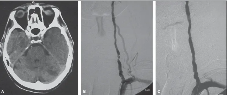
Patient 4 – A 63-year-old man with his-tory of hypertension and hyperlipidemia and episodes of transient ischemic attacks manifested by deficiency in the visual field. The patient was conservatively treated with dual antiplatelet therapy, but two months later developed a stroke affecting the left cerebellar hemisphere and the left
occipi-tal lobe, which left the patient with residual quadrantanopia (Figure 1A). The patient was submitted to magnetic resonance an-giography that revealed the presence of a lesion in the left vertebral artery. Pre-angioplasty digital angiography demon-strated an eccentric and irregular atheroma-tous plaque determining about 95% steno-sis (Figure 1B). A 6F guide-catheter was inserted into the subclavian artery, and a 3.5
× 16 mm Taxus stent (Boston Scientific,
Irvine, CA, USA) was implanted over the lesion. Angiography revealed marked im-provement of the flow to the distal verte-bral artery and towards the posterior circu-lation, without residual stenosis (Figure 1C). The patient was discharged, remain-ing under antiplatelet therapy and asymp-tomatic along the 26-month neurologic follow-up.
Patient 5 – A 54-year-old man with coronary artery disease presented to the emergency department hospital with ver-tiginous syndrome associated with vomit-ing. Cranial magnetic resonance imaging revealed punctate ischemic lesions in both cerebellar hemispheres, suggesting the embolic origin of the lesions (Figure 2A). Doppler ultrasonography study of the ca-rotid and vertebral arteries suggested the presence of severe stenosis > 90% in the right vertebral artery. Pre-angioplasty digi-tal angiography findings corroborated such
diagnosis (Figure 2B), demonstrating an extensive atheromatous lesion determining 90% stenosis in the left vertebral artery ostium. Angioplasty was performed with a
3.0 × 16 mm Taxus stent (Boston
Scien-tific, Irvine, CA, USA) implantation, with excellent angiographic outcome, and with-out evidence of post-angioplasty residual lesion (Figure 2C). At the 12th month, fol-low-up with computed tomography an-giography demonstrated asymptomatic, minimum intra-stent stenosis, estimated to be 15%.
Patient 14 – A 62-year-old man, 50 pack year history of smoking, and history of systemic arterial hypertension, diabetes mellitus and peripheral vascular disease, presented with transient ischemic attacks with manifestation of vertiginous syn-drome and diplopia. The patient was sub-mitted to diagnostic digital angiography that demonstrated a lesion affecting 95% of the left vertebral artery ostium associated with 85% stenosis of the subclavian artery proximal to the origin of the vertebral ar-tery (Figure 3A). The left vertebral arar-tery was the dominant artery. Left subclavian artery catheterization was performed and a
4 × 16 mm Taxus stent (Boston Scientific,
Irvine, CA, USA) was implanted in the ostium of the left vertebral artery (Figure 3B). Later, balloon angioplasty of the
sub-clavian artery was performed [7 × 20 mm
Table 1 Angiographic characteristics of the lesions, with stenosis degree before and after angioplasty with drug-eluting stent. Long term stenosis degrees and angiographic follow-up method.
Patient’s number 1 2 3 4 5 6 7 8 9 10 11 12 13 14 Stent diameter (mm) 4.0 4.0 4.5 3.5 3.0 5.0 4.0 4.0 3.0 4.0 4.0 4.5 5.0 4.0 Stent length (mm) 12 12 16 16 16 12 12 16 16 16 16 16 12 16 Pre-angioplasty stenosis (%) 75 83 50 95 90 80 70 80 85 90 80 60 70 95 Post-angioplasty residual stenosis (%) 0 10 0 15 0 0 0 0 20 10 25 0 0 0
Stenosis at 24 month-follow-up (%) 0 20 0 20 15 0 20 0 30 20 60 20 0 10 Follow-up method CT angio CAG CT angio CT angio CT angio CT angio CAG CT angio CT angio CAG CAG CT angio CT angio CT angio Risk factors SAH Hch SAH, Smk Hch SAH, Hch Smk SAH, DM — SAH, DM SAH, Smk SAH SAH DM Hch, Smk
Powerflex balloon (Cordis; Warren, NJ, USA)] (Figure 3C). Post-angioplasty an-giography revealed the resolution of the stenosis at the origin of the left subclavian vertebral artery, with excellent anterograde flow in this artery and residual stenosis in the subclavian artery (Figure 3D). The pa-tient presented immediate improvement of symptoms after the procedure and re-mained asymptomatic over the 18-month follow-up, presenting minimum intra-stent stenosis, estimated to be 10%.
DISCUSSION
The analysis of the present study popu-lation identifies hypertension as the most common risk factor for severe
atheroscle-rotic involvement of the vertebrobasilar system, followed by smoking and hyperc-holesterolemia.
The mechanism of vertebrobasilar is-chemia may be embolic or hemodynamic. Embolic causes of vertebrobasilar ischemia are distal embolization of plaques or le-sions of the subclavian, vertebral and/or
basilar arteries(3,7,11). Hemodynamic
symp-toms onset depends on the presence of sub-stantial disease in both vertebral arteries; and an incomplete Willis polygon. Al-though such mechanism is not completely understood, analysis of microemboli present in the posterior circulation demon-strates that the majority of microemboli result from coexistent heart disease, with no correlation with vertebrobasilar system
system(2). Additionally, most of the causes
related to posterior circulation ischemia are associated with hemodynamic compro-mise, in contrast with carotid artery disease, where the emboli represent the main source of stroke in the anterior circulation. Alter-natively, hemodynamic ischemia with stenosis of the proximal subclavian artery may occur, leading to a subclavian steal syndrome. Some studies question the ben-efits of endovascular treatment of vertebral artery stenosis as compared with drug
therapy(12). In the clinical practice,
vascu-lar reconstruction either by surgical or endovascular means has already been con-sidered as a reasonable option to improve blood supply in patients with vertebro-basilar insufficiency. However, currently
Figure 1. A: Cranial computed tomography demonstrating hypodense lesions in both cerebellar hemispheres, the largest lesion in the left hemisphere. B: Digital angiography of left vertebral artery demonstrating an atherosclerotic lesion at the V2 segment estimated at 95%. C: Post-angioplasty digital angiography of left vertebral artery, without residual stenosis.
A B C
A B C
there is no good reason to refuse the mini-mally invasive procedure for patients with symptomatic vertebral stenosis refractory to drug treatment.
The present study demonstrates that the use of drug-eluting stents in patients with vertebral artery stenosis is feasible and ben-eficial to prevent recurrent vertebrobasilar ischemia and vascular restenosis. The as-sessment of the clinical value of angio-plasty with stent placement in the manage-ment of vertebral artery stenosis would re-quire a comparison with drug therapy in a larger randomized and controlled study. The current experience with angioplasty and stent placement in the management of vertebral stenosis is almost exclusively based on the utilization of conventional
stents(1,3–9) (Table 2). Restenosis of the
ves-sel in which the stent was placed, in the angiographic follow-up is generally de-fined as the degree of luminal narrowing of
at least 50%(13).
A retrospective study developed by
Gupta et al.(14) reported the use of
drug-eluting stents in the endovascular treatment of 27 vertebral artery lesions. Two types of
stents were utilized: Cypher® (Cordis;
Mi-ami, FL, USA) and Taxus Express®
(Bos-ton Scientific; Ca, USA). After four-month follow-up, 7% of the lesions presented
restenosis of at least 50%(14). In the present
study, the significant restenosis rate was 7.1% at the 24th month of clinical and angiographic follow-up. Apparently, such results were significantly superior to results
that would have been obtained in case con-ventional stents had been utilized.
Undoubtedly, drug-eluting stents are considered as a significant progress for the clinical practice. The drug released by such stents reduces the risk for restenosis, inhib-iting macrophage accumulation and prolif-eration of smooth muscle cells around the
stent. However, as such actions also inhibit
the reendothelialization on the stent sur-face(15,16), it was feared that such effects
would increase the risk for late thrombo-sis of the stent, but it seems that the treat-ment with antiplatelet drug plays a relevant role in the reduction of the risk for such
thrombosis(17,18). Additionally, studies on
long term clinical outcomes in patients have contributed to confirm the benefits for the survival of drug-eluting coronary stents
as compared with conventional stentsin up
to five years of follow-up, and have not
demonstrated any difference in the risk for
late thrombosis of the stent(17-20).
It is known that morphological factors related to vertebral artery lesions may af-fect the result from the therapy with angio-plasty and stent placement. For example, the lesion extent affects the restenosis rate(3,21) and excessive vascular tortuosity
may hinder the stent implant. In the present study, any lesion length was included and vascular tortuosity was not considered as an exclusion criterion.
The present study presents some limi-tations, particularly regarding the small number of included patients, but it is suf-ficient as a feasibility study. A higher rate of late restenosis was observed, particularly in cases where residual stenosis was de-tected at the end of the procedure. Thus, the presence of residual stenosis immediately after stent implantation in vertebral artery stenosis seems to be a predictor of resteno-sis at the follow-up, although the relevance of such finding in the clinical outcome is still to be determined.
Even though the dose of the drug re-leased in the circulation is small with no evidences of paclitaxel-induced neurotox-icity, the potential neurotoxicity risk result-ing from the implantation of a paclitaxel-eluting stent in a neurovascular territory still remains a concern that must be
care-fully evaluated(22–24).
Extracranial vertebral artery disease is difficult to diagnose and follow-up by means of non-invasive methods such as
Figure 3. A: Digital angiography of left subclavian artery demonstrating ostial lesion estimated at 95% in the left vertebral artery associated with obstructive lesion of the subclavian artery. B: Final angiographic appearance after angioplasty of the ostium vertebral and subclavian arteries.
A B
Table 2 Intra-stent restenosis rates in the main post-angioplasty studies with conventional stents.
Authors
Chastain et al.(4)
Albuquerque et al.(5)
Lin et al.(3)
SSYLVIA study(6)
Weber et al.(7)
Akins et al.(8)
Taylor et al.(9)
Hatano et al.(1)
Number of treated
lesions
50
80
32
6
26
7
44
117
Percentage of intra-stent
restenosis*
11.1%
28%
25%
66.7%
46.1%
43%
48%
9.6%
Doppler ultrasonography, and its manage-ment is not so well defined as the treatmanage-ment for carotid vascular disease. For initial evaluation, therefore, computed tomogra-phy angiogratomogra-phy or magnetic resonance imaging was utilized for screening, before the performance of digital angiography, in order to avoid the unnecessary costs and exposure of the patients to angiography risks. However, the use of computed to-mography angiography would not be sus-ceptible of resulting in a patient selection bias, as such method tends to exaggerate
the degree of vascular stenosis(25).
Al-though the utilization of clopidogrel for a whole year has been recommended for pa-tients with coronary disease following
drug-eluting stent implantation, it has been
a common practice in the author´s institu-tion to administer aspirin and clopidogrel for six months, and after that, aspirin alone continuously for patients who received stents in the vertebral carotid artery or in intracranial location.
The surgical correction of lesions at the origin of the vertebral artery and subclavian artery by means of ostial vertebral endart-erectomy, subclavian endartendart-erectomy, or vertebral artery or carotid artery reimplan-tation in the subclavian artery is technically difficult. In a series of 325 surgically treated lesions in 290 patients, Thevenet et
al.(26) have reported a low mortality rate
(0.6%), but a relatively high rate (28%) of post-operative Horner’s syndrome, a com-plication that is not observed in cases of percutaneous therapy. Although good long term outcomes have been reported, they are associated with a relatively high rate of
perioperative morbidity(3,26).
CONCLUSIONS
The results of the present retrospective clinical series suggest that angioplasty with placement of paclitaxel-eluting stents in symptomatic patients with vertebral artery
stenosis is feasible and promising in terms of safety potential and effectiveness for preventing recurrent ischemia and resteno-sis. Although a long-term clinical and angiographic follow-up is required to evaluate the durability of the stent perme-ability, the clinical progression in this group of patients suggests a continuous benefit from the process. A larger random-ized and controlled study would be re-quired for such purpose.
REFERENCES
1. Hatano T, Tsukahara T, Miyakoshi A, et al. Stent placement for atherosclerotic stenosis of the ver-tebral artery ostium: angiographic and clinical outcomes in 117 consecutive patients. Neuro-surgery. 2011;68:108–16.
2. Caplan LR, Amarenco P, Rosengart A, et al. Embolism from vertebral artery origin occlusive disease. Neurology. 1992;42:1505–12. 3. Lin YH, Juang JM, Jeng JS, et al. Symptomatic
ostial vertebral artery stenosis treated with tubular coronary stents: clinical results and restenosis analysis. J Endovasc Ther. 2004;11:719–26. 4. Chastain HD 2nd, Campbell MS, Iyer S, et al.
Extracranial vertebral artery stent placement: in-hospital and follow-up results. J Neurosurg. 1999; 91:547–52.
5. Albuquerque FC, Fiorella D, Han P, et al. A reappraisal of angioplasty and stenting for the treatment of vertebral origin stenosis. Neuro-surgery. 2003;53:607–16.
6. SSYLVIA Study Investigators. Stenting of Symptomatic Atherosclerotic Lesions in the Ver-tebral or Intracranial Arteries (SSYLVIA): study results. Stroke. 2004;35:1388–92.
7. Weber W, Mayer TE, Henkes H, et al. Efficacy of stent angioplasty for symptomatic stenoses of the proximal vertebral artery. Eur J Radiol. 2005;56: 240–7.
8. Akins PT, Kerber CW, Pakbaz RS. Stenting of vertebral artery origin atherosclerosis in high-risk patients: bare or coated? A single-center consecu-tive case series. J Invasive Cardiol. 2008;20:14– 20.
9. Taylor RA, Siddiq F, Suri MF, et al. Risk factors for in-stent restenosis after vertebral ostium stenting. J Endovasc Ther. 2008;15:203–12. 10. Virmani R, Guagliumi G, Farb A, et al. Localized
hypersensitivity and late coronary thrombosis secondary to a sirolimus-eluting stent: should we be cautious? Circulation. 2004;109:701–5.
11. Burnay J, Pietri J. Vertebral and subclavian reim-plantation into the common carotid artery with widening angioplasty on the vertebral ostium. Presse Med. 1986;15:580.
12. Wehman JC, Hanel RA, Guidot CA, et al. Atherosclerotic occlusive extracranial vertebral artery disease: indications for intervention, endovascular techniques, short-term and long-term results. J Interv Cardiol. 2004;17:219–32.
13. Biria M, Tadros P, Gupta K. Subclavian-vertebral artery bifurcation stenting using drug-eluting stents: a report of two cases using different techniques. J Invasive Cardiol. 2007;19:E156–9.
14. Gupta R, Al-Ali F, Thomas AJ, et al. Safety, feasibility, and short-term follow-up of drug-eluting stent placement in the intracranial and extracranial circulation. Stroke. 2006;37:2562–6.
15. Vajda Z, Miloslavski E, Güthe T, et al. Treatment of stenoses of vertebral artery origin using short drug-eluting coronary stents: improved follow-up results. AJNR Am J Neuroradiol. 2009;30:1653– 6.
16. Yu SC, Leung TW, Lam JS, et al. Symptomatic ostial vertebral artery stenosis: treatment with drug-eluting stents—clinical and angiographic results at 1-year follow-up. Radiology. 2009;251: 224–32.
17. Regar E, Serruys PW. The Ravel trial. Zero percent restenosis: a cardiologists dream comes true! Rev Esp Cardiol. 2002;55:459–62. 18. Dabus G, Gerstle RJ, Derdeyn CP, et al.
Endovas-cular treatment of the vertebral artery origin in patients with symptoms of vertebrobasilar ischemia. Neuroradiology. 2006;48:917–23. 19. Du B, Wong EH, Jiang WJ. Long-term outcome
of tandem stenting for stenoses of the intracranial vertebrobasilar artery and vertebral ostium. AJNR Am J Neuroradiol. 2009;30:840–4.
20. Morice MC, Serruys PW, Barragan P, et al. Long-term clinical outcomes with sirolimus-eluting coronary stents: five-year results of the RAVEL trial. J Am Coll Cardiol. 2007;50:1299–304.
21. Zhou Z, Yin Q, Xu G, et al. Influence of vessel size and tortuosity on in-stent restenosis after stent implantation in the vertebral artery ostium. Cardiovasc Intervent Radiol. 2011;34:481–7. 22. Pace A, Bove L, Aloe A, et al. Paclitaxel
neuro-toxicity: clinical and neurophysiological study of 23 patients. Ital J Neurol Sci. 1997;18:73–9. 23. Pace A, Nisticò C, Cuppone F, et al. Peripheral
neurotoxicity of weekly paclitaxel chemotherapy: a schedule or a dose issue? Clin Breast Cancer. 2007;7:550–4.
24. van Gerven JM, Moll JW, van den Bent MJ, et al. Paclitaxel (Taxol) induces cumulative mild neurotoxicity. Eur J Cancer. 1994;30A:1074–7. 25. Yoo WJ, Lim YS, Ahn KJ, et al. Assessment of vertebral artery stents using 16-slice multi-detector row CT angiography in vivo evaluation: comparison of a medium-smooth kernel and a sharp kernel. Eur J Radiol. 2009;70:362–8. 26. Thevenet A, Ruotolo C. Surgical repair of