RBCCV 44205-1511 DOI: 10.5935/1678-9741.20130089
Use of a stent-graft and vascular occlude to treat
primary and re-entry tears in a patient with a
Stanford type B aortic dissection
O uso de endoprótese e oclusor vascular para tratar ruptura primária e de re-entrada em paciente com
dissecção aórtica tipo B de Stanford
Huihua Shi
1, Min Lu
1, Mier Jiang
11Hospital Afiliated Shanghai Jiaotong University School of Medicine, Shanghai, China.
No inancial support.
Work carried out at Hospital Afiliated Shanghai Jiaotong University School of Medicine, Shanghai, China.
Correspondence address: Min Lu
Shanghai Ninth People’s Hospital Afiliated Shanghai Jiaotong University
School of Medicine
Zhizaoju road, 639, Shanghai, the People’s Republic of China - Zip code: 200011
E-mail: [email protected]
Article received on December 6th, 2012 Article accepted on September 2nd, 2013 Abstract
Thoracic endovascular aortic repair for aortic dissections is recognized as an effective treatment. We herein report the case of a 72-year-old male with a Stanford type B aortic dissection. A stent-graft and double-disk vascular occluder was used to repair the primary and re-entry tears, respectively. At 3 month post-operatively, computed tomographic angiography revealed no endoleaks, the stent-graft and vascular occluder to be in optimal positions, the false lumen was almost completely thrombosed, and the visceral arteries were patent. This case illustrates that it is feasible to treat re-entry tears with a vascular occluder after primary proximal stent-graft repairs.
Descriptors: Vascular diseases. Vascular surgical proce-dures. Aortic diseases.
Resumo
Reparação endovascular de aorta torácica para dissecção
aór-tica é reconhecida como um tratamento eicaz. Relatamos o caso
de um homem de 72 anos de idade, com dissecção aórtica tipo B de Stanford. A endoprótese e oclusor duplo disco vascular foi usado para reparar as rupturas primária e de re-entrada,
respectivamen-te. Aos três meses de pós-operatório, angiotomograia computado -rizada não revelou vazamentos, o oclusor e a endoprótese vascular estavam em posições melhores, a falsa luz foi quase completamente trombosada, e as artérias viscerais estavam patentes. Esse caso de-monstra que o tratamento de rupturas na re-entrada com endopró-tese vascular após reparos proximais primários é viável.
INTRODUCTION
Aortic dissection is the most common acute emergency involving the aorta, and often results in death. The incidence of aortic dissection has been reported to be 2,000 new cas-es per year in the United Statcas-es and 3,000 in Europe [1-4].
The eficacy and safety of thoracic endovascular aortic repair
(TEVAR) for acute [5-7] and chronic [8-10] aortic dissec-tions has been shown in a many studies. As our experience with TEVAR has increased, the importance of re-entry sites (secondary tears) has drawn attention [11,12].
Herein we report a case in which we applied a stent-graft and double-disk vascular occluder to repair the primary and re-entry tears, respectively, in a patient with Stanford type B aortic dissection.
Written informed consent was obtained from the patient for publication of this case report and any accompanying images. This study was approved by the Institutional Review Board
of Shanghai Ninth People’s Hospital Afiliated Shanghai Ji -aotong University School of Medicine (number is 201293).
CASE REPORT
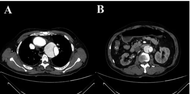
A 72-year-old male was admitted with a complaint of chest discomfort for 1 month. Computed tomography re-vealed an aortic dissection with entry and re-entry tears (Figure 1). Angiography then demonstrated a Stanford type B aortic dissection with the primary tear distal to the left
sub-Abbreviations, acronyms & symbols
TEVAR Thoracic endovascular aortic repair
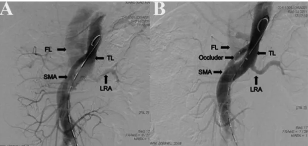
clavian artery, and a re-entry tear below the superior
mesen-teric artery oriice (Figure 2A). The right renal artery was not visualized, the kidney was atrophic and low was from the
false lumen.
The patient was taken to the operating room within 48 hours of the computed tomography angiography. After in-duction of general anesthesia a 5F sheath was inserted into the left axillary artery, and a centimeter sizing 5F pigtail catheter (Cook, USA) was introduced into the ascending aorta through the left subclavian artery. A 5F pigtail catheter was introduced into the ascending aorta through the femo-ral artery. Angiography was performed in two projections, left anterior oblique and anteroposterior. First, the 5F pigtail
catheter was conirmed to be in the true lumen, and then the precise location of the primary tear was identiied to be 2 cm
distal to the left subclavian artery. By using the centimeter sizing pigtail catheter, the diameter of the landing zone was measured and compared to that determined by computed to-mography angiography. Before the deployment of the stent-graft, heparin (1 mg/kg) was given intravenously.
An extra-stiff guidewire (Lunderquist, Cook, USA) was threaded into the ascending aorta through the pigtail cathe-ter, and the delivery system was introduced to the appropri-ate position over the guidewire. A tube-shaped stent-graft (Zenith TX2 32×160 mm, Cook, USA) was deployed under
luoroscopy. Angiography was performed to conirm the
correct position and that there were no endoleaks. A 10 mm wide re-entry tear was found below the superior mesenteric
artery oriice and opposite to left renal artery. Because the
re-entry tear and false lumen were so large, and right renal artery was atrophic, we decided to use an occluder to seal the re-entry tear.
A 9F long sheath with a Cobra-shaped tip (SFA9F
Occlud-er Transmission System; Lifetech Scientiic Co. Ltd, Shen -zhen, China) was advanced over the guidewire to the false lumen through the re-entry tear. Sized to exceed the 10 mm diameter re-entry tear by 2 mm, the waist of the 12-mm
dou-ble-disk symmetrical occluder (SearCare; Lifetech Scientiic
Co. Ltd) was connected to the tip of the delivery cable by a
microscrew ixed to the posterior disk, and collapsed into a
loader. The collapsed device was then advanced into the sheath
by pushing the delivery cable. Under luoroscopic guidance,
the anterior disk (26 mm) was deployed in the false lumen
against the dissection lap after passing through the rupture,
and the waist of the occluder was placed in the re-entry tear,
which was both felt and observed by luoroscopy. Then, the
posterior disk (22 mm) was deployed by further withdrawal of the sheath. The position of the occluder within the re-entry tear was determined to be in a secure and stable position by
gentle pushing and pulling of the delivery cable. The occluder was released by unscrewing; the conveyor was rotated
coun-terclockwise to separate after angiography had veriied its po -sition and ruled out interference with aortic branch vessels. On completion angiography, the device was in an optimal position and the re-entry tear was covered. There was no leakage into the false lumen and the superior mesenteric artery and left re-nal artery were patent (Figure 2B).
The patient recovered uneventfully and no complications occurred. She was discharged 2 weeks later in good condi-tion (in China, hospital stays are routinely much longer than in other countries). At 3 month postoperatively, computed to-mography revealed thrombosis in the descending false lumen and thrombosis in the abdominal false lumen (Figure 3). No endoleaks were noted, the stent-graft and vascular occluder were in optimal positions, and the visceral arteries (except the right renal artery) were patent.
Fig. 3 - Computed tomography (CT) images of the false lumen. A) A large false lumen was identiied in the abdominal aorta before surgery. B) At 3 month postoperatively CT revealed thrombosis in the descending false lumen and (C) thrombosis in the abdominal false lumen
Author’s roles & responsibilities
HS Performed research/study, managed the literature searches and analyses, wrote the irst draft of the manuscript
ML Designed the study and wrote the protocol, performed research/ study, critically reviewed the manuscript
MJ Designed the study and wrote the protocol DISCUSSION
The ideal results after TEVAR include aortic reconstruc-tion and false lumen thrombosis or resolureconstruc-tion. Adequate seal-ing of primary entry tears in the descendseal-ing thoracic aorta after stent-graft placement can reduce pressure in the false lu-men to avoid further dilatation or rupture. In acute onset aor-tic dissections, if there are no endoleaks or re-entry tears the false lumen will be completely obliterated within 6 months after stent-graft placement [13].
Compared with acute aortic dissections, chronic dissec-tions may have one or more re-entry tears in the abdominal aorta and a un-thrombosis abdominal false lumen originating
from persistent low or pressure through the re-entry tear
[8-10]. In our case of subacute dissection, because the re-entry site diameter was large it was unlikely to seal spontaneously. The false lumen would progressively dilate as a result of a patent re-entry site in the abdominal aorta, and the risk of rupture would persist. A study by Dias et al. [14] in which endovascular treatment was used to treat 11 patients with chronic type B aortic dissections found that although stent-graft deployment was technically successful in all patients
false lumen lows persisted in the thorax in 27% of the pa
-tients and in the abdomen in 82%, and that aortic diameter
was not decreased postoperatively. The authors concluded that endovascular treatment of chronic type B dissections is not effective as it does not decrease aortic diameter. Other studies, however, have indicated that endovascular treatment is effective for chronic aortic dissections [8-10]. Jia et al. [8] reported lower aorta-related mortality in patients with chron-ic type B dissections treated with stent-grafting as compared to those treated with medical management and a decrease of thoracic aorta diameter from a mean of 42.4 mm to 37.3 mm in the TEVAR group. Andacheh et al. [9] reported expansion of the thoracic true lumen and regression of the false lumen in patients following TEVAR, and similarly Parsa et al. [10] found depressurization of the false lumen after TEVAR.
As re-entry tears in the abdominal aorta tend to be locat-ed near the branch vessels, they are generally unfavorable for exclusion with a stent-graft because the branch ostia may be partially covered, which may lead to ischemia of the spinal cord, liver, intestine, gallbladder, or kidney. In our case, we believe that the atrophy of the right kidney was a result of the dissection. Though hybrid techniques that combine traditional surgery to place bypass grafts between the visceral arteries and abdominal aorta before endovascular intervention to expand the applicability of TEVAR are used, we have found this
strat-egy signiicantly increases surgical trauma and dificulty. Few
studies have reported the combined use of stent-grafts and oc-cluders for the treatment of chronic type B dissections. Chang et al. [15] reported the endovascular repair of a type B aortic dissection in which the proximal entry tear was 5 mm distal to
the oriice of the left subclavian artery. Ascending aorta-left
common carotid artery - left subclavian artery bypass was per-formed to treat the dissection. Then, the proximal entry tear was obliterated with a ventricular septal defect occluder. Tang et al. [11] reported the case of a 34-year-old female in which a type I endoleaks and a patent reentry tear above the celiac
ar-tery oriice was noted 6 months after stent-graft repair of a type
B aortic dissection. The reentry tear was successfully treated with a double-disk vascular occluder.
Compared with other occluders, the SearCare self-ex-panding nitinol double-disk device uses its short
connect-ing waist to dock at the re-entry tear, forcconnect-ing blood to low through the access illed with thrombogenic polytetraluoro -ethylene material. The device is symmetrical in design, the centers of the anterior and posterior disks are on the same axis, and the anterior disk is larger than the posterior disk, which can be customized to seal the re-entry tear and avoid covering the adjacent branch vessels. We chose a device size 2 to 3 mm larger than the size of the re-entry tear. Sheath size depends on the size of the occluder chosen for closure. In this patient, the re-entry (10 mm) was below the superior
mesenteric artery oriice, so a 12 mm symmetrical device and
a 9F long sheath were used.
Selecting the type and size (waist diameter) of the occluder should be planned based on the computed tomography
angi-ography prior to the procedure, and then conirmed by intraop -erative angiography. An excessively large waist may tear the
intima, inluence the inal coniguration of the occluder, and
even interfere with the hemodynamics of adjacent visceral ar-teries. The re-entry may take an acute angle off the longitudinal
aortic axis, causing some dificulties in guidewire engagement
during the procedure. One solution, which we adopted in this case, is to shape the guidewire and sheath with a long pre-shaped Cobra sheath to successfully engage the re-entry tear.
Occluders generally yield good results with few
proce-dural dificulties; however, complications that have been
reported include device migration [16,17] and inaccurate placement [18].
The primary limitation of this report is that long-term fol-low-up is lacking.
CONCLUSION
The double-disk vascular occluder is a minimally inva-sive option compared with hybrid surgery. Our experience
suggests that the use of this occluder is feasible, eficacious,
REFERENCES
1. Mehta RH, Suzuki T, Hagan PG, Bossone E, Gilon D, Llovet A, et al; International Registry of Acute Aortic Dissection (IRAD) Investigators. Predicting death in patients with acute type a aortic dissection. Circulation. 2002;105(2):200-6.
2. Hagan PG, Nienaber CA, Isselbacher EM, Bruckman D, Karavite DJ, Russman PL, et al. The International Registry of Acute Aortic Dissection (IRAD): new insights into an old disease. JAMA. 2000;283(7):897-903.
3. Wheat MW Jr. Acute dissecting aneurysms of the aorta: diagnosis and treatment:1979. Am Heart J. 1980;99(3):373-87.
4. Nienaber CA, Fattori R, Mehta RH, Richartz BM, Evangelista A, Petzsch M, et al; International Registry of Acute Aortic Dissection. Gender-related differences in acute aortic dissection. Circulation. 2004;109(24):3014-21.
5. Nienaber CA, Rousseau H, Eggebrecht H, Kische S, Fattori R, Rehders TC, et al; INSTEAD Trial. Randomized comparison of strategies for type B aortic dissection: the INvestigation of STEnt Grafts in Aortic Dissection (INSTEAD) trial. Circulation. 2009;120(25):2519-28.
6. Fioranelli A, Razuk Filho A, Castelli Júnior V, Karakhanian W, Godoy JM, Caffaro RA. Mortality within the endovascular treatment in Stanford type B aortic dissections. Rev Bras Cir Cardiovasc. 2011;26(2):250-7.
7. Hughes GC, Andersen ND, McCann RL. Management of acute type B aortic dissection. J Thorac Cardiovasc Surg. 2013;145(3 Suppl):S202-7.
8. Jia X, Guo W, Li TX, Guan S, Yang RM, Liu XP, et al. The results of stent graft versus medication therapy for chronic type B dissection. J Vasc Surg. 2013;57(2):406-14.
9. Andacheh ID, Donayre C, Othman F, Walot I, Kopchok G, White R. Patient outcomes and thoracic aortic volume and morphologic changes following thoracic endovascular aortic repair in patients with complicated chronic type B aortic dissection. J Vasc Surg. 2012;56(3):644-50.
10. Parsa CJ, Williams JB, Bhattacharya SD, Wolfe WG, Daneshmand MA, McCann RL, et al. Midterm results with thoracic endovascular aortic repair for chronic type B aortic dissection with associated aneurysm. J Thorac Cardiovasc Surg. 2011;141(2):322-7.
11. Tang X, Fu W, Xu X, Yang J, Shi Y, Yan Z, et al. Use of a vascular occluder to treat a re-entry tear in a patient with Stanford type B aortic dissection: acute and 1-year results. J Endovasc Ther. 2008;15(5):566-9.
12. Hausegger KA, Tiesenhausen K, Schedlbauer P, Oberwalder P, Tauss J, Rigler B. Treatment of acute aortic type B dissection with stent-grafts. Cardiovasc Intervent Radiol. 2001;24(5):306-12.
13. Qin YL, Deng G, Li TX, Jing RW, Teng GJ. Risk factors of incomplete thrombosis in the false lumen after endovascular treatment of extensive acute type B aortic dissection. J Vasc Surg. 2012;56(5):1232-8.
14. Dias RR, Judas G, Oliveira MA, Malbouisson LM, Fiorelli AI, Stolf NA. Is the endovascular procedure an option for treatment of chronic type B aortic dissections? Rev Bras Cir Cardiovasc. 2007;22(4):441-7.
15. Chang G, Wang H, Chen W, Yao C, Li Z, Wang S. Endovascular repair of a type B aortic dissection with a ventricular septal defect occluder. J Vasc Surg. 2010;51(6):1507-9.
16. Goel PK, Kapoor A, Batra A, Khanna R. Transcatheter retrieval of embolized AMPLATZER Septal Occluder. Tex Heart Inst J. 2012;39(5):653-6.
17. Vottero GV, Niclauss L, Marcucci C, Hurni M, von Segesser LK. Late migration of percutaneous bio-absorbable devices--a word of caution. J Card Surg. 2012;27(2):183-5.