ABSTRACT
Scanning Electron Microscopic study of
Piper betle
L. leaves extract effect against
Streptococcus
mutans
ATCC 25175
Zubaidah Haji Abdul RAHIM1, Nalina THURAIRAJAH2
1- BSc Hons, PhD, Department of Oral Biology, Dental School, University of Malaya, Kuala Lumpur, Malaysia. 2- BSc Hons, MSc, PhD, UCSI University, Dental School, University of Malaya, Kuala Lumpur, Malaysia.
Corresponding address: Zubaidah Hj Abd Rahim (PhD) - Department of Oral Biology, Faculty of Dentistry - University of Malaya, 50603 - Kuala Lumpur - Malaysia - Phone: 603-79674851 - Fax: 603-79674536 - e-mail address: [email protected]
!
I
ntroduction: Previous studies have shown that Piper betle L. leaves extract inhibitsthe adherence of Streptococcus mutans to glass surface, suggesting its potential role
in controlling dental plaque development. Objectives: In this study, the effect of the
Piper betle L. extract towards S. mutans (with/without sucrose) using scanning electron
were determined. Material and Methods: S. mutans were allowed to adhere to glass beads
suspended in 6 different Brain Heart Infusion broths [without sucrose; with sucrose; without
sucrose containing the extract (2 mg mL-1 and 4 mg mL-1); with sucrose containing the
extract (2 mg mL-1 and 4 mg mL-1)]. Positive control was 0.12% chlorhexidine. The glass
beads were later processed for SEM viewing. Cell surface area and appearance and, cell
population of S. mutans adhering to the glass beads were determined upon viewing using
the SEM. The glucosyltransferase activity (with/without extract) was also determined. One- and two-way ANOVA were used accordingly. Results: It was found that sucrose
increased adherence and cell surface area of S. mutans (p<0.001). S. mutans adhering to
100 μm2!"#
extracellular appearance and cell population in the presence of the Piper betle L. leaves
extract. It was also found that the extract inhibited glucosyltransferase activity and its
inhibition at 2.5 mg mL-1 corresponded to that of 0.12% chlorhexidine. At 4 mg mL-1 of
the extract, the glucosyltransferase activity was undetectable and despite that, bacterial $&'*
inhibitory effects of the Piper betle L. leaves extract towards cell adherence, cell growth and
extracellular polysaccharide formation of S. mutans visually. In bacterial cell adherence,
other factors besides glucosyltransferase are involved.
Key words: Bacterial adhesion. Glucosyltransferases. Streptococcus mutans. Scanning
electron microscope.
INTRODUCTION
Plants belonging to the genus Piper are reputed in the Indian Ayurvedic system of medicine for their medicinal properties9,31 and in folklore medicine of Latin America and West Indies. Piper betle L. (Piperaceae) leaves have a strong pungent aromatic flavour. In medicine, the leaves are useful in catarrhal and pulmonary infections31. The practice in India is to chew the leaves alone or with areca
Other plant extracts like that of Syzygium a r o m a t i c u m2 1, P o l y g o n u m c u s p i d a t u m2 7,
Andrographis paniculata, Cassia alata, Chinese black tea, Guava, Harrisonia perforata10, Propolis Type 63 and cacao bean18 also have an antiadherence effect. The adhering property could contribute to the virulence activity of the bacteria11,12. The ability to adhere to oral surfaces so successfully is one of the factors that afford these bacteria a critical role in the formation of dental plaque which, eventually lead to oral diseases such as caries, gingivitis and periodontitis1,13,15. In previous studies, it has been reported that the crude aqueous extract of Piper betle L. leaves exhibit antivirulence and antibacterial activities towards S. mutans16. It was suggested that the antimicrobial activity is attributed to the hydroxychavicol component of the extract25.
Streptococcus mutans has been implicated as one of the primary causative agents of dental caries in human and experimental animals6,19. The bacteria after the initial colonization of the pellicle will attach strongly to the tooth surface by the extracellular polysaccharides (EPS). These extracellular polysaccharides are synthesized by the S. mutans in the presence of sucrose via the enzymatic action of one or more glucosyltransferases (GTFs). GTF activity may represent the virulence factor of mutans Streptococci7and can be used as an index for determining the antivirulence activity of the extract. The presence of GTF-isoforms B, C and D as seen under electron microscopy within all layers of the in situ formed pellicle7
+$* obtained from previous work17 have indicated the
Piper betle L. extract affects the adhering capacity of S. mutans via inhibition of the GTF activity and hence the extracellular polysaccharide formation. This conclusion was based on biochemical and microbiological studies. The objectives of this study were to use scanning electron microscopy (SEM) to demonstrate visually the effect of the aqueous extract of Piper betle L. leaves in the presence and absence of sucrose on the cell adherence, cell growth and extracellular polysaccharide formation of S. mutans and to relate the effect of the extract on the GTF activity with bacterial adherence.
MATERIAL AND METHODS
Preparation of crude aqueous extract of the leaves of Piper betle
The leaves of Piper betle L. were obtained from one source in Mentakab, Pahang. Crude aqueous extract of the leaves was prepared according to Nalina and Rahim17 (2006). Before use the dried pellets were weighed, dissolved and diluted to the required concentrations using deionized distilled $ * ! =$>µm
"?"@"J@ which is for sterilization process26.
Preparation of bacterial suspension
Streptococcus mutans ATCC 25175 [American Type Culture Collection (ATCC), Manassas, VA 201808, USA] suspension was prepared according to Nalina and Rahim17 (2006). The bacterial stock which was kept frozen in glycerol at -70°C before use was thawed at room temperature to revive the bacteria. The thawed stock was then dispersed in 30 mL Brain Heart Infusion (BHI) broth (Oxoid, Hampshire, England) before incubating it at 37°C for 18-20 h. The number of bacterial cells in the suspension used in the study was standardized by adjusting the absorbance of the bacterial suspension spectrophotometrically (OD550 nm) to 0.144. This absorbance is equivalent to 106 cells mL-1 23. To ensure that only pure culture of the stock are used in the study, the revived bacteria were consistently tested for purity by culturing them on BHI plates containing 5% blood.
SEM analysis on the adherence capacity and growth of S. mutans
SEM analysis was done to determine a) the effect of sucrose on the growth and adherence capacity of S. mutans to glass surface and b) the effect of Piper betle L. leaves extract on the adherence capacity and growth of S. mutans on the glass surface in the presence and absence of sucrose. The adherence capacity was determined from the bacterial cell population adhering to the surface of glass beads and the growth from the size and dividing appearance of the bacterial cells. The extracellular surface appearance of bacterial cells as viewed by SEM was also noted.
a) Effect of sucrose on the growth and adherence capacity of S. mutans to glass surface
population, cell growth and extracellular surface appearance of S. mutans. The assay was carried out in triplicate.
For blank control, the assay was repeated using BHI without bacterial inoculation. The blank control was to validate that the assay is devoid of bacterial contamination. For negative control, the experiment was repeated using BHI broth without sucrose.
b) Effect of Piper betle L. leaves extract on the adherence capacity and growth of S. mutans on the glass surface in the i) absence and ii) presence of 1% sucrose
The assay procedure in a) was repeated with the addition of the Piper betle extract at the respective concentrations (2 mg. mL-1 and 4 mg. mL-1) to the inoculated BHI broth. In the determination of i) the effect of plant extract in the absence of sucrose, the BHI broth devoid of sucrose was used and (ii) the effect of plant extract in the presence of sucrose, the BHI broth containing 1% sucrose was used.
Sterile deionized distilled water and 0.12% chlorhexidine were used in place of the plant extract for negative and positive controls respectively and the assays were also carried out using the BHI broth with or without sucrose. The beads from the experiments were also processed for the SEM study. All of the above assays were carried out in triplicates. There were 9 beads to be viewed under SEM for each experiment with the respective broths.
Preparation of the glass beads for SEM analysis
*!! glutaraldehyde were then processed for SEM analysis according to the following steps. First, the glass beads were washed with sodium cacodylate (Sigma @&$!=$XYZ$K in 2% (w/v) osmium tetroxide (Merck) in the buffer solution overnight. The next day, the beads were gently washed in distilled water twice for 15 min and then dehydrated in an ascending series of ethanol (BDH Laboratories Supplies; VWR International, Lutterworth, Leicestershire, UK) concentrations (10, 20, 30, 40, 50, 60, 70, 80, 90 and 95%) for
15 min for each step. Following this, the beads were further dehydrated in 100% ethanol twice for 15 min and subsequently in ethanol:acetone mixture " "'^'XX_" then ethanol:acetone (1:1) for 15 min and followed by ethanol:acetone (BDH) (1:3) for 15 min. Finally, the beads were treated 3 times with pure acetone for 15 min.
The beads were then processed for critical point drying for about 2 h. Once the beads were completely dry, they were mounted onto metal stubs with double sided tape and then coated with gold. Three beads from each assay were then used for viewing using a scanning electron microscope (Phillips, Eindhoven, Netherlands).
Determination of the number of bacterial cells adhering to the glass surface
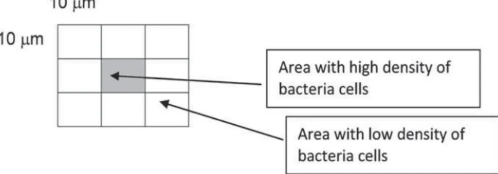
The number of cells adhering to the glass surface on a 100 µm2 area of the glass bead viewed at a
_"=== number of cells was determined from an area of 1 mm2 of the glass surface. The area with high density of bacteria cells was used in the calculation of the number of cells (Figure 1). In total, 9 beads from 3 different assays per experiment were processed and analyzed.
Determination of bacteria cell surface area * ! X="===$@"` $ The size of the individual adhered bacterial cell was measured according its length (L) and width (W) in {|}~ ~{2 of the total number of adhered cells from 9 beads used in the determinations.
Determination of the appearance of the extracellular surface
This was carried out by comparison with the appearance of the extracellular surface of S. mutans
adhering to hydroxyapatite beads28.
Figure 1- Diagram showing the calculation for the number of bacterial cells on 100 Pm2 area in a total area of 1 mm2 of
Preparation of partially purified cell-associated GTF of S. mutans
Crude GTF was isolated from S. mutans ATCC 25175 strain in the research laboratory, Oral Biology Department, University of Malaya using the method described by Ooshima, et al.18 (2000) with 17,21. The crude enzyme was then ! ! (Macrosep Omega membrane - mw cut off=300 kd; Pall Life Science, Exton, PA, USA). The crude enzyme (Macrosep Omega membrane; Pall Life Science) at 5,000 g for 20 min to remove residual cells and the supernatant collected was then referred as partially ` $* enzyme was kept in the freezer (-20°C) to be used for further analyses.
Determination the effect of the extract WRZDUGV SDUWLDOO\ SXUL¿HG FHOODVVRFLDWHG GTF (CAG) activities
The reaction mixture used in the determination * was adapted from the method described by Mukasa,
et al.14XZ
carried out in this study. The reaction mixture consists of 0.01% of thimerosal (Sigma Aldrich Co.), 1.4 g of sucrose, 34.3 mg of exogenous dextran (ICN Biomedicals, Aurora, OH, USA) and mixed in 100 mL of 5 mM sodium phosphate (Sigma Chemical Co.) buffer (pH 6.5). 100 mL of the partially * > }$ The mixture was then incubated at 37°C. After 18 h (to allow the enzyme to catalyze the formation + " the reaction was terminated by placing the tube in a boiling water bath for 5 min. After 5 min, the mixture was centrifuged (Sigma 302K, Osterode, Germany) at 2,000 g for 10 min. The pellet obtained after centrifugation was collected and used in the determination of water-insoluble (WIS) extracellular polysaccharide content while the supernatant was discarded.
The pellet was washed 3 times each with 5 mL of distilled water and then uniformly suspended in 1 mL of distilled water by agitation. 50 mL of the WIS extracellular polysaccharide suspension was then pipetted into a clean test tube and assayed for glucose content.
The experiment was repeated by adding the plant extract at the respective concentrations (0–4.0 mg mL-1) to the enzyme reaction mixtures. Comparison ! CAG treated with plant extract and that without the treatment. The activity of the enzyme in the presence of 0.12% chlorhexidine (Sigma Aldrich Co.) was also determined using the same procedure.
Determination of glucose content of the WIS glucan
The determination of glucose was carried out according to the method by Dubois, et al.4 (1956). To 50 mL of the WIS glucan suspension, 50 mL of deionized distilled water was added and this was followed by the addition of 50 mL of phenol (Sigma Aldrich Co.) (80% w/v). The mixture was vortexed and subsequently 2 mL of concentrated sulfuric acid (R&M Chemicals, Essex, UK) was added to the mixture. This mixture was then allowed to stand for 10 min at room temperature before its optical density at 490 nm was measured. For the standards, a range of volumes (0–100 mL) of the stock glucose (Sigma Aldrich Co.) solution (1 mg mL
-1
from 0 to 1 mg mL-1. For the blank control, 100 mL of deionized distilled water was used instead. The determinations were carried out in triplicates.
The glucose content determined corresponds to the amount of extracellular polysaccharide formed ! * which was then used in the calculation of the GTF activity. The enzyme activity is expressed as µmol
glucose in the extracellular polysaccharide produced per min (µmole glucose min-1)21.
Statistical analysis
Normality tests for all the data obtained were carried out using the Shapiro-Wilks analysis. The data for the comparison of the cell population and cell growth in the respective experiments were analyzed using a two-way ANOVA. For the &@ between the different experiments, a one-way ANOVA was used.
RESULTS
Effect of sucrose on the adherence capacity and growth of S. mutans on glass surface
to the glass surface and they are in aggregates. The extracellular surface of the cells appeared to have # surface of the glass bead (Figure 2C).
The quantitative measurement of the bacterial cell population was carried out during the SEM _"===$ the number of cells adhering to the glass surface increases in the presence of sucrose and the increase was 6-fold higher than that in the absence (p<0.0001) (Table 1).
The cell surface area of the bacterial cells which was calculated based on the SEM viewing at 10,000x *!>$ there was an increase (33%) in the cell surface area of the bacterial cells when grown in the presence of sucrose compared to that grown in the absence (p<0.0001).
Effect of Piper betle L. leaves on the adherence capacity and growth of S. mutans
in the presence and absence of 1 % sucrose The SEM micrographs in Figure 3 show the effect of the different concentrations of the Piper betle L. leaves extract on bacterial cells grown in the BHI broths without sucrose and with 1% sucrose based X="=== sucrose in the presence of 2 mg mL-1 extract - Figure 3A; with 1% sucrose in the presence of 2 mg mL-1 extract - Figure 3B; without sucrose in the presence of 4 mg mL-1 extract - Figure 3C; and with 1% sucrose in the presence of 4 mg mL-1 extract - Figure 3D). The effect of the extract appeared to be concentration dependent where the number of cells adhering to the glass surface decreases with the increase in the concentration of the extract. It was shown that sucrose also has an effect on the growth and adherence of the bacterial cells.
Effect of the Piper betle L. leaves extract on the adherence capacity of the S. mutans
It was found that the addition of the Piper betle
extract to the BHI broths reduces the cell population of S. mutans adhering to the glass surface. The reduction was observed both in the absence and presence of sucrose. The number of cells adhering to the glass surface reduced by 27% (p<0.0001) when grown without sucrose and in the presence of 2 mg mL-1 of the extract compared to that in the absence of the extract. A similar reduction in the number of cell adhering to the glass surface was observed for those grown in the presence of sucrose. Increasing the extract concentration to 4 mg mL-1 resulted in a further decrease of the cell population, with a reduction of more than 80% both in the absence and presence of sucrose (Table 1). No bacterial cells were observed to be adhering to the glass surface when chlorhexidine was used instead of the extract.
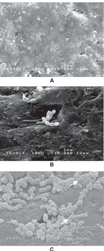
Figure 2- Scanning electron microscope micrographs of
S. mutans cells grown on glass beads in the absence and
presence of 1% sucrose. A: Blank control (10,000x). Note the absence of bacteria on the glass bead’s surface. B: S.
mutans cells grown in the absence of sucrose (10,000x).
There were not many cells adhering to the glass surface and they appeared to be in the dividing state. C: S. mutans grown in the presence of sucrose (1%) (10,000x). There were many cells adhering to the glass surface. The cells appeared to be covered by thick sticky extracellular layer,
A
B
Effect of the Piper betle L. leaves extract on the growth of S. mutans with and without sucrose
The cell surface area of the bacterial cells grown in the presence of the extract, with and without
sucrose based on the SEM viewing at 10,000x *! >$ that the bacterial cells grown in the presence of 2 mg mL-1 of the extract without sucrose exhibited
Cell surface area (μm2)
Experiments Absence of sucrose Presence of sucrose
Negative control (without the CA) 0.209±0.018a,b 0.278±0.026a
2 mg mL-1 CA 0.216±0.016b,x 0.244±0.018d,x
4 mg mL-1 CA 0.146±0.010c,y 0.145±0.009e,y
Positive control (0.12% chlorhexidine) nd nd
The number of cells adhered to the glass beads /100 μm2 was expressed as the mean ± standard deviation from nine
determinations
a – p<0.0001 comparing number of adhering cells in the presence and absence of sucrose at 0 mg/mL of extract
b – p<0.0002 comparing number of adhering cells in the presence of CA 2 mg mL-1 (without sucrose) and negative control
(without sucrose)
c – p<0.0001 comparing number of adhering cells in the presence of CA 4 mg mL-1 (without sucrose) and negative control
(without sucrose)
d – p<0.0001 comparing number of adhering cells in the presence of CA at the two concentrations (with sucrose) and negative control (with sucrose)
x- p<0.0001 comparing number of adhering cells in the presence of CA at the two concentrations (without sucrose) and CA (with sucrose)
y – p<0.0001 comparing number of adhering cells in the presence of CA at the two concentrations (without sucrose) and chlorhexidine (without sucrose)
z – p<0.0001 comparing number of adhering cells in the presence of CA at the two concentrations (with sucrose) and chlorhexidine (with sucrose)
nd – not detectable
Table 2- Comparison of the changes in the size of S. mutans ATCC 25175 cells when grown in different concentrations of the crude aqueous Piper betle L. extract (CA), in the absence and presence of sucrose (1%). (Statistical analysis; two-way ANOVA; scanning electron microscope at 10,000x)
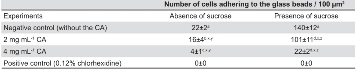
Number of cells adhering to the glass beads / 100 μm2
Experiments Absence of sucrose Presence of sucrose
Negative control (without the CA) 22±2a 140±12a
2 mg mL-1 CA 16±4b,x,y 101±11d,x,z
4 mg mL-1 CA 4±1c,x,y 22±2d,x,z
Positive control (0.12% chlorhexidine) 0±0 0±0
The number of cells adhered to the glass beads was expressed as the mean ± standard deviation from nine determinations a – p<0.0001 comparing number of adhering cells in the presence and absence of sucrose at 0 mg/mL of extract
b – p<0.0002 comparing number of adhering cells in the presence of CA 2 mg mL-1 (without sucrose) and negative control
(without sucrose)
c – p<0.0001 comparing number of adhering cells in the presence of CA 4 mg mL-1 (without sucrose) and negative control
(without sucrose)
d – p<0.0001 comparing number of adhering cells in the presence of CA at the two concentrations (with sucrose) and negative control (with sucrose)
x- p<0.0001 comparing number of adhering cells in the presence of CA at the two concentrations (without sucrose) and CA (with sucrose)
y – p<0.0001 comparing number of adhering cells in the presence of CA at the two concentrations (without sucrose) and chlorhexidine (without sucrose)
z – p<0.0001 comparing number of adhering cells in the presence of CA at the two concentrations (with sucrose) and chlorhexidine (with sucrose)
from those grown in the absence of the extract. Increasing the concentration of the extract to 4 mg mL-1 decreases the cell surface area by 30% (p<0.0001).
It is shown in Table 2 that the cell surface area of the bacterial cells grown in the presence of 1% sucrose and 2 mg mL-1 of the extract is reduced by 12% compared to that of those grown without the extract (p<0.0078). The bacterial cells also appeared to aggregate and exhibit # ! compared to those grown in the absence of the extract (Figure 3B versus Figure 2C). There was an almost 50% reduction in the cell surface area when the bacterial cells were grown in the presence of a higher extract concentration (4 mg mL-1) (p<0.001).
It was observed that the effect of the different concentrations of the extract is not greatly # ! $ * surface area of the bacteria in the presence of 1% sucrose and 2 mg mL-1 extract only increased by 12% compared to the cell surface area of those in the absence of sucrose (p<0.0002). At 4 mg mL-1 concentration of the extract, the bacterial cells attained almost similar size both in the presence and absence of sucrose.
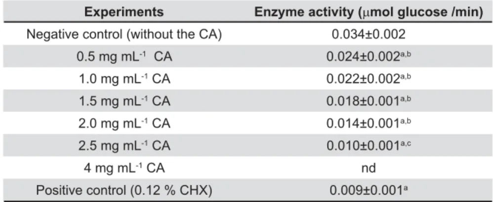
Effect of the Piper betle L. leaves extract RQ WKH DFWLYLW\ RI SDUWLDOO\ SXUL¿HG FHOO associated GTF (CAG)
Table 3 shows the effect of the extract on &@ $ *
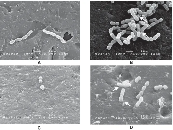
Figure 3- Scanning electron microscope micrographs of S. mutans cells treated with crude aqueous Piper betle L. extract (CA) on the glass beads in the absence and presence of 1% sucrose. A: S. mutans cells grown in the absence of 1%
sucrose and presence of 2 mg mL-1 !!
the glass surface and they appeared to be in dividing state. B: S. mutans cells were grown in the presence of 1% sucrose
and 2 mg mL-1 ! !!!
appeared to be in the dividing state. Fluffy extracellular surface appearance is also observed (arrow). C: S. mutans cells
grown in the absence of 1% sucrose and presence of 4 mg mL-1 " !
seen adhering to the surface compared to that in Figure 3A and they also appeared to be in dividing state. D: S. mutans
cells grown in the presence of 1% sucrose and 4 mg mL-1 " ! !
adhering to the surface compared to that in Figure 3B
A
B
was expressed as μmol glucose content of the extracellular polysaccharide per minute and was found to decrease in the presence of the extract. The inhibitory effect was found to be concentration concentrations of the extract. At 0.5 mg mL-1 of " &@ reduced by 29% (p<0.0001) when compared with the negative control (without the plant extract). At a higher concentration of the extract (1 mg mL-1), the activity was reduced by 35% (p<0.0001) and at 2.5 mg mL-1 of the extract, the decrease was 70% (p<0.0001), the latter being almost similar to that observed with chlorhexidine (p>0.05). Increasing the concentration of the extract to 4
mg mL-1!
CAG activity.
DISCUSSION
In this study, the SEM was used to determine both visually and quantitatively i) the effect of sucrose on growth and adhering property of S. mutans and ii) the effect of Piper betle extract on S. mutans in the presence and absence of sucrose. The SEM study can also be used to show the appearance of the extracellular surface of the cells in the presence of the extract with or without sucrose. Previous studies2,8,24,30 had employed spectrophotometric technique that allows for quantitative determination of cell population (CFU mL-1) to relate bacterial population and growth.
The increase in cell population adhering to glass surface in the presence of sucrose compared with those in the absence indicates that sucrose has a role in the adhering property30 and growth of S. mutans2,8,24,30,32. S. mutans is a secondary
colonizer of dental plaque featuring cariogenic properties including cell adhesion, cell surface hydrophobicity and glucan synthesis activities21. The initial adherence or attachment of bacteria to the tooth surfaces may involve hydrophobic and ionic interactions between the receptors on the host surface and the adhesins on the cell surface of the bacteria11,12. The extracellular polysaccharide (glucan) which is formed favorably in the presence sucrose may be responsible in enhancing the adherence of more bacterial cells to the host surface.
* ` ! # ! the cell surface area. It was found to be increased by 33% in the presence of sucrose. Sucrose is a good substrate for GTF as its hydrolysis produces ` in the formation of glucan, an extracellular polysaccharide. Thus its presence in the BHI broth helps to promote the activity of this enzyme. This explains for the formation of the extracellular # extracellular surface of S. mutans when grown in the presence of sucrose.
For all the experiments to show the effect of the extract of Piper betle L. leaves towards S. mutans, sub-minimal inhibitory concentrations (sub-MICs) (2 mg mL-1 and 4 mg mL-1) were used. These concentrations were used to ensure that the observed reduced number of adhering cells was not attributed to cell death. This parameter has been used as a reference for bacteriostatic effect of plant extract20.
It was shown that the Piper betle L. leaves extract reduces adherence of the bacterial cells to "! the calculated cell population visualized by SEM.
a - p<0.0001 comparing plant extract-treated glucosyltransferase samples to negative control b - p<0.0001 comparing plant extract-treated glucosyltransferase samples to positive control c - p<0.0675 comparing CA 2.5 mg mL-1-treated glucosyltransferase samples to positive control
nd- not detectable
Experiments Enzyme activity (Pmol glucose /min)
Negative control (without the CA) 0.034±0.002
0.5 mg mL-1 CA 0.024±0.002a,b
1.0 mg mL-1 CA 0.022±0.002a,b
1.5 mg mL-1 CA 0.018±0.001a,b
2.0 mg mL-1 CA 0.014±0.001a,b
2.5 mg mL-1 CA 0.010±0.001a,c
4 mg mL-1 CA nd
Positive control (0.12 % CHX) 0.009±0.001a
Table 3- The effect of crude aqueous Piper betle# $& $&'*!
It was thought that the reduced adherence is attributed to extracellular polysaccharide not being " * activity in the presence of the extract.
In this study, it was shown that there are bacteria though few in number adhering to the glass surface despite the undetectable GTF activity in the presence of 4 mg mL-1 of the extract. In the presence of 0.12% chlorhexidine there were no bacterial cells adhering to the glass surface and at that concentration, the GTF activity corresponds to that of 2.5 mg mL-1 of the extract and, cell adherence was observed in the presence of the later. This suggests that GTF is not involved and that other factors such as hydrophobic and ionic interactions maybe involved in the adherence or attachment of bacteria to the tooth surfaces which, is in agreement with what had been reported in previous work11,12. However, it has been shown that the cell surface hydrophobicity of S. mutans is reduced in the presence of the plant extract16. The crude Piper
betle }$ # adhesion between the cell surface of the bacteria and the host surface via ionic interaction which could be responsible for the adherence effect demonstrated at 4 mg mL-1 extract concentration.
It was also shown that the Piper betle L. leaves # (size) of the bacterial cells. The presence of sucrose appears not to have effect on the cell surface area of bacterial cells in the presence of the 4 mg mL-1 extract as with or without sucrose, the bacterial cells appear to attain similar cell surface area. This indicates that bacteria grown in the presence of the extract could experience environmental stress, # !
! 5,29. It
has been reported that the aqueous Piper betle L. extract exhibited similar effect towards other oral bacteria for example, S. sanguinis, S. mitis and
Actinomyces sp.5.
CONCLUSION
The SEM analysis provided visual (qualitative) and quantitative evidence that the Piper betle
L. leaves extract has a reducing effect on cell adhesion, cell growth and extracellular appearance of S. mutans. The effect of the Piper betle L. leaves extract on the GTF activity and adherence capacity of S. mutans suggests the involvement of other factors in the adherence of bacterial cells to the glass surface.
ACKNOWLEDGEMENTS
This project was supported by research grants (Vote F and IRPA) provided by the Malaysian
government. The authors would like to acknowledge Ms Zubaidah Abu Hassan of the Electron Microscopy Unit, Medicine School, University of Malaya for their assistance in the electron microscopy procedure.
REFERENCES
X?&"@" ""!$? surface properties and protein expression in oral Streptococcus sanguis. Arch Oral Biol. 2004;49:295-304.
2- Cury JA, Francisco SB, Del Bel Cury AA, Tabchoury CP. In situ
study of sucrose exposure, mutans streptococci in dental plaque and dental caries. Braz Dent J. 2001;12:101-4.
3- Duarte S, Koo H, Bowen WH, Hayacibara MF, Cury JA, Ikegaki M, et al. Effect of a novel type of propolis and its chemical fractions on glucosyltransferases and on growth and adherence of mutans streptococci. Biol Pharm Bull. 2003;26:527-31.
4- Dubois M, Gilles K, Hamilton JK, Rebers PA, Smith F. A colorimetric method for determination of sugars and related substances. Anal Chem. 1956;28:350-6.
5- Fathilah AR, Yusoff M, Rahim ZHA. The effect of Psidium guajava
and Piper betle extracts on the morphology of dental plaque bacteria. Pak J Med Sci. 2009;25:928-33.
6- Hamada S, Slade HD. Biology, immunology and cariogenicity of Streptococcus mutans. Microbiol Review. 1980;44:331-84. 7- Hannig C, Ruggeri A, Al-Khayer B, Schmitz P, Spitzmüller B, Deimling D, et al. Electron microscopic detection and activity of glucosyltransferase B, C and D in the in situ formed pellicle. Arch Oral Biol. 2008;53:1003-10.
8- Karjalainen S, Karjalainen M, Söderling E. Effect of sucrose # $ Caries Res. 1993;27:38-42.
9- Kirtikar KR, Basu BD. Indian Medicinal Plants. Allahabad: Lalit Mohan Basu Publications; 1933. v. III, p.2128.
10- Limsong J, Benjavongkulchai E, Kuvatanasuchati J. Inhibitory effect of some herbal extracts on adherence of Streptococcus mutans. J Ethnopharmacol. 2004;92:281-9.
XX$+!!$&$ 2004;38:204-11.
12- Marsh PD, Martin MV. Oral Microbiology. 5th ed. Philadelphia:
Churchill Livingstone; Elsevier Ltd; 2009.
13- Matsumoto M, Minami T, Sasaki H, Sobue S, Hamada S, Ooshima T. Inhibitory effects of oolong tea extract on caries inducing properties of mutans streptococci. Caries Res. 1999;33:441-5.
14- Mukasa H, Shimamura A, Tsumor H. Effect of salts on water-insoluble glucan formation by glucosyltransferase of Streptococcus mutans. Infect Immun. 1979;23:564-70.
15- Murchison H, Larrimore S, Curtiss R 3rd. Isolation and
characterization of Streptococcus mutans mutants defective in adherence and aggregation. Infect Immun. 1981;34:1044-55. 16- Nalina T, Rahim ZH. The crude aqueous extract of Piper betle
L. and its antibacterial effect towards Streptococcus mutans. Amer J Biotech Biochem. 2007;3:10-5.
17- Nalina T, Rahim ZH. The effect of Piper betle L. leaf extract on the virulence activity of Streptococcus mutans. Pak J Biol Sci. 2006;9:1470-5.
18- Ooshima T, Osaka Y, Sasaki H, Osawa K, Yasuda H, Matsumura M, et al. Caries inhibitory activity of cacao bean husk extract in
in vitro and animal experiments. Arch Oral Biol. 2000,45:639-45. 19- Ooshima T, Yoshida T, Hamada S. Detection of caries-inducing microorganisms in hyposalivated rats without infection of mutans streptococci. Microbiol Immunol. 1994;38:39-45.
20- Prabu GR, Gnanamani A, Sadulla S. Guaijaverin – a plant
# + Streptococcus
mutans. J Appl Microbiol. 2006;101:487-95.
22- Ramji N, Ramji N, Iyer R, Chandrasekharan S. Phenolic antibacterials from Piper betle in the prevention of halitosis. J Ethnopharmacol. 2002;83:149-52.
23- Razak FA, Rahim, ZH. The anti-adherence effect of Piper betle L. and Psidium guajava extracts on the adhesion of early settlers in dental plaque to saliva-coated glass surfaces. J Oral Sci. 2003;45:201-6.
24- Seppä L, Pöllänen L, Hausen H. Streptococcus mutans counts obtained by a dip-slide method in relation to caries frequency, #$&$X>>'>>$ 25- Sharma S, Khan IA, Ali I, Ali F, Kumar M, Kumar A, et al. !"# activities of hydoxychavicol for its potential use as an oral care agent. Antimicrob Agents Chemother. 2009;53:216-22.
26- Smith JR, Laudicinia RJ, Rufi RD. Learning guides for microbiology laboratory. New York: John Wiley & Sons; 1985. 27- Song JH, Yang TC, Chang KW, Han SK, Yi HK, Jeon JG. In vitro
effects of a fraction separated from Polygonum cuspidatum root ! "!"! of mutans streptococci. J Ethnopharmacol. 2007;112:419-25.
28- Staat RH, Langley SD, Doyle RJ. Streptococcus mutans
adherence: presumptive evidence for protein-mediated attachment followed by glucan-dependent cellular accumulation. Infect Immun. 1980;27:675-81.
29- Tao L, Tanzer JM, MacAlister TJ. Bicarbonate and potassium regulation of the shape of Streptococcus mutans NCTC 10449S. J. Bacteriol. 1987;169:2543-7
30- Wan Nordini HW, Razak FA, Rahim ZH. The role of sucrose ! $ Online J. Biol Sci. 2006;6:51-5.
31- The Wealth of India. A dictionary of Indian raw materials and industrial products. New Delhi: Publications and Information "&X$ v. 8.