RELATIONSHIP BETWEEN THE ELECTRICAL ACTIVITY
OF SUPRAHYOID AND INFRAHYOID MUSCLES DURING
SWALLOWING AND CEPHALOMETRY
Relação da atividade elétrica dos músculos supra e infra-hióideos
durante a deglutição e cefalometria
Maria Elaine Trevisan(1), Priscila Weber(2), Lilian G.K. Ries(3), Eliane C.R. Corrêa(4)
(1) Physical Therapist; Professor at Physical Therapy
Depart-ment (Master Degree) and doctoral student at Graduate Program of Human Communication Disorders, Federal Uni-versity of Santa Maria – UFSM, RS, Brazil.
(2) Physical Therapist; Graduate Program Human
Commu-nication Disorders (Master Degree), Federal University of Santa Maria – UFSM, RS, Brazil.
(3) Physical Therapist; Professor at Physical Therapy
Depart-ment (PhD), University of the Santa Catarina State – UDESC , SC, Brazil.
(4) Physical Therapist; Professor at Physical Therapy
Depart-ment (PhD) and Graduate Program of Human Communica-tion Disorders, Federal University of Santa Maria – UFSM, RS, Brazil.
Conlict of interest: non-existent
INTRODUCTION
The deglutition process is a complex sensory-motor mechanism that involves, in a sequence, excitation and inhibition of different levels of the Central Nervous System (CNS)1,2. It is
charac-terized by three phases: oral, pharyngeal and esophagic; which demand coordinated movements of mouth, tongue, larynx and esophagus and they are independent of each other 1-3. Nevertheless, the
generation of the CNS patterns controls the time of these events and the peripherical manifestations of these phases that depend on peripherical sensory ABSTRACT
Purpose: to investigate the inluence of the habitual head posture, jaw and hyoid bone position on the supra and infrahyoid muscles activity of the muscles during swallowing of different food textures. Method: an observational, cross-sectional study, with women between 19 and 35 years, without myofunctional swallowing disorders. The craniocervical posture, position of the mandible and hyoid bone were evaluated by cephalometry. The electromyographic activity of the supra and infrahyoid muscles was collected during swallowing water, gelatin and cookie. Results: sample of 16 women, mean age 24.19 ± 2.66 years. At rest, there were negative/moderate correlations between the electrical activity of the suprahyoid muscles with NSL/CVT (head position in relation to the cervical vertebrae) and NSL/OPT (head position in relation to the cervical spine) postural variables, and positive/moderate with the CVA angle (position of lexion/extension of the head). During swallowing the cookie, the activity of infrahyoid muscles showed a negative/moderate correlation with NSL/OPT angle. It was found higher electrical activity of the suprahyoid muscles during swallowing of all foods tested, and of the infrahyoid muscles at rest. There was difference on the muscle activity during swallowing of foods with different consistencies, which was higher with cookie compared to water and gelatin. Conclusion: the head hyperextension relected in lower activity of the suprahyoid muscles at rest and of the infrahyoid muscles during swallowing. The consistency of food inluenced the electrical activity of the suprahyoid and infrahyoid muscles, with greater muscle recruitment in swallowing solid food.
stimulus3. The oral phase of the deglutition is a
voluntary event, while the pharyngeal is involuntary and independent1. However, the deglutition always
occurs in the same sequence, being the pharynx and esophagus responses dependent on the food bolus properties1-3
The complexity of the pharyngeal phase must be pointed out once it requires the concomitance of a series of events, including the antero-posterior displacement of the hyoid bone and the thyroid cartilage; epiglottis closing; vocal cord closing and superior esophagic sphincter opening. The hyoid displacement to upward and forward occurs at the moment in which the bolus crosses the pharyngeal cavity and depends on the tongue basis and the supra-hyoid muscle contraction4-6.
For an eficient swallowing function, the mandible adopts a ix and stable position, by the intercuspal of the occlusal surfaces, immediately before the tongue impulses the food bolus to the oropharynx7. On the
other hand, the mandibular stabilization allows the suprahyoid muscle contraction and, consequently, the hyoid bone and larynx antero-superior traction assuring a safe deglutition7,8.
There are evidences that the mandibular rest position suffers alterations due to occlusal interfer-ences, temporomandibular, stress, nasal obstruction and head posture9. Considering the established
relations between the craniocervical posture and craniofacial morphology, body posture changes, especially in the head, tend to modify the activity of the muscles that take part in the positioning of the mandibular rest 10. Suprahyoid muscles are
directly involved in the mandible stabilization during intercuspal and food grinding, as well in the active elevation of the hyoid bone and the larynx during swallowing, having close association between the functions that involve the mandible posture and the supra and infra-hyoid muscle action11.
Forward head posture is a commonly observed postural change that leads to compensations such as cranium and upper cervical spine hyperextension and lower cervical curvature lexion. It also produces alterations in the mandible, hyoid and tongue position, modifying the craniocervicomandibular biomechanical relations and, consequently, the mandibular rest position. The mandible in a more retruded and elevated position pulls the supra-hyoid musculature12.
Harmony and balance between form and function are essential to determine the system stomatognathic health condition. Therefore, under-standing the relation between the craniocervical posture, mandible and hyoid bone position and the supra and infra-hyoid muscle activity may elucidate the biomechanical changes that, occasionally,
affect the stomatognathic functions, in particular, the deglutition.
Recent studies have investigated the supra and infra-hyoid muscle behavior in different body and head positioning during the swallowing function7,13-15.
Differently, this study aimed to investigate the inluence of the usual head posture, mandibular and hyoid bone position on the supra and infra-hyoid muscles during the swallowing of three different types of food.
METHOD
The present experiment was approved by the Research Ethics Committee of the local institution, under protocol CAAE number 0281.0.243.000-08. The volunteers were included in the research after signing the Consent Term.
It consists of a cross-sectional observational study, with quantitative data analysis. The partici-pants were evaluated by an experienced speech language pathologist in orofacial motricity, according to the Myofunctional Evaluation with Scores Protocol (AMIOFE)16, prior to participating in the study.
The inclusion criteria were: female gender, age from 19 to 35 years old, without myofunctional alterations during the masticatory and swallowing functions.
The exclusion criteria were: facial trauma, craniomandibular or cervical orthopedic surgical procedures, musculoskeletal deformities, temporo-mandibular disorder (TMD), Angle Class II and III occlusions, tooth loss, anterior and posterior open bite, cross bite, edge-to-edge bite and overbite, as well as being wearing braces. The TMD presence was investigated by only one examiner according to the Research Criteria for Temporomandibular Disorder (RDC/TMD)17. The occlusion was evaluated
through intra-oral photographies observed by an Orthodontist.
The craniocervical posture, mandible and hyoid position were evaluated using cephalometric analysis. The volunteers have undergone a right lateral radiography of the cranium and cervical spine in standing position, without any instruction for aligning it. In order to reproduce the natural head position, they were oriented to keep staring at the relection of their eyes in a mirror placed at one-meter distance18,19. The radiography was performed on the
Orthophos Plus (Siemens, Germany) equipment, with 1.52 m of focus-ilm distance.
CVA- lexion/extension head position and CPL/ Horizontal line – forward head posture (Figure 3) 18,20-22. The mandibular and hyoid bone spatial
position were determined, respectively, by NSL/ML (cranium basis inclination related to the mandible)18
and by the linear distance from the hyoid to mentum (HY/ME), to mandible (HY/ ML) and to third cervical vertebra (HY/C3) (Figure 4)
4,22.
The cephalograms were manually traced, by the same examiner, on acetate paper with the aid of a mechanical pencil with 0.3mm tip, tape, soft rubber, with the radiographs placed on a negatoscope in order to allow a better visualization of the structures. A protractor, for the angular measurements, and a millimeter ruler for the linear measures were used.
CV2tg – the tangent point at the superior posterior extremity of the odontoid process of the second cervical vertebra (C2); CV4ip – the most infero-posterior point on the body of the fourth cervical vertebra; CV6ip – the most infero-posterior point on the sixth cervical vertebra.
Figure 1 – Anatomical points utilized in the cephalometric analysis of the CVT/EVT angle
N (nasion) – anterior point at fronto-nasal suture; S (Sella) – center of sella turcica CV2tg – the tangent point at the supe-rior postesupe-rior extremity of the odontoid process of the second cervical vertebra (C2); CV2ip – the most infero-posterior point on the body of the second cervical vertebra; CV4ip – the most infero-posterior point on the body of the fourth cervical vertebra.
Figure 2 – Anatomical points utilized in the cephalometric analysis of the NSL/OPT and NSL/CVT angles
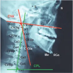
sp – anterior nasal spine; pm – posterior nasal spine; O – basi--occiput; CV2Ap – tangent point to the apex of the C2 dente; CV2ia – the most infero-anterior point on the body of the second cervical vertebra; Ce1 a Ce7 – central points of the vertebral bodies from C1 to C7.
Figure 3 – Anatomical points utilized in the cephalometric analysis of the CVA and CPL angles
N (nasion) – anterior point at fronto-nasal suture; S (Sella) – cen-ter of sella turcica; H – most ancen-terosuperior point of the Hyoid bone; RGn (retrognathion) – the most inferior posterior point at the mandibular symphysis; Me (Mentum) – most inferior point at the mandibular symphysis.
The craniovertebral angle (CVA) gradually classiies the antero-posterior cranium position related to the cervical spine: CVA between 96-106 corresponds to the head natural position, head extension < 96, and head lexion > 10620.
From the CPL angle, the individuals with the measure lower and higher than 80º, that is, presenting more or less forward head posture22.
For the electromyographic signal (EMG) acqui-sition of the supra and infra-hyoid muscular groups, the individuals were instructed to comfortably seat in a chair, with eyes open and head oriented in the Frankfort Plan position. The signals were acquired at least three times for each of the tests in search of a better signal quality23.
The room temperature was maintained at approx-imately 25ºC and the possible noises that could interfere in the EMG acquisition were controlled. The Miotool 400 (Miotec, Porto Alegre, Brasil) was used for the electromyographic evaluation, with four channels, 14 bit resolution, 2000 Hz sample frequency per channel, Butterworth ilter and band pass with 20-500Hz cutoff frequency. Electrodes Ag/AgCl double type (Hal Indústria e Comércio Ltda) connected to pre-ampliiers active sensors were positioned in the supra and infra-hyoid area. A reference electrode unipolar (Meditrace 100) was placed on the sternum aiming to reduce interference and/or noise during the EMG acquisition24.
The EMG data acquisition was carried out during the swallowing of 20ml of water, 20 ml of gelatin and half cookie (BONO®). The bolus size of the ine
liquids were based on a previous study25 and the
sequence water, gelatin and cookie was at random and maintained for all participants. Depending on the food texture/consistency, after chewing, if necessary, the volunteer was instructed to swallow the entire volume offered in one single gulp, under the evaluator verbal command. For each texture/ consistency, there were three attempts with one-minute rest intervals between them, totaling nine swallows for each volunteer.
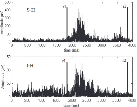
The signals were analyzed by the Matlab software (The MathWorks®, version 5.3). To detect the onset of the swallowing activity (t1) 20 Hz high-pass and 50 Hz low-pass ilters were used26.
t1 was determined as the point where the supra-hyoid muscle signal amplitude became higher than three standard-deviations (SD) that were observed in the mean amplitude (basal activity) before the swallowing activity of each subject. The end of the swallowing activity (t2) was determined after 2000 ms from t1 (t1+2000ms = t2). The supra and
infrahyoid muscle signals were aligned and the t1 and t2 points were the same for both muscles.
In order to determine the integral of the rectiied electromyographic signal (SEMG), 20 Hz high-pass and 400Hz low-pass ilters were used. The S EMG during the swallowing test were demarcated between t1 and t2. These SEMG were corrected by SEMG of base line, being calculated between 50 and 150 ms after the beginning of the data acquisition, according to the equation bellow:
S EMG is the integral of the EMG signal in the time space determined for the water, gelatin and cookie swallowing activities less 20 times 100 ms of the EMG base recorded between 50 and 150 ms after the beginning of the acquisition (igure 5). The SEMG during resting (SEMG) were demarcated between t1 and t2 comprehending medial time spaces of 2000 ms (t1 + 2000ms= t2), according to the equation bellow:
The normalized SEMG
(SEMG%) of each activity was expressed as the maximum value obtained from three repetitions of the cookie swallowing, for each muscle and subject.
The participants were characterized by descriptive statistics (mean, standard-deviation) and for each EMG variable, the aritmethic mean of the three repetitions was considered. The data for normality and homocedasticity were tested by Shapiro-Wilk and Levene tests, respectively.
The repeated measures variance analysis were carried out to test the effect of the ix factor muscles (supra and infrahyoid), the ix factor swallowing (water, gelatin, cookie) and the interaction of these factors in the quantitative dependent muscular variable.
In all analysis, Tukey’s HSD post hoc test was used. To analyze the difference between the supra and infrahyoid muscle activity at rest, the t test was used for paired data.
was considered very low for r < 0.2; low for 0.2 < r < 0.3; moderate for 0.4 < r < 0.69; high for 0.7 < r < 0.89 and very high for 0.9 < r < 127. The Statistical
Package for the Social Sciences (SPSS) version 17.0 for Windows (SPSS) statistics software was used for the analysis and P values <0.05 (bi-tailed) were considered statistically signiicant.
RESULTS
The study group was composed of 16 women with 24.19±2.66 years old and CMI of 23.89±4.83 kg/cm2.
The angular cephalometric variables (mean and standard-deviation) related to the head and cervical spine position were: NSL/CVT (103±5.7°); NSL/OPT (100±6.9°); EVT/CVT (4.4±5.9°); CVA (96.9±7.5°) e CPL (78.2±3.6°). Regarding the spatial position of the hyoid bone, the variables were HY/C3 (43.6±4.3
mm), HY/ML (14.1±5.5) and HY/Me (55.8±7.1 mm). The mean of NSL/ML angle, related to the mandibular position was 30.9±6.8°.
Table 1 shows the correlations between the craniocervical and electromigraphic variables. At rest, it was observed moderate negative correlations between the suprahyoid activity and NSL/CVT and
NSL/OPT angles, as well as a moderate positive correlation with CVA angle. There was a moderate negative correlation between infrahyoid muscles and NSL/OPT angle during cookie swallow.
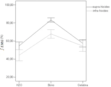
Figure 6 illustrates the results of the supra and infrahyoid muscle activity during water, gelatin and cookie swallow.
The repeated measure variance analysis showed a signiicant difference between the supra and infrahyoid muscles (F=4.32, p=0.04) during swallowing. Additionally, the suprahyoid muscles showed a higher activity during all food swallowing.
In contrast, the infrahyoid presented signiicant higher activity (44.28±19.42) than the suprahyoid muscles (14.08±4.67) at rest ( p=0.00). Using Tukey`s HSD test, it was observed differences in the cookie swallow compared to the water (p=0.00) and to the gelatin ( p=0.00) . As there was no inter-action between the muscles and the swallowing (F=1.01, p=0.37) it is possible to generalize the effect of the different food swallowing on both evaluated muscles.
Therefore, it can be inferred that the cookie swallow demanded higher activity of both muscles compared to the water and gelatin swallow.
The signal from t1 to t2 (t1+2000ms) was used for the integral EMG calculation.
DISCUSSION
Considering the inverse relation that the angles have, biomechanically, between them, it can be stated that: the more the NSL/CVT and NSL/OPT angles increase, the less the CVA reduces, charac-terizing a posterior cranial tilt on the upper cervical spine20.
In this study, the correlation among the NSL/ CVT, NSL/OPT and CVA with the electromyographic activity of the suprahyoid muscles at rest can be attributed to the postural changes in the mandibular
segment as result of the posterior rotation of the head.
Due to the interaction between cervical and craniomandibular systems, the hyperextension of the head produces a mandibular plane elevation, with consequent activation of the masseter muscle in order to keep mouth closed, what may relect on the lower activation of suprahyoid muscles28.
The modiication of the mandibular positioning interferes in the muscular iber length, resulting in electromyographic activity changes in both the masseter and the suprahyoid muscles28. In this
condition, a higher activation of the masseter muscle
Suprahyoid Infrahyoid
water cookie gelatin Resting water cookie Gelatin Resting
NSL/CVT -0.30 -0.03 -0.30 -0.59* 0.06 -0.50 -0.09 0.30
NSL/OPT -0.09 -0.04 -0.25 -0.55* 0.22 -0.55* 0.09 -0.31
EVT/CVT -0.15 0.23 0.38 0.17 -0.23 -0.01 -0.25 -0.11
CVA 0.20 -0.03 0.27 0.57* -0.16 0.34 0.22 -0.36
CPL 0.30 -0.04 0.00 0.46 0.11 0.37 0.35 -0.00
NSLML 0.09 -0.24 -0.34 0.08 -0.09 -0.38 -0.03 -0.07
HyC3 -0.42 -0.09 0.05 -0.40 -0.39 -0.27 -0.40 0.05
HyML -0.23 0.25 -0.40 -0.48 -0.37 -0.39 -0.38 0.03
HyMe -0.21 0.02 0.11 -0.30 0.46 -0.41 0.27 0.42
Table 1 – Correlation between craniocervical posture, mandible and hyoid position and the electromyographic activity of suprahyoid and infrahyoid muscles during resting and swallowing
NSL/OPT; NSL/CVT (cranium inclination in relation to C2 and in relation to the cervical spine); CVT/EVT (cervical curve); CVA (lexion/ extension head position); CPL (forward head posture); NSL/ML (cranium basis inclination related to the mandible); HY/ME (linear dis-tance from the hyoid to mentum); HY/ML (linear disdis-tance from the hyoid to mandible) e HY/C3 (linear distance from the hyoid to third
cervical vertebra). Results expressed in r (Pearson correlation coeficient),* p<0,05.
Figure 6 – Mean and standard-deviation of integral signal % between t1 e t2 (t1+2000ms) of the suprahyoid and infrahyoid muscles during the water, gelatin and cookie swallowing
pulls the mandible upward, causing a passive tension of the suprahyoid muscles, decreasing their activation at rest. On the other hand, the infrahyoid muscle tension increases at rest in order to keep the hyoid bone stability29. Considering that, the
main muscles that displace the hyoid bone upward (milohyoid) and forward (geniohyoid) are originated in the mandible, its adopted position directly inter-feres on these muscles due to the length-tension relation5.
The muscular synergism of the craniocervico-mandibular system has been previously demon-strated. Studies have shown the hyperextension of the head as the most commonly postural alter-ation that modiies the mandible and hyoid bone positioning12,20,30.
In a recent study22, based on the interpretation
of the correlation for NSL/OPT, NSL/CVT and CVA angles with the mandibular and hyoid bone position, it was concluded that the hyperextension of the cranium causes the mandible elevation. Consequently, there is an increase in the hyoid to mentum distance, placing the suprahyoid muscle at a disadvantage to exert its function.
However, it was not observed signiicant corre-lation between the craniocervical posture and the suprahyoid muscle activity during swallowing. Only one inverse and signiicant correlation was found between NSL/OPT angle and the infrahyoid muscle activity during cookie swallow. It is believed that this result may be due to the small number of subjects in this study, as well as their craniocervical posture, with values close to the normality for the CVA angle. Based on the repercussion of the cranium hyperex-tension on the supra-hyoid action by the cranioman-dibular interdependence, the correlation between these structures during the muscular action may not have been evidenced due to the normality condition of the craniocervical posture observed in the partici -pants in this study.
The results of the present study also demon-strated a signiicant higher electrical activity of the suprahyoid muscles during the water, gelatin and cookie swallowing. Such inding is explained by the fact that the swallowing act is, essentially, exerted by the suprahyoid muscles, whose action promotes the forward and upward dislocation of the hyoid bone7.
It must be pointed out the importance of such dislocation of the hyoid bone, since when the suprahyoid muscle action becomes reduced, it may have a smaller opening of the upper esophagic sphincter, penetration and/or aspiration of food, besides the permanence of pharyngeal residues post swallowing31.
Finally, it was evidenced that the cookie swallowing demanded higher muscular activity of the supra and infrahyoid muscles compared to water and gelatin. It is known that the viscosity has a considerable effect on the swallowing and, bolus with greater viscosity tends to have a lower swallowing velocity due to the higher resistance to the movement and therefore with a higher activity of the muscles responsible for swallowing32. Ishida
et al. 33 observed greater upward and forward
hyoid excursion during the solid food swallowing compared to liquid consistencies in young subjects, conirming the need for higher muscular activity for the solid swallowing.
CONCLUSION
REFERENCES
1. Ertekin C, Aydogdu I. Neurophysiology of swallowing. Clin. neurophysiol. 2003;114:2226-44. 2. Lang IM. Brain stem control of the phases of swallowing. Dysphagia. 2009;24(3):333-48.
3. Butler SG, Stuart A, Castell D, Russel GB, Koch K, Kemp S. Effects of age, gender, bolus condition, viscosity, and volume on pharyngeal and upper esophageal sphincter pressure and temporal measurements during swallowing. J. speech lang. hear. res. 2009;52(1):240-53.
4. Sheng CM, Lin LH, Su Y, Tsai HH. Developmental changes in pharyngeal airway depth and hyoid bone position from childhood to young adulthood. Angle orthod. 2009;79(3):284-90.
5. Pearson WG Jr, Langmore SE, Zumwalt AC. Evaluating the structural properties of suprahyoid muscles and their potential for moving the hyoid. Dysphagia. 2011;26(4):345-51.
6. van der Kruis JG, Baijens LW, Speyer R, Zwijnenberg I. Biomechanical analysis of hyoid bone displacement in videoluoroscopy: a systematic review of intervention effects. Dysphagia. 2011;26(2):171-82.
7. Perry JL, Bae Y, Kuehn DP. Effect of posture on deglutitive biomechanics in healthy individuals. Dysphagia. 2012;27(1):70-80.
8. Monaco A, Cattaneo R, Spadaro A, Giannoni M. Surface electromyography pattern of human swallowing. BMC oral health. 2008;8(6):2-11.
9. Minagi S, Ohmori T, Sato T, Matsunaga T, Akamatsu Y. Effect of eccentric clenching on mandibular deviation in the vicinity of mandibular rest position. J. oral rehabil. 2000;27:175-9.
10. MacKay E, Tingey DDS, Peter H. Buschang MA, Gaylord S. Mandibular rest position: A reliable position inluenced by head support and body posture. Am. j. orthod. dentofacialorthop. 2001;120:614-22.
11. Miralles R, Gutiérrez C, Zucchino G, Cavada G, Carvajal R, Valenzuela S et al. Body position and jaw posture effects on supra- and infrahyoid electromyographic activity in humans. Cranio. 2006;24(2):98-103.
12. Biasotto-Gonzalez DA. Abordagem interdisci-plinar das disfunções temporomandibulares. São Paulo: Manole; 2005.
13. Tsukada T, Taniguchi H, Ootaki S, Yamada Y, Inoue M. Effects of food texture and head posture on oropharyngeal swallowing. J. Appl. oral physiol. 2009;106:1848-57.
14. Inagaki D, Miyaoka Y, Ashida I, Yamada Y. Inluence of food properties and body position on swallowing-related muscle activity amplitude. J. oral rehabil. 2009;36: 176-83.
RESUMO
Objetivo: investigar a inluência da postura habitual da cabeça, da posição mandibular e do osso hióide na atividade dos músculos supra e infra-hióideos durante deglutição de diferentes consistên-cias de alimentos. Método: estudo observacional, transversal, com mulheres entre 19 e 35 anos, sem alterações miofuncionais de deglutição. A postura craniocervical, posição da mandíbula e osso hióide foram avaliados pela cefalometria. A atividade eletromiográica dos músculos supra e infra-hióideos foi coletada durante a deglutição de água, gelatina e biscoito. Resultados: amostra com 16 mulhe-res, média de idade 24,19±2,66 anos. No repouso, observaram-se correlações negativas/moderadas entre a atividade elétrica dos músculos supra-hióideos com as variáveis posturais NSL/CVT (posição da cabeça em relação às vértebras cervicais) e NSL/OPT (posição da cabeça em relação à coluna cervical) e positiva/moderada com o ângulo CVA (posição de lexão/extensão da cabeça). Durante a deglutição do biscoito, a atividade dos músculos infra-hióideos apresentou correlação negativa/mode-rada com o ângulo NSL/OPT. Constatou-se maior atividade elétrica dos músculos supra-hióideos durante a deglutição de todos os alimentos testados e, dos músculos infra-hióideos, no repouso. Os supra-hióideos foram mais ativos que os infra-hióideos durante a deglutição, entretanto, houve aumento da atividade eletromiográica em ambos os grupos musculares durante a deglutição do bis-coito, comparado com a deglutição de água e gelatina. Conclusão: a hiperextensão da cabeça reper-cutiu na menor atividade dos músculos supra-hióideos no repouso e, dos músculos infra-hióideos, na deglutição. A consistência do alimento inluenciou na atividade elétrica dos músculos supra e infra--hióideos, havendo maior recrutamento muscular na deglutição de alimento sólido.
15. Sakuma T, Kida I. Relationship between ease of swallowing and deglutition-related muscle activity in various postures. J. oral rehabil. 2010;37(8):583-9. 16. Felício CM, Ferreira CLP. Protocol of orofacialmyofunctional evaluation with scores. Int. j. pediatr. otorhinolaryngol. 2008;72(3):367-75.
17. Dworkin SF, Leresche L. Research diagnostic criteria for temporomandibular disorders: review, criteria, examinations and speciications, critique. J. craniomandib. disord.1992;6(4):301-55.
18. Solow B, Sonnesen L. Head Posture and Malocclusion. Eur. j. ortho. 1998;20(6):685-93. 19. Rosa LP, Moraes LC. Estudo comparativo da inluência do método de posicionamento convencional e natural de cabeça para obtenção de radiograias laterais cefalométricas utilizando análise crânio-cervical. Ciênc. odontol. bras. 2009;12(1):56-62.
20. Rocabado M. Biomechanical Relationship of the Cranial, Cervical and hyoid Regions. J. craniomandib. pract. 1983;1(3):61-6.
21. Tecco S, Tete S, Festa F. Relation between cervical posture on lateral skull radiographs and electromyographic activity of masticatory muscles in Caucasian adult women: a cross-sectional study. J. oral rehabil. 2007;34(9):652-62.
22. Weber P, Corrêa ECR, Bolzan GP, Ferreira FS, Soares JC,Silva AMT. Relationship between craniocervical posture, mandible and hyoid bone and inluence on alimentary functions. Braz. j. oral sci. 2012;11(2):141-7.
23. Corrêa ECR, Bérzin F. Eficacy of physical therapy on cervical muscle activity and on body posture in school-age mouth breathing children. Int. j. pediatr. otorhinolaryngol. 2007;71(10):1527-35.
24. Ries IGK, Bérzin F. Ativação assimétrica dos músculos temporal e masseter em crianças com paralisia cerebral. Fisioter. mov. 2009;22(1):45-52. 25. Miyaoka Y, Ashida I, Kawakami S, Tamaki Y, Miyaoka S. Activity patterns of the suprahyoid muscles during swallowing of different luid volumes. J. oral rehabil. 2010; 37: 575-82.
26. Solnik S, Lnik S, Devita P, Rider P,Long B,Hortobágyi T. Teager–Kaiser Operator improves the accuracy of EMG onset detection independent of signal-to-noise ratio. Acta Bioeng Biomech. 2008;10(2):65-8.
27. Pestana MH, Gageiro JN. Análise de Dados para Ciências Sociais – A Complementaridade do SPSS.Lisboa: Edições Silabo; v.2, 2000.
28. Ballenberger N, von Piekartz H, Paris-Alemany A, La Touche R, Angulo-Diaz-Parreño S. Inluence of different upper cervical positions on electromyography activity of the masticatory muscles. J. manip. physiol. ther. 2012;35(4):308-18. 29 .Forsberg CM, Hellsing E, Linder-Aronson S, Sheikholeslam A. EMG activity in neck and masticatory muscles in relation to extension and lexion of the head. Eur J Orthod 1985;7:177-84. 30. Corrêa ECR, Bérzin F. Temporomandibular disorder and dysfunctional breathing. Braz. j. oral sci. 2004;3(10):498-502.
31. Steele CM, Bailey GL, Chau T, Molfenter SM, Oshalla M, Waito AA, et al. The relationship between hyoid and laryngeal displacement and swallowing impairment. Clin. otolaryngol. 2011;36:30-6.
32. O’Leary M, Hanson B, Smith CH. Variation of the apparent viscosity of thickened drinks. Int. j. lang. commun. disord. 2011;46:17-29.
33. Ishida R, Palmer JB, Hiemae KM. Hyoid motion during swallowing: factors affecting forward and upward displacement. Dysphagia. 2002;17:262-72.
Received on: November 14, 2012 Accepted on: June 17, 2013
Mailing address: Maria Elaine Trevisan
Universidade Federal de Santa Maria
Avenida Roraima 1000 – Prédio 26, Sala 1308 – Cidade Universitária – Bairro Camobi
Santa Maria – RS – Brasil CEP: 97105-900