AN EVALUATION AND PARTIAL CHARACTERIZATION OF A BACTERIOCIN PRODUCED BY LACTOCOCCUS
LACTIS SUBSP LACTIS ST1 ISOLATED FROM GOAT MILK
Parinaz Taheri1*, Nasrin Samadi², Mohammad Reza Ehsani1, Mohammad Reza Khoshayand2, Hossein Jamalifar2
¹Department of Food Science and Technology, Faculty of Agriculture and Natural Resources, Science and Research Branch, Islamic Azad University, Tehran, Iran; ²Department of Drug and Food Control, Faculty of Pharmacy and Pharmaceuticals Quality
Assurance Research Center, Tehran University of Medical Sciences, Tehran, Iran.
Submitted: March 24, 2011; Returned to authors for corrections: April 23, 2012; Approved: June 07, 2012.
ABSTRACT
A bacteriocin-like inhibitory substance producing Lactococcus lactis subsp lactis strain, ST1, isolated from goat milk of Iranian origin and with broad spectrum of activity and desirable technical properties was used for evaluating some futures of bacteriocin inhibitory activity. Cell growth and bacteriocin production studies were carried out in MRS medium incubated statically under uncontrolled pH condition. The antibacterial activity presented a primary metabolite pattern and showed a rapid decrease at the stationary phase. Microaerobiosis and capnophily growth conditions resulted in higher bacteriocin production while aerobiosis showed negative effect on both cell growth and bacteriocin production. Bacteriocin production, on the other hand, was favored in MRS broth (pH; 6.5) inoculated with 0.1 ml l-1 fresh culture when incubation was carried out at 30 °C. This indicated that the conditions resulted in higher levels of growth were frequently favoring bacteriocin production by ST1 as well. Decrease in activity, at the stationary growth phase, was much pronounced in favored growth condition. Nutrient depletion, deferent effect of low pH on bacteriocin production and/or protein degradation seemed more responsible for this phenomenon. The study also provided further data on new method for bacteriocin release from the cell wall of producer. It was clearly shown that both heating and ultrasound shock for 5 min at pH 2 could increase bacteriocin activity significantly. The release was more pronounced in the presence of 0.5% Tween80.
Key words: Lactococcus lactis subsp lactis ST1, Bacteriocin production, Environmental factors, Bacteriocin release, Ultrasound shock
INTRODUCTION
The growing demands of less-processed and more natural and safe food products have led to considerable interest in the application of natural antimicrobial substances as food
preservatives. Bacteriocins, the focus of abundant studies in this concept, are defined as ribosomally synthesized, extracellular, antibacterial peptides that are generally active against closely related species to the producer microorganisms (11). Although many bacteria are reported as potent bacteriocin
producers, bacteriocins produced by lactic acid bacteria (LAB) are of particular interest (11, 24). Among all, several strains of
Lactococcus lactis subsp lactis produced nisin with desirable stability and a broad spectrum of activity not only against closely related species but also towards some food-borne pathogens such as Listeria monocytogenes. Nisin has been approved as a GRAS food preservative and became as the most widely studied bacteriocin (1, 10, 20).
For effective application and the highest production of bacteriocins understanding the relation between cell growth and bateriocin production sound necessary. It has been reported that nisin biosynthesis occurs during the exponential growth phase affected by several cultural and environmental factors (1, 23, 26). Additionally, a dramatic decline of activity at the final stages of exponential growth phase was demonstrated for nisin and some other bacteriocins (1, 10, 22, 32, 33). As a result, investigating the metabolic features of bacteriocin biosynthesis and understanding the most effective factors on bacteriocin production is significant in order to improve the production rate. Additionally, creating the optimum conditions for the maintenance of the activity seem to be economically significant.
Studies have demonstrated that the bacteriocin molecules are, in general terms, adsorbed by the producer cell (5, 36). Releasing bacteriocin molecules from the surface of the producer is an important preliminary step to achieve high quantity of purified bacteriocins for production or further characterizing. Accordingly, various researches based on different techniques had been investigated. Acid extraction (5, 36) was reported as effective method for bacteriocin release from the producer cells. The positive effect of ethanol-treatment and Tween-ethanol-treatment during fermentation on desorption of bacteriocin molecules was reported by Callewaert et al. (8) and Aymerich et al. (2). Furthermore, a combination of pH treatment with heat treatment after fermentation, seem responsible for an increase in soluble activity (2).
Apart from effective parameters on the bacteriocin
connections to the cell wall of the producer, each change in the structure and functionality of cell wall can be effective on release of bacteriocin. Dmitriev et al. (15) claimed that the functionality of the cell wall of bacteria can be influenced by every kind of shock that can be due to improvement or damage of its normal physiological and vital activities. Ultrasound has been introduced as an effective factor in damaging the cell wall of the micro-organisms by causing cavitations in aqueous solutions (28). This has inspired and partly assisted the hypothesis for evaluation the effect of ultrasound shock on the bacteriocin release.
In our study the kinetics of cell growth and bacteriocin production by an isolated LAB strain from Iranian milk samples are studied. It is shown that bacteriocin production and maintenance is affected by different environmental factors such as atmospheric growth condition. Furthermore, the effect of different treatments such as ultrasound shock on bacteriocin desorption from the cell wall of the producer is evaluated, and the best method for bacteriocin release was introduced.
MATERIALS AND METHODS
Bacterial strains and culture media
In previous studies, bacteriocin producing strains were detected in fermented and non-fermented dairy samples collected from central regions of Iran and the isolated strain showing prominent inhibitory spectrum with highlighted technical properties (heat stability, stability over a wide range of pH and viability over time) was genetically identified based on the sequence of 16S rDNA and selectedfor further studies.
agar and soft agar afterward. All chemical reagents and enzymes were gained from Sigma-Aldrich (St. Louis, MO, USA), and the culture media used in the experiment were supplied by MERCK (Germany).
Cell growth and Bacteriocin production
Bacterial growth and BLIS production were studied at 37 °C in 250-ml Erlenmeyer flasks containing 100 ml of MRS broth medium prepared from single ingredients (12). Overnight culture of ST1 was inoculated into MRS broth (1 ml l−1) to reach the initial cell density of approximately 106 c.f.u ml−1. Incubation was then carried out statically under uncontrolled pH condition.
Taking atmospheric experiments into account, aerobiosis as well as microaerobiosis (MART system, 5% CO2, 5.9% O2,
7.2% H2, 79% N2) and capnophily (candle jar) conditions were
tested. Samples were taken aseptically every 2 h and changes in bacteriocin activity (AU ml−1), cell count (c.f.u ml−1) and pH (pH 211, HANNA instrument, Woonsocket, RI, USA) were determined for 20 h.
Bacteriocin activity of the cell-free supernatants (CFS) was assayed by agar well diffusion assay (AWDA) (3). Accordingly, cultures were centrifuged at 5,000g for 15 min, and the obtained cell-free culture supernatants were, first, adjusted to pH 6.5 with 1 mol l−1 NaOH, then, treated with catalase (1 mg ml−1, Bovine Catalase, EC1.11.1.6), and finally sterilized by microfiltration (0.22 -µl size; Millipore Co., Bedford, MA, USA). The samples were transferred into holes of 6mm in diameter while they were drilled into soft CASO agar plates which were seeded with M. luteus suspension (equivalent to 1.5 108 c.f.u ml−1) using a sterile cotton swab. For the pre-diffusion phase, the plates were initially kept at 4°C for 2 h, and then incubated for 16 h at 30 °C. Antimicrobial activity was detected by a clear zone around a test well. Proteinase K, trypsin and pepsin were afterwards used to test the protein nature of the antimicrobial activity. Each one was added to the cell-free supernatant at a final concentration of 1 mg/ml. After 2 h incubation at 37 °C, the reaction was stopped
by heating at 100 °C for 3 min. The inhibitory activity was then assayed by AWDA.
Antimicrobial activity was expressed as arbitrary units (AU) per ml and for this assessment the resulting supernatant sample was serially diluted twofold with sterilized phosphate buffer (0.1 mol l−1, pH 6.5) and the antimicrobial activity of each diluted sample was detected according to AWDA. One AU has been defined as the reciprocal of the highest dilution showing a clear zone of growth inhibition.
For evaluating the cell counts (c.f.u ml−1), plate count method was used through the application of MRS agar incubated at 30°C for 48 h.
Effects of inoculum size, pH and incubation temperature on
cell growth and BLIS production
The kinetic of growth and bacteriocin production of L. lactis subsp lactis ST1 was studied in separate set of experiments in MRS flask cultures and the effects of initial pH ranges (between 4.5 and 8.5); and different inoculum levels (0.1, 1.0 and 10.0 ml l−1) were investigated.
To study the effect of pH, the initial pH of MRS medium was adjusted in the pH range of 4.5 to 8.5 with 5 mol l−1 HCl or 5 mol l−1 NaOH. An overnight culture of ST1 strain was used to inoculate 200 ml of MRS broth medium (106 c.f.u ml−1), and incubation was carried out at 37 °C.
To evaluate the effect of inoculum size on the level of bacteriocin production 0.1, 1.0 or 10.0 ml l−1 inoculum from an overnight culture of ST1 strain in MRS broth (with 109 c.f.u ml−1 cell concentration) was added into 200 ml MRS broth (pH 6.2) to reach a final concentration of 105, 106 or 107 c.f.u ml−1, respectively and the cultures were incubated at 37 °C.
To estimate the effect of temperature, MRS broth (pH 6.2) was inoculated with the producer suspension and incubated at 25, 30 and 37 °C.
Bacteriocin release from the surface of producer cells
Studies have shown that the fermentation broth can be best collected as soon as maximum activity is reached (13). Therefore, for evaluating the effect of ultrasonic waves, heat treatment and Tween80 at different pH conditions on physical desorption of the bacteriocin from the cell wall of the producer (ST1), a 14 h culture was obtained as described above and split up into two parts; Tween80 (0.5 %) was added into one part and the other remained intact. Each sample was then divided in 10 ml aliquots, adjusted to pH 2.0, 4.0 and 6.0 using sterile 0.02N HCl and heat treated at 100 °C or exposed to the ultrasound shock for 5, 10 and 15 min in the constant frequency of 28 KHz (Starsonic 60, GALLAY, Melbourn) at room temperature.
Thus, both the initial bacteriocin activity of ST1 culture before treatment (BT) and the activity of treated sample after each treatment (AT) were determined according to AWDA. AWDA assessment was performed after removing the producer cells from the culture and readjustment of the pH to 6.5. Thus, the percentage of desorption was calculated as: [(AT−BT)/ BT] × 100.
Statistical analysis
For estimating the effect of different parameters on cell count, bacteriocin activity and changes in pH, each experiment was repeated three times and the mean was reported. ANOVA with a 99% confidence levels was used for analyzing the variances and detecting the significant or nan-significant differences. For gaining and measuring the regression equation and its related statistical results Microsoft Excel Software 7 was applied.
RESULTS AND DISCUSSION
Screening and identification of antagonistic bacterium Among all five LAB possessed bacteriocin like inhibitory activities isolated from Iranian milk products in previous studies, Lactococcus lactis subsp lactis ST1 from goat milk
showed prominent antimicrobial activities in preliminary tests. The activities included the broad spectrum of activity against Gram-positive (such as S. aureus and L. monocytogenes) and some Gram-negative indicators (like E. coli and S. typhimurium), stability over a wide range of pH (2-11), stability towards different heat treatment (partially inactivated at 100 °C for 60 min) and good viability over time (100% active after 4 weeks storage at -20 °C). The antimicrobial activity of cell free supernatant ST1 was completely inactivated towards all three proteolytic enzymes tested. This indicated the proteinaceous nature of the antimicrobial compound produced by ST1. The strain was further deposited in GenBank under the accession number GU5235466 as a bacteriocin producer strain which was applied in this study.
Cell growth and bacteriocin production
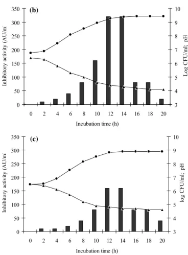
Figure 1 illustrates cell growth, pH variation, and bacteriocin production for L. lactis subsp lactis ST1 in MRS broth under different atmospheric growth condition.
In all tested conditions, the exponential phase started after 2 h and the maximum biomass was achieved after about 14 h of incubation which remained constant up to the end of incubation time. In this context, the pH decreased rapidly during 12 h of exponential growth phase and the rate of pH drop decreased at the early stationary phase. In addition to these findings, the bacteriocin production started as soon as the cells entered the exponential phase. The bacteriocin activity rose rapidly and the maximum activity was attained after about 12−14 h incubation. As shown in Figure 1 the maximum bacteriocin production was achieved at the end of exponential growth phase when the intensity of cell growth and pH drop decreased.
recorded as 9.62 and 9.42 c.f.u ml−1 in microaerophilic and capnophilic condition, respectively. The maximum activity was also detected after 12 h of incubation in candle jar as well as microareophilic growth conditions by 320 AU ml−1 (Fig 1a and b, respectively), but under aerobic condition the activity just reached 160 AU ml−1 after 12 h of incubation (Fig 1c). Incubation under aerobic condition resulted in final pH 4.6 after 20 h incubation that is much higher than 3.9 and 4.1 in the case of microaerophilic and capnophilic condition. Oxidative stress could, in general, slightly impact the growth of LAB, since they are defined as aerotolerant microorganisms though different behavior had been detected for bacteriocin production in various atmospheric incubation conditions (7, 17, 19, 27). Hirsch (17) suggested strict anaerobiosis as the suitable conditions for bacteriocin production. Leroy et al. (19) also reported no interfering effect of oxygen on cell growth and bacteriocin production of E. faecium RZS C5. It is in contrast to Cabo et al. (7) who found that aeration is essential for optimum bacteriocin production. Sousa et al. (27) also showed the negative effect of anaerobiosis on antagonism expression. The differences between various tested strains may be the reason for these discrepancies.
The specified pattern of cell growth and bacteriocin production for ST1 indicated that the produced bacteriocin is a primary metabolite. Similar results have been reported for bacteriocins produced by Lactobacillus helveticus G51 (6), and
Lactococcus lactis subsp. lactis (4). The activity remained constant for about 2h in all tested conditions and decreased afterwards when the cells entered the stationary phase. A similar decrease in activity was shown in previous studies for nisin (1) and some other bacteriocins such as bacteriocin A164 (10), bacteriocin GM005 (22), bacteriocin AMA-K (32) and bacteriocin ST13BR (33). The reduction in activity was clearly shown in all incubation conditions. Since higher intensity in this reduction of activity was observed in microaerophilic and capinophilic growth condition (around 88 and 93 % reduced activity, respectively), it can be concluded that favorable growth condition may be responsible for this phenomenon. On
the other hand, incubation under aerobiosis preserved the activity with only 75 % reduction, up to the end of incubation time.
This loss of activity has been related to proteolytic
degradation, protein aggregation, and/or adsorption to cell surface
of the producer while adsorption phenomenon has been considered
more responsible by some authors (13, 20, 22, 32). But,
bacteriocin release from the surface of producer at acidic
conditions has been reported previously (5, 36) and on the basis of
these findings it is expected that by reducing the pH at the end of
fermentation, bacteriocin desorption should be favored and hence
a higher activity would be detected. However, reduction of activity
was observed at the end of fermentation and by higher
acidification under microaerophilic and capinophilic growth
conditions lower bacteriocin activity was observed at the end of
fermentation. Thus, decrease in activity cannot be explained by
bacteriocin adsorption. On the other hand, there is no conclusive
study indicating the presence of nisin-specific protease (nisinase)
in L. lactisthat can describe the reduction phenomenon (1). This
reduction in activity might be related to the deterrent effect of low
pH or nutrient depletion on bacteriocin production. Zamfir et al.
(37) also related the reduction of cell growth and bacteriocin
production of L. acidophilus IBB 801 to the lactic acid
accumulation and hence low pH or an exhausted energy source at
the end of fermentation. Wolf-Hall et al. (35) also claimed that the
shortage of certain nutrients critical for bacteriocin production
could be responsible for this phenomenon during the latter stages
of fermentation.
(a)
0 50 100 150 200 250 300 350
0 2 4 6 8 10 12 14 16 18 20 Incubation time (h)
In
h
ib
ito
ry
activ
ity
(
A
U
/m
l
3 4 5 6 7 8 9 10
lo
g
C
F
U
/m
l;
p
(b) 0 50 100 150 200 250 300 350
0 2 4 6 8 10 12 14 16 18 20
Incubation time (h)
In h ib ito ry a ctiv ity (A U /m l 3 4 5 6 7 8 9 10 L o g C F U /m l; p H (c) 0 50 100 150 200 250 300 350
0 2 4 6 8 10 12 14 16 18 20
Incubation time (h)
Inhi bi to ry a ct ivi ty (A U /m l 3 4 5 6 7 8 9 10 log C F U /m l; p H
Figure 1. Time course of growth and BLIS production by L. lactis subsp lactis ST1, in MRS broth at 37 °C under microaerobic (a); capnophilic (b) and aerophilic (c) growth condition. Viable cell numbers (log c.f.u ml−1, ――); BLIS activity (AU ml−1, ――); pH (――). Data are average values of at least three replicates.
Cell growth and bacteriocin production at different initial
pHs, inoculum sizes, and incubation temperatures
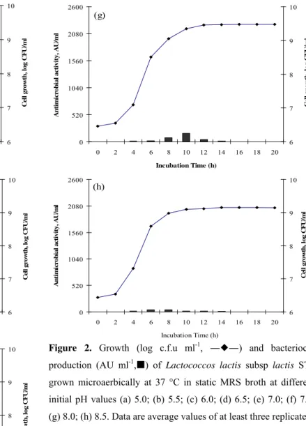
As shown in Figure 2, increasing the initial pH from 4.5 to 6.0 enhanced cell growth and bacteriocin production. Maximum bacteriocin activity (640 AU/ml) was obtained when the pH reached 6.5, followed by when it reached 7.0 and 7.5. Slight reductions in growth and activity were detected when the pH reached 8.5. The broadest range of stability in antimicrobial activity was achieved at pH 6.5 and cell growth was also among the highest (Fig. 2d) while, no activity was recorded at pH 4.5 (data not shown). By increasing the initial
pH, the exponential growth phase was reduced.
From the results obtained and other surveys by Aymerich
et al. (2) (enterocins A and B), Todorov and Dicks (31) (bacteriocin ST23LD) and Todorov and Dicks (29) (bacteriocin ST34BR) it can be concluded that bacteriocin production was mostly achieved at initial pH values between 6.5 and 7.5. There are some evidences also demonstrating the inhibitory effect of low initial pH on both cell growth and bacteriocin production by Aymerich et al. (2), Todorov and Dicks (31), and Powell et al. (23). Despite similar reports for the effect of initial pH on bacteriocin production Mitra et al. (21) observed a distinct behaviour for L. lactis CM1 and claimed that under pH 11.0 the activity of the nisin production was more significant than the initial pH of 6.5.
(a) 0 520 1040 1560 2080 2600
0 2 4 6 8 10 12 14 16 18 20
Incubation Time (h)
A n ti m ic robi a l ac ti vi ty , A U /m l 6 7 8 9 10 C el l gr ow th , l o g C F U /m l (b) 0 520 1040 1560 2080 2600
0 2 4 6 8 10 12 14 16 18 20
Incubation Time (h)
(c) 0 520 1040 1560 2080 2600
0 2 4 6 8 10 12 14 16 18 20
Incubation Time (h)
A n ti m ic ro b ia l ac ti vi ty, A U /m l 6 7 8 9 10 C el l gr ow th , l og C F U /m l (d) 0 520 1040 1560 2080 2600
0 2 4 6 8 10 12 14 16 18 20
Incubation Time (h)
A n ti m ic rob ia l ac ti vi ty, A U /m l 6 7 8 9 10 C el l gr ow th , l og C F U /m l (e) 0 520 1040 1560 2080 2600
0 2 4 6 8 10 12 14 16 18 20
Incubation Time (h)
A n ti m ic rob ia l ac ti vi ty, A U /m l 6 7 8 9 10 C el l gr ow th , l og C F U /m l (f) 0 520 1040 1560 2080 2600
0 2 4 6 8 10 12 14 16 18 20
Incubation Time (h)
A n ti m ic rob ial ac ti vi ty, A U /m l 6 7 8 9 10 Ce ll g ro w th , l o g CF U/ m l (g) 0 520 1040 1560 2080 2600
0 2 4 6 8 10 12 14 16 18 20
Incubation Time (h)
An ti m ic ro b ia l a ct iv it y , AU/ m l 6 7 8 9 10 C el l gr ow th, l o g C F U /m l (h) 0 520 1040 1560 2080 2600
0 2 4 6 8 10 12 14 16 18 20
Incubation Time (h)
A n ti m ic rob ia l ac ti vi ty, A U /m l 6 7 8 9 10 Ce ll g ro wt h , l o g CF U/ m l
Figure 2. Growth (log c.f.u ml-1, ――) and bacteriocin
production (AU ml-1,) of Lactococcos lactis subsp lactis ST1
grown microaerbically at 37 °C in static MRS broth at different
initial pH values (a) 5.0; (b) 5.5; (c) 6.0; (d) 6.5; (e) 7.0; (f) 7.5;
(g) 8.0; (h) 8.5. Data are average values of at least three replicate.
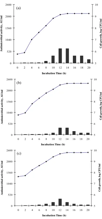
Based on the results reported in Figure 3, inoculum level
significantly affected bacteriocin production by ST1, while in all
the samples, cell growth entered stationary phase after about 14 h
of incubation. Maximum bacteriocin activity and stability (640
AU/ml) were obtained at the lowest inoculum level (Fig. 3a).
Aside from lower BLIS production, reduction in activity during
the stationary phase was also achieved in the case of high initial
cell density (Fig. 3c). These results are contradictory to those
reported for Camobactetium piscicola LV17 (25), Lactobacillus
plantarum 17.2b (14). The positive effect of inoculum size on
bacteriocin production was reported for these producer strains.
Saucier et al. (25) and Himelbloom et al. (16) explained this
inoculum. While, Himelbloom et al. (16) claimed that inoculum
size had no significant effect on the production of bacteriocin by
L. acidophilus LF221 and C. piscicola A9b, respectively.
Nevertheless, differences in the bacterial strains and production
medium, as well as incubation conditions may be responsible for
the inconsistency of these results.
(a) 0 520 1040 1560 2080 2600
0 2 4 6 8 10 12 14 16 18 20
Incubation Time (h)
A n ti m ic ro bi al a ctiv ity, A U /m l 5 6 7 8 9 10 C el l gr ow th , l og C F U /m l (b) 0 520 1040 1560 2080 2600
0 2 4 6 8 10 12 14 16 18 20
Incubation Time (h)
A n tim ic rob ial ac ti vit y, A U /m l 5 6 7 8 9 10 Ce ll g ro wt h , l o g CF U/ m l (c) 0 520 1040 1560 2080 2600
0 2 4 6 8 10 12 14 16 18 20
Incubation Time (h)
A n tim ic rob ial ac ti vit y, A U /m l 5 6 7 8 9 10 C el l gr ow th
, log C
F
U
/m
l
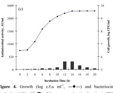
The effect of different temperatures (25, 30 and 37 °C) on growth and bacteriocin production by ST1 is shown in Figure 4. The highest cell density was achieved at 37 °C (9.57 log CFU/ml), while the optimum temperature for bacteriocin production was recorded as 30 °C. The inhibitory activity was reduced much further at 37 °C, which may be related to an increase in proteolytic degradation or nutrition depletion. Maximum bacteriocin production at suboptimal growth temperatures has also been observed in other surveys (9, 14, 30).
The results of bacteriocin production by L. lactis subsp.
lactis ST1, under different incubation temperature are consistent with the previous studies by Cheigha et al., (9) and Todorov and Dicks (29) which showed the maximum nisin activity and maintenance at 30 °C for Lactococcus lactis subsp.
lactis A164 and Lactococcus lactis subsp. lactis ST34BR, respectively. (a) 0 520 1040 1560 2080 2600
0 2 4 6 8 10 12 14 16 18 20
Incubation Time (h)
A n ti m ic ro b ia l a cti v ity , A U /m l 5 6 7 8 9 10 C el l gr ow th , lo g C F U /m l (b) 0 520 1040 1560 2080 2600
0 2 4 6 8 10 12 14 16 18 20
Incubation Time (h)
A n ti m ic ro b ia l a cti v ity , A U /m l 5 6 7 8 9 10 C el l gr ow th , l og C F U /m l
Figure 3. Growth (log c.f.u ml-1, ――) and bacteriocin
production (AU ml-1, ) of Lactococcos lactis subsp lactis
ST1 grown microaerbically at 37 °C in static MRS broth
(c)
0 520 1040 1560 2080 2600
0 2 4 6 8 10 12 14 16 18 20
Incubation Time (h)
A
n
ti
m
ic
ro
b
ia
l a
ctiv
ity
, A
U
/m
l
5 6 7 8 9 10
Ce
ll
g
ro
w
th
, l
o
g
CF
U/
m
l
Figure 4. Growth (log c.f.u ml-1, ――) and bacteriocin
production (AU ml-1, ) of Lactococcos lactis subsp lactis ST1
grown microaerbically in static MRS broth incubated at different
temperature (a) 25 °C; (b) 30 °C; (c) 37 °C. Data are average
values of at least three replicates.
Effect of Ultrasound, heat treatment, Tween and pH on
desadsorption
The effect of heat treatment and ultrasonic waves as well as
Tween80 at different pH conditions on the release of adsorbed
bacteriocin from the cell wall of L. lactis subsp lactis ST1 is
reported in Table 1. Alkaline pH was not tested because of its
slight inactivating effect on the bacteriocin, as it has been
determined in preliminary tests (data not shown).
Adjusting the pH of CFS to all acidic conditions, without any
further treatment, did not influence the bacteriocin activity.
Release was achieved after 5 and 10 min, in all treated samples at
pH 2 and 4. No release was achieved when the samples were
heated or exposed to ultrasonic waves at pH 6. Increasing
treatment period from 10 to 15 min was not favorable in all cases
and resulted in lower activity that could be related to the
inactivation of inhibitory compound during these conditions.
In whole, heating at acidic conditions was more effective
than ultrasound shock. Heating at 100 C in the presence of 0.5%
Tween80 resulted in 200% increase in bacteriocin activity but just
133.3% increase in the activity was observed after the ultrasound
shock in the presence of Tween80.
Furthermore, Tween80 played an important positive role in
bacteriocin release and it caused higher activity after heating and
ultrasound shock. It was obviously found that pH 2 and 0.5%
Tween80 resulted in higher soluble activities while by increasing
the pH of supernatants and omitting Tween80 from the CFS,
release was reduced which was more pronounced in the case of
ultrasonic shock.
In agreement with results of this study, it has been
demonstrated that the solubility, stability and biological activity of
nisin are dependent on the pH of the solution, and they increase
drastically with the lowering of pH to 2 (1). Aymerich et al. (2)
also introduced a combination of pH treatment with heat treatment
essential for an increase in soluble activity. Yang et al. (36), on the
other hand, indicated the influence of pH on the adsorption of
bacteriocins onto cells and introduced pH 6.0 for high adsorption
and pH 2.0 for maximum release. Hurst and Dring (18) also
claimed that at a pH below 6.0, more than 80% of nisin, produced
by L. lactis subsp. lactis was present in the culture supernatant
fluid.
In this report, the positive effect of heating at pH 2 in the
presence of 0.5% Tween80 on bacteriocin release from the
producer cell has been clearly shown. Aymerich et al. (2) and
Todorov (34) also reported the positive effect of Tween on the
adsorption reduction of bacteriocin on the bases of its capacity to
prevent the adsorption of bacteriocin molecules.
Table 1. Effect of heat treatment, ultrasonic shock and Tween80 at different pH conditions on the release of adsorbed nisin from the cell wall of L. lactis subsp lactis ST1.
Bacteriocin activity increase (%)
CFS pH 2 4 6
Time Courses (min) 5 10 15 5 10 15 5 10 15
Treatment
No treatment 0 0 0 0 0 0 0 0 0
Heating (100° C) 133.3 100.0 ― 133.3 33.3 ― 0 0 ―
Heating+Tween80 200.0 166.7 0 166.7 100.0 ― 0 0 ―
Ultrasound (28kHz) 100.0 100.0 33.3 66.7 33.3 ― 0 0 ―
Ultrasound+Tween80 133.3 66.7 ― 100.0 66.7 ― 0 ― ―
CONCLUSION
Lactococcus lactis subsp lactis ST1, isolated from goat milk, was tested for bacteriocin production kinetics at different growth conditions. This study indicated that the conditions resulting in higher levels of growth frequently favor bacteriocin production by ST1. Higher cell growth and inhibitory activity was detected in capnophilic and microaerophilic growth conditions while aerobiosis resulted in lower cell growth and bacteriocin production. On the other hand, higher bacteriocin activity was detected at suboptimal growth temperatures and the lowest initial cell count resulted in the highest bacteriocin production. The study also demonstrated bacteriocin activity increase after heating the culture at acidic condition and after ultrasound shock. This could be described by the release of adsorb bacteriocin from the cell wall of the producer, particularly in the presence of 0.5% Tween80. Thus, ultrasound shock can be used as an alternative method for heating in order to release bacteriocin from the producer cell wall. Bacteriocin activity reduced at stationary growth phase in all test conditions. This reduction was more pronounced in favorable growth condition. On the other hand, as the culture became more acidic at such growth phase and conditions, our finding expected higher levels of release and inhibitory activity in these conditions while the expectation did not come true, and a considerable reduction was detected instead. Thus, this decrease in activity cannot be explained by bacteriocin desorption, and the situatuation needs to be analyzed and observed through a different base. Based on the highest cell growth and bacterial activity under desirable growth conditions, this reduced inhibitory activity could be related to nutrition depletion, protein degradation and/or deferent effect of low pH on bacteriocin production.
REFERENCES
1. Arauza, L.J.; Jozalaa, A.F.; Mazzolab P.G.; Penna, T.C.V. (2009). Nisin biotechnological production and application: a review. Trends. Food. Sci. Tech. 20, 146−154
2. Aymerich, T.; Artigas, M.G.; Garriga, M.; Monfort, J.M.; Hugas, M. (2000). Effect of sausage ingredients and additives on the production of enterocins A and B by Enterococcus faecium CTC492 Optimization of in vitro production and anti-listerial effect in dry fermented sausages. J. Appl. Microbiol. 88, 686−694.
3. Batdorj, B.; Dalgalarrondo, M.; Choiset, Y.; Pedroche, J.; Metro, F.; Prevost, H.; Chobert, J.M.; Haertle, T. (2006). Purification and characterization of two bacteriocins produced by lactic acid bacteria isolated from Mongolian airag. J. Appl. Microbiol. 101, 837–848. 4. Benkerroum, N.; Oubel, H.; Zahar, M.; Dlia, S.; Filali-Maltouf, A.
(2000). Isolation of a bacteriocin-producing Lactococcus lactis subsp.
lactis and application to control Listeria monocytogenes in Moroccan jben. J. Appl. Microbiol. 89, 960−968.
5. Bhunia, A.K.; Johnson, M.C.; Ray, B.; Kalchayanand, N. (1991). Mode of action of pediocin AcH from Pediococcus acidilactici H on sensitive bacteria strains. J. Appl. Bacteriol. 70, 25−33.
6. Bonade, A.; Murelli, F.; Vescovo, M.; Scolari, G. (2001). Partial characterization of a bacteriocin produced by Lactobacillus helveticus. Lett. Appl. Microbiol. 33, 153−158
7. Cabo, M.L.; Murado, M.A.; Gonza´lez, M.P.; Pastoriza, L. (2001). Effects of aeration and pH gradient on nisin production. A mathematical model. Enzyme. Microb. Tech. 29, 264–273.
8. Callewaert, R.; Holo, H.; Devreese, B.; Van Beeumen, J.; Nes, I.; De Vuyst, L. (1999). Characterization and production of amylovorin L471, a bacteriocin purified from Lactobacillus amylovorus DCE 471 by a novel three-step method. Microbiology. 145, 2559–2568.
9. Cheigh C.I.; Choia, H.J.; Parka, H.; Kima, S.B.; Kooka, M.C.; Kimb, T.S.; Hwanga, J.K..; Pyuna, Y.R. (2002). Influence of growth conditions on the production of a nisin-like bacteriocin by Lactococcus lactis subsp.
lactis A164 isolated from kimchi. J. Biotechnol. 95, 225–235.
10. Choi, H.J.; Cheigh, C.I.; Kim, S.B.; Pyun, Y.R. (2000). Production of a nisin-like bacteriocin by Lactococcus lactis subsp. lactis A164 isolated from Kimchi. J. Appl. Microbiol. 88, 563−571.
11. Cleveland, J.; Montville, T.J.; Nes, I.F.; Chikindas, M.L. (2001). Review article; Bacteriocins: safe, natural antimicrobials for food preservation.
Int. J. Food. Microbiol. 71, 1–20
12. De Man, J.D.; Rogosa, M.; Sharpe, M.E. (1960). A medium for the cultivation of lactobacilli. J. Appl. Bact.23, 130−135.
13. De Vuyst, L.; Vandamime, E.J. (1992). Influence of the carbon source on nisin production in Lactococcus lactis ubsp. lactis batch fermentations. J. Gen. Microbiol. 138, 571−578.
14. Delgado, A.; Arroyo L´opez, F.N.; Brito, D.; Peres, C.; Fevereiro, P.; Garrido-Fern´andez, A. (2007). Optimum bacteriocin production by
Lactobacillus plantarum 17.2b requires absence of NaCl and apparently follows a mixed metabolite kinetics. J. Biotechnol. 130, 193–201. 15. Dmitriev, B.; Toukach, F.; Ehlers, S. (2005). Towards a comprehensive
16. Himelbloom, B.; Nilsson, L.; Gram, L. (2001). Factors affecting production of an antilisterial bacteriocin by Carnobacterium piscicola
strain A9b in laboratory media and model fish systems. J. Appl. Microbiol, 91, 506-513.
17. Hirsch, A. (1951). Growth and nisin production of a strain of
Streptococcus lactis. J. Gen. Microbiol. 5, 208 –221.
18. Hurst, A.; Dring, G.J. (1968). The relation of the length of lag phase of growth to the synthesis of nisin and other basic proteins by Streptococcus lactis grown under different cultural conditions. J. Gen. Microbiol. 50, 383−390.
19. Leroy, F.; Vankrunkelsven, S.; De Greef, J.; De Vuyst, L. (2003). The stimulating effect of a harsh environment on the bacteriocin activity by
Enterococcus faecium RZS C5 and dependency on the environmental stress factor used. Int. J. Food. Microbiol. 83, 27–38.
20. Mitra, S.; Chakrabartty, P.K.; Biswas, S.R. (2005). Production and Characterization of Nisin-Like Peptide Produced by a Strain of
Lactococcus lactis Isolated from Fermented Milk. Curr. Microbiol. 51, 183–187.
21. Mitra, S.; Chakrabartty, P. K.; Biswas, S. R. (2007) Production of Nisin Z by Lactococcus lactis Isolated from Dahi, Appl. Biochem. Biotechnol. 143, 41–53.
22. Onda, T.; Yanagida, F.; Tsuji, M.; Shinohara, T.; Yokotsuka, K. (2003). Production and purification of a bacteriocin peptide produced by
Lactococcus sp. strain GM005, isolated from Miso-paste. Int. J. Food. Microbiol. 87, 153 – 159.
23. Powell, J.E.; Witthuhn, R.C.; Todorov, S.D.; Dicks, L.M.T. (2007). Characterization of bacteriocin ST8KF produced by a kefir isolate
Lactobacillus plantarum ST8KF. Int. Dairy J. 17, 190–198.
24. Rodgers, S. (2001). Preserving non-fermented refrigerated foods with microbial cultures — a review, Trends. Food .Sci. Tech. 12, 276–284. 25. Saucier, L.; Poon, A.; Stiles, M.E. (1995). Induction of bacteriocin in
Carnobacterium piscicola LV17. J. Appl. Bacteriol, 78, 684–690. 26. Settanni, L.; Valmorri, S.; Suzzi, G.; Corsetti, A. (2008). The role of
environmental factors and medium composition on bacteriocin like inhibitory substances (BLIS) production by Enterococcus mundtii
strains. Food Microbiol. 25, 722– 728.
27. Sousa, M.N.B.; Mendes, E.N.; Apolonio, A.C.M.; Farias, L.D.M.;
Magalha, E.S. (2010). Bacteriocin production by Shigella sonnei isolated from faeces of children with acute diarrhea. APMIS. 118, 125–135 28. Tabatabaie, F.; Mortazavi, A. (2008). Studying the Effects of Ultrasound
Shock on Cell Wall Permeability and Survival of Some LAB in Milk, World. Appl. Sci. J. 3, 119-121.
29. Todorov, S.D.; Dicks, L.M.T. (2004). Influence of growth conditions on the production of a bacteriocin by Lactococcus lactis subsp. lactis
ST34BR, a strain isolated from barley beer, J. Basic. Microb. 44, 305– 316.
30. Todorov, S.D.; Dicks, L.M.T. (2004). Screening of lactic-acid bacteria from south African barley beer for the production of bacteriocin-like compounds, Folia Microbiol, 49, 406–410.
31. Todorov, S.D.; Dicks, L.M.T. (2006). Screening for bacteriocin-producing lactic acid bacteria from boza, a traditional cereal beverage from Bulgaria: comparison of the bacteriocins. Process. Biochem. 41, 11–19.
32. Todorov, S.D.; Nyati, H.; Meincken, M.; Dicks, L.M.T. (2007). Partial characterization of bacteriocin AMA-K, produced by Lactobacillus plantarum AMA-K isolated from naturally fermented milk from Zimbabwe. Food Control. 18, 656–664.
33. Todorov, S.D.; van Reenen, C.A.; Dicks, L.M.T. (2004). Optimization of bacteriocin production by Lactobacillus plantarum ST13BR, a strain isolated from barley beer. J. Gen. Appl. Microbiol. 50, 149–157 34. Todorov, S.D. (2008). Bacteriocin Production by Lactobacillus
plantarum AMA-K Isolated from AMASI, a zimbabwean Fermented Milk Product and Study of the Adsorption of Bacteriocin AMA-K to
Listeria SP. Braz. J. Microbiol. 39, 178-187.
35. Wolf-Hall, C. E.; Gibbons W. R.; ; Bauer, N. A. (2009). Development of a low-cost medium for production of nisin from Lactococcus lactis
subsp. lactis. World. J. Microbiol. Biotechnol. 25, 2013–2019.
36. Yang, R.; Johnson, M.C.; Ray, B. (1992). Novel method to extract large amounts of bacteriocins from lactic acid bacteriat. Appl. Environ. Microb. 58, 3355–3359.
37. Zamfir, M.; Callewaert, R.; Cornea, P.C.; De Vuyst, L. (2000). Production kinetics of acidophilin 801, a bacteriocin produced by
Lactobacillus acidophilus IBB 801. FEMS Microbiol. Lett. 190, 305– 308.