Case Report
Key Words
Pericardial effusions; chylopericardium idiopathic primary.
The accumulation of chyle in the pericardial space, or chylopericardium, is a condition occurring most frequently after trauma, cardiac and thoracic surgery, or in association with tumors, tuberculosis or lymphangiomatosis. When its precise cause cannot be identified, it is called primary or idiopathic chylopericardium. This is a rare clinical entity. We report the case of a surgically treated 20-year-old female patient. A brief review of the literature and comments on the clinical presentation, etiopathogenesis, ancillary diagnostic tests and treatment options are also presented.
Primary Idiopathic Chylopericardium - Case Report
Marcos Augusto de Moraes Silva, Antonio Sérgio Martins, Nelson Leonardo K. Leite de Campos, Rubens Ramos de
Andrade, Leonardo Massato Tohi, João Carlos Hueb
Faculdade de Medicina de Botucatu da Universidade Estadual Paulista (Unesp), Botucatu, SP - Brazil
Mailing address: Marcos Augusto de Moraes Silva•
Disciplina de Cirurgia Cardiovascular do Departamento de Cirurgia e Ortopedia da Faculdade de Medicina de Botucatu - UNESP - Distrito de Rubião Jr. s/n, Botucatu – 18618-970 – São Paulo, SP - Brazil E-mail: [email protected]
Manuscript received January 29, 2008; revised manuscript received June, 24, 2008; accepted July 11, 2008.
Introduction
Isolated chylopericardium was first described by Hasebrock in an autopsy case1, in 1888. This is a rare clinical entity in which chyle accumulates in the pericardial cavity1-3. It may be caused by surgical trauma, irradiation, tuberculosis, caval obstruction, and primary or metastatic mediastinal tumors1,3-5. The pathophysiology common to all these conditions seems to be thoracic duct obstruction without development of collateral drainage4. Congenital lymphangiomatosis and lymphangiectasia may also be the cause of chylopericardium6,7.
However, the precise etiology cannot be established in many cases. In order to describe these cases, Groves and Effler8, in 1954, introduced the terms “primary” or “idiopathic” chylopericardium.
We report the case of a surgically treated idiopathic or primary chylopericardium.
Case Report
A 20-year-old female patient from the city of Lençóis Paulista (State of SP) diagnosed with a large pericardial effusion was referred to our hospital in May 10, 2005.
History of present illness: mild dyspnea on heavy exertion for six months, which progressed to moderate exertion felt
for two months. She denied edema, dizziness, palpitations or chest pain.
Physical examination
Good general state of health, mucous membranes moist and pink, no cyanosis; P=HR= 96 bpm; BP = 110/60 mmHg. No jugular venous distension or lower limb edema. Spleen negative percussion sign, nonpalpable. Apical impulse not visible, not palpable; no thrills; heart sounds with normal rhythm and slightly muffled; no heart murmurs.
Ancillary tests
Normal blood count, Na, K, Mg, Ca, blood glucose, BUN, creatinine, protein profile, LDH, AST, and ALT; negative latex text; normal PCR, and negative serological tests for hepatitis B, C and HIV. Chest radiography: enlarged cardiac silhouette (Figure 1).
Echocardiogram - significant pericardial effusion with no signs of restrictive diastolic filling.
Computed tomography scan of the chest - enlarged heart with signs of pulmonary venous congestion.
Hospital course
In May 17, 2005, the patient underwent pericardial drainage via the subxiphoid approach using Marfan’s technique; 700 ml of a thick milky chylous fluid were drained. A pericardial biopsy was performed and fluid and blood samples were collected for tests. The total volume drained in the postoperative period was of approximately 500 ml, and the pericardial catheter was removed on day 4, 48 hours after drainage had ceased. The patient was discharged on postoperative day 7, and lymphoscintigraphy was scheduled to be performed on an outpatient basis.
Pericardial biopsy - fibrous thickening and mesothelial reaction.
Laboratory tests:
Blood - total cholesterol 140 mg/dl; LDL-cholesterol 62 mg/dl; triglycerides 70 mg;dl.
Pericardial fluid - triglycerides 1,420 mg/dl; culture-sterile; Gram-staining- absence of bacteria, rare leukocytes; cytology – global count: erythrocytes 2.6 x 10³/mm3
;leukocytes 3.2 x 10³/mm3; differential count: lymphocytes 3%; monocytes 8%; segmented neutrophils 89%.
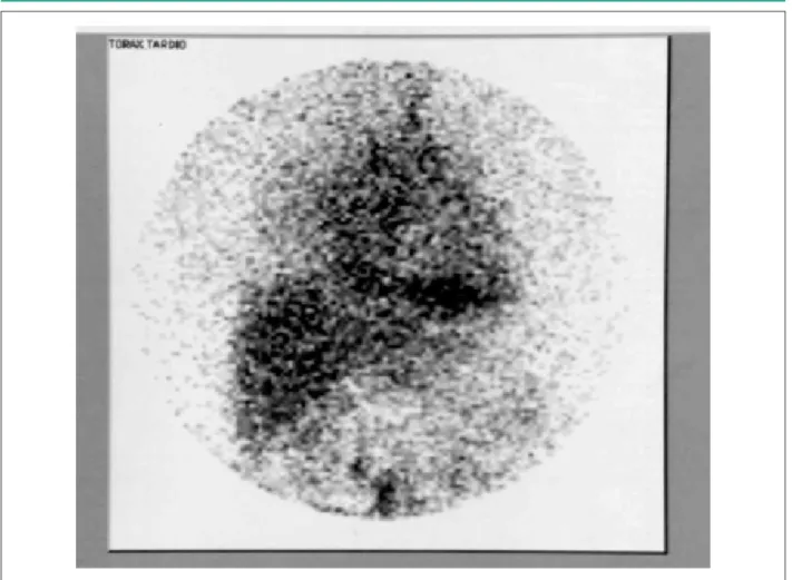
Lymphoscintigraphy performed 14 days after pericardial drainage showed lower limbs with normal aspect and abnormal thoracic radiopharmaceutical accumulation, which
Case Report
Silva et al Primary idiopathic chylopericardium
Arq Bras Cardiol 2009; 92(6) : e40-e43
Figure 1 -Chest radiography: enlarged cardiac silhouette.
could represent the presence of lymph in the pericardial or pleural space, secondary to an obstructive process or partial aplasia of the thoracic duct (Figure 2).
Twenty days after discharge, the patient was rehospitalized with significant pericardial effusion and complaint of dyspnea on moderate exertion. She was operated on via midsternal thoracotomy, and approximately 1,000 ml of a thick milky chylous fluid were removed. Partial pericardiectomy associated with pericardioperitoneal shunt was performed. The pleural and mediastinal cavities were drained.
In the postoperative period, the volume drained remained between 150 and 400 ml/day (mean of 240 ml/day) for 15 days, to further decrease until it ceased completely on day 30.
Discussion
From Groves and Effler’s8 description in 1954, Dunn1, in 1975, reported 22 cases of “primary” or “idiopathic” chylopericardium described in the literature. Up to 1992,
Akamatsu et al6 reported 79 cases and, up to 1997, Yüksel et al2 reported 89 cases. From 1997, we found 25 new cases reported, in a total of 114 cases described up to 2007. In the Brazilian literature we only found one case reported by Fernandes et al3 in 1998.
The pathophysiology of primary chylopericardium may be related to an abnormal connection between the thoracic duct and the pericardium, with the presence of fistulas, chyle reflux associated with lymphatic hypertension with loss of the valve mechanism and increased permeability of lymphatic vessel walls3.
Clinical manifestations may vary from the absence of symptoms to signs of cardiac tamponade. The most common symptoms are dyspnea, fatigue and cough5.
The differential diagnosis includes all causes of pericardial effusion, and in most cases chylopericardium is only confirmed by pericardiocentesis, with the finding of a chylous pericardial fluid containing chylomicrons and high triglyceride levels. When the diagnosis is confirmed, study of the lymphatic
Case Report
Silva et al
Primary idiopathic chylopericardium
Arq Bras Cardiol 2009; 92(6) : e40-e43
Figure 2 - Lymphoscintigraphy: abnormal thoracic radiopharmaceutical accumulation
vessels using lymphoscintigraphy is indicated, because it helps identify lymphopericardial fistulas, anatomical variations or partial aplasia of the thoracic duct 3,5,9. Computed tomography scan may be useful to rule out lymphangiomatosis.
Conservative treatment for chylopericardium consists of adopting a low-fat diet with medium-chain triglycerides, which are absorbed via the portal vein rather than via the lymphatic vessels, as occurs with long-chain triglycerides3. However, in contrast to post-traumatic chylopericardium, the conservative treatment of primary idiopathic chylopericardium is hardly ever successful5.
In these cases, surgical treatment is indicated, including pericardioperitoneal window, pericardiectomy, and ligation with resection of the thoracic duct just above the diaphragm6,7.
Akamatsu et al6 reported that of the 79 cases described in the literature up to 1992, conservative treatment was chosen for 10 (13%), and recurrence of the effusion was observed in six (60%) of these patients. The remainder 69 patients (87%) were surgically treated; 21 (27%) using a pericardial window, seven (9%) using ligation plus resection of the thoracic duct, and 41 (52%) using ligation plus resection of the thoracic duct associated with pericardial window.
Thoracic duct ligation and resection just above the
diaphragm associated with partial pericardiectomy has been the treatment used in most cases6. More recently, this procedure has also been performed by means of thoracoscopy4.
With the management adopted in the present case – partial pericardiectomy associated with pericardioperitoneal window, after more than two years of follow-up the patient is well, refers no complaints, and her follow-up was uneventful except for a small right pleural effusion. Thus, we believe the patient outcome was favorable, despite the inconvenience of the high volume of fluid drained in the first postoperative days and the long hospital stay.
Potential Conflict of Interest
No potential conflict of interest relevant to this article was reported.
Sources of Funding
There were no external funding sources for this study.
Study Association
This study is not associated with any post-graduation program.
Case Report
Silva et al Primary idiopathic chylopericardium
Arq Bras Cardiol 2009; 92(6) : e40-e43
References
1. Dunn RP. Primary chylopericardium: a review of the literature and illustrated case. Am Heart J. 1975; 89 (3): 369-77.
2. Yüksel M, Yildizeli B, Zonüzi F, Batirel HF. Isolated primary chylopericardium. Eur J Cardiothorac Surg. 1997; 12: 319-21.
3. Fernandes F, Arteaga E, Carvalho MSS, Ianni BM, Fernandes PP, Mady C. Quilopericardio idiopático. Arq Bras Cardiol. 1998; 71 (2): 131-4.
4. Furrer M, Holp M, Ris HB. Isolated primary chylopericardium: treatment by thorascoscopic thoracic duct ligation and pericardial fenestration. J Thorac Cardiovasc Surg. 1996; 112 (4): 1120-1.
5. Svedjeholm R, Jansson K, Olin C. Primary idiopathic chylopericardium – a
case report and review of the literature. Eur J Cardiothorac Surg. 1997; 11: 387-90.
6. Akamatsu H, Amano J, Sakamato T, Suzuki A. Primary chylopericardium. Ann Thorac Surg. 1994; 58 (1): 262-6.
7. Mahon NG, Nölke L, MacCann H, Sugrue D, Hurley J. Isolated chylopericardium. Surg J R Coll Surg Edinb Irel. 2003; 1: 236-8.
8. Groves LK, Effler DB. Primary chylopericardium. N Engl J Med. 1954; 250 (12): 520-3.
9. Mewis C, Külkamp V, Sokiranski R, Karsch KR. Primary chylopericardium due to partial aplasia of the thoracic duct. Eur Heart J. 1997; 18: 880-1.