Complete removal of the spinal nerve sheath
tumors. Surgical technics and results from a
series of 30 patients
Remoção completa dos tumores da bainha dos nervos raquianos. Técnica cirúrgica e
resultados de uma série de 30 pacientes
Rudi Lenck Fernandes1, José Carlos Lynch1, Leonardo Welling1, Mariangela Gonçalves2, Rodrigo Tragante1, Vicente Temponi1, Celestino Pereira1
ABSTRACT
Objective:Observe whether a microsurgical gross total removal (GTR) of a spinal nerve sheath tumors (SNSTs) is safe and decreases the
tumor recurrence.Method:We identify 30 patients with 44 SNSTs.Results:We operated upon 15 males and 15 females patients; mean age 40 years. GTR was achieved in 29 (96.6%) instances. Surgical mortality was 3.3% and the recurrence rate was 3.3%. The median follow-up time was 6.2 years.Conclusion:The surgical approach used in this group of patients afford that the great majority of tumors could be totally removed with low mortality and low recurrence rates, proving to be safe and effective.
Keywords:microsurgery, neurofibroma, schwannoma, spinal nerve sheath tumors, surgical treatment.
RESUMO
Objetivo:Observar se a ressecção microcirúrgica completa dos shwannomas ou neurofibromas raquianos é uma técnica segura e efetiva.
Método:Foram operados 30 pacientes com 44 schwannomas ou neurofibromas intrarraquiano.Resultados:A remoção total da lesão
ocorreu em 27 casos (96.6%). A taxa de mortalidade cirúrgica observada nesta série foi de 3.3%. O tempo médio de seguimento foi de 6.2 anos.Conclusão:A estratégia microcirúrgica empregada com esses pacientes propiciou a remoção total dos tumores na maioria dos pacientes, com baixa mortalidade e recidiva tumoral, mostrando ser segura e efetiva.
Palavras-chave:microcirurgia, neoplasia raquiana, neurofibroma, tumors da bainha dos nervos, schwannoma raquiano.
The annual incidence rates for primary spinal neoplasms are reported to be in the range of 1.3 to 10.0 per 100,0001. One-third is nerve sheet tumors1,2,3. The spinal nerve sheet tumors (SNSTs) comprise the schwannomas and neurofibro-mas1,2,3,4. Nevertheless little attention has been paid to this subject within Brazilian settings, which motivated us to review and present the cases of this pathological condition treated by our medical team, and to assess the safety and efficiency of microsurgical removal of those lesions.
Various terms have been used for this tumor: neuroma, neurinoma, neurilemoma, perineurial fibroblastoma, schwannoma gliome peripherique, schwannoma and neuro-fibroma2,4. The SNSTs are benign, usually solitary, slow-growing, encapsulated neoplasms composed of differen-tiated neoplastic Schwann cells. The most common origin of these tumors is the posterior spinal nerve root1,2,3,4,5,6.
METHOD Data collection
The 30 patients registered in this retrospective study with SNSTs were operated at the Hospital Federal dos Servidores do Estado (RJ) and at Rede D’Or São Luiz between 1986 and 2012. The medical charts, pre and postoperative imaging, and pathological reports were retrospectively reviewed for each patient to confirm the diagnosis of SNSTs; creating a database from which information pertin-ent to the prespertin-ent study was collected. The intraoperative videos of 11 patients were analyzed for nuances of the microsurgical technique. The need for informed consent was waived due to the retrospective character of the study. Detailed neurological examinations were performed in the neurosurgery and/or neurology department. Radiographs
1Departamento de Neurocirurgia, Hospital Federal dos Servidores do Estado, Rio de Janeiro RJ, Brazil;
2Rede D
’Or São Luiz, Rio de Janeiro RJ, Brazil.
Correspondence:José Carlos Lynch; Rua Jardim Botânico 600, sala 605; 22461-000. Rio de Janeiro, RJ. Brasil; E-mail: [email protected]
Conflict of interest:There is no conflict of interest to declare.
Received 15 October 2013; Received in final form 09 December 2013; Accepted 06 January 2014.
(XRs), computed tomography (CTs) and magnetic resonance imaging (MRIs) scans were reviewed with the radiology department. The postoperative images controls were done at hospital discharge and with 3 and/or by 12 months fol-low-up. Neurophysiological monitoring was not routinely used in this series.
Surgical technique
In all cases the same microsurgery technique was used following these general steps: After intubation, and appropri-ate arterial and venous access, the patient is turned to the prone position. The neck is maintained in a slightly flexed position with a Mayfield head clamp for the cervical lesions and high thoracic lesions and resting in the horseshoe for tumors in others locations. The involved spine region was shaved, prepared, and draped in a sterile fashion. The procedure starts with the use of a 4.5x loupe and co-axial lightning.
Exposing: A rectilinear midline incision is made start-ing in the superior level of the tumor and extendstart-ing to the inferior level. In the cases that we are planning a partial laminectomy, the skin incision is being place about 2 cm lateral of midline. The thoracolumbar fascia is opened in the midline with a 10 scalpel; the paravertebral muscles are subperiostlly desinserted and laterally retracted with a periosteum elevator. A unilateral subperiosteally expo-sure of the posterior spinal elements is performed only in the affected side. A bilateral muscle dissection is per-formed if the tumor is bilateral situated. The exposure is maintained with the appropriate autostatic retractors. At this point, the spinous processes and laminas of the involved level were well visualized. In small tumors, lami-nectomy can be limited to one single vertebral level and also limited to one side. Drilling does not need to be per-formed beyond the midline. That is done with the aid of air drill using cutting and/or diamond burr of 2 to 5 mm dia-meter. Laksell rongeur and Kerrinson forceps are also employed to finish the bone work. If the tumor is a dumb-bell shape, a unilateral facetectomy and foraminotomy must be necessary. During the whole procedure, careful hemostasis is obtained with bipolar forceps to keep the field bloodless and to prevent adhesions that can be induced around the spilled blood. At this moment, the sur-gical microscope is introduced in the field. The dura mater is sectioned in the midline with a 15 blade. The free border of the dura mater is sutured, using 4.0 threads, in the para-vertebral musculature.
Removing the lesion:In most of the cases, the lesion can be identified under the arachnoidal membrane. The ara-chnoidal covering the nerves roots is then opened in the posterior aspect of the exposure with micro scissors and gently dissected from the tumor surface to the sides using micro-surgery instruments and left intact to protect de
medulla and roots from the surgical dissection. At this point, the tumor is brought completely to view and a high mag-nification of the operating microscope is to be beneficial. The border between the tumor and ventral nerves roots and or spinal cord is established and the roots are protected with small cottonoids that are placed between the tumor and the roots. The surface of the schwannoma is coagulated with a low voltage bipolar under continue saline irrigation and sectioned with micro scissors. The interior of the lesion is penetrated and progressive debulked. After this, the tumor is shrinked and can be rolled on and we can visualize and identify with certainty the proximal root that originated the tumor that is cut with micro scissors. The surgeon should be aware, that in large lesions several roots appear to give origin to the tumor, these passage roots should be speared. When dealing with large lesions, that compress and deform the spinal cord or/and the roots, these tumors should be dissect from and not against the cord, and when free of adhesions, remove from the surgical field. We only attempted en block resection in small tumors that came out easily, without traction or compression of neural struc-tures. After removing the tumor, the subarachnoid space is carefully irrigated and the involved nerves roots are brought to this normal position. At this point, the blood pressure should retour to his normal levels. The dura is closed in a water tight fashion. Biological glue is added on the suturing line.
The suture of paravertebral musculature and fascia is performed in 3 planes to prevent muscle atrophy and resid-ual pain; the skin is sutured with 3.0 nylon.
ILUSTRATIVE CASES Case 1
A 61-year-old woman came to our attention complaining of backache and irradiating pain to the anterior aspect of the left thigh for 6-month duration. The neurological examina-tion where within normal limits, with the excepexamina-tion of a mild paresis of the extension of the left thigh. She did not exhibit signs compatible with neurofibromatosis. MRI depicted a dumbbell shape lesion involving from the 3thto the 4th lum-bar vertebrae with intense enhancement. (Figure 1A) The lesion was completed removed via a unilateral laminectomy and facetectomy. (Figure 1B) During the early postoperative period the patient developed a neuropatic pain that disap-peared completely after 3 months.
Case 2
T1 MRI demonstrated a medium size intradural L4 schwan-noma. (Figure 2A) We obtained GTR using microsurgical techniques and a L4 hemilaminectomy. (Figure 2B) The postoperative was uneventfully and he was pain relieved.
RESULTS
To the best of our knowledge, this is the largest series of SNSTs published in Brazil. As this is a retrospective study, it has inherent biases and drawbacks that only a multicenter, prospective study can overcome. This sample is composed of 15 (50%) females and 15 males (50%) and 44 SNSTs; ran-ging from 8 to 70 years (mean 40 years). We detected 2 cases with obvious features of Recklinghausen’s disease. The patient’s demographics and clinical characteristics are sum-marized in the Table 1.
Clinical outcome:In our series, one surgical death (3.3%) occurred. During the follow up period, three individuals died. One patient present with lumbar melanotic SNST. It was completely removed but recurred in the pelvic region. She died 2 years after surgery despite adjuvant therapy. Another death was of a 16-years-old boy with Neuro-fibromatosis 2 (NF-2). He presented almost completely para-plegic, with approximately 30 tumors located in several levels of the spinal cord. We removed 2 dumbbell C2 Schwannomas involving both sensory and motor rootlets
with critical compression of the spinal-medullary junction. (Figure 3A and 3B) He improved and was discharged home stable, but unfortunately, 3 months later, he was admitted to another hospital with acute respiratory distress and died due from aspiration pneumonia and sepsis. The third patient present with a rare intramedullary schwannoma, he was paraplegic at admission, with sphincters compromise. He did not improve after surgery and died two years later due to repetitive urinary tract infection. Four patients were lost in the follow up and were not included in this study. We detected a single tumor recurrence that was reoperated with good result. At the time of the last follow-up, 25 (82.3%) patients were alive. Excellent or good outcome was achieved in 24 patients (80%). Temporary postoperative complications including infection, CSF fistula, deep venous thrombosis, and pulmonary complications, occurred in 4 patients (10%).
Figure 1.Case 1. Preoperative axial enhanced T1 MR imaging demonstrating a medium size left dumbbell L3-L4 schwannoma (A). Postoperative axial enhanced T1 imaging demonstrating total removal of the tumor and postoperative changes (B).
Figure 2.Case 2: Preoperative axial enhanced T1 MR imaging demonstrating a medium size right intradural L4 schwannoma (A). Postoperative axial enhanced T1 imaging demonstrating completed removal of the tumor through a hemi laminectomy (B).
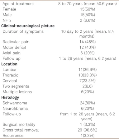
Table 1.Characteristics of 30 patients treated with spinal nerve sheet tumors.
Age at treatment 8 to 70 years (mean 40.6 years)
Female 15(50%)
Male 15(50%)
NF 2 2 (6.6%)
Clinical-neurological picture
Duration of symptoms 10 day to 2 years (mean, 8.4 months)
Radicular pain 14 (46%)
Motor deficit 12 (40%)
Axial pain 6 (20%)
Follow up 1 to 26 years (mean, 6.2 years)
Location
Lumbar 11(36.6%)
Thoracic 10(33.3%)
Cervical 7(23.3%)
Two segments 2(6.6)
Multiple lesions 6(20%)
Histology
Schwannoma 24(80%)
Neurofibroma 6(20%)
Follow-up from 1 to 26 years (mean, 6.2 years)
Surgical mortality 1 (3.3%)
Gross total removal 29 (96.6%)
Extension of resection and recurrence
Gross total removal (GTR) was achieved in 29 (96.6%) patients, confirmed by surgeon impression and/or post-operative images. GTR was obtained through a laminectomy for large lesions or a hemilaminectomy in 9 patients (26.6%) with small or middle size tumors (Figures 1A, 1B, 2A and 2B). Recurrence was observed once (3.3%); reoperation was performed 2.5 years later, with complete tumoral resection. In 28 patients, the root that originated the lesion could not be separated from the tumor and was divided. Eight patients noticed persistence of mild radicular deficits that did not cause any disturbance in their routine lives. Surgery in the spinal canal may cause atrophy of the cord or arachnoiditis. This was seen in one patient.
DISCUSSION
To the best of our knowledge, this is the largest series of SNSTs published in Brazil. As this is a retrospective study, it
has inherent biases and drawbacks that only a multicenter, prospective study can overcome. The age distributions in our study and that by Seppälä et al.1 were similar, with a peak incidence between 40 and 60 years of age,2,3,6,7. Like in the present study, previously authors had reported equal incidence in males and females1. Our study confirms that axial or radiating pain is the most common presenting symptom in patients with SNST1,2,3,4,5,6,7. Similar to others published series, in our sample, the duration of symptoms also varied widely, ranging from a few days to many years1-6. This paper confirms that most SNSTs are located intradural, followed by dumbbell shape and extradural locations1-7. The size of SNST varies from a small intradural tumor invol-ving a single rootlet to a large mass involinvol-ving several motors and sensory roots1,2,3,4,5,6,7. (Figure 4A, 4B and 4C) A conven-tional laminectomy or partial laminectomy was sufficient for the treatment of most lesions as proposed by several sur-geons1,2,6. In case of intradural
–extradural and large
extra-dural tumors, a laminotomy was combined with unilateral partial facetectomy2.
Mortality, gross total tumor removal and recurrence
The mortality in surgery of SNSs is usually low. In pub-lished series it’s varies from zero to 6%1,2,3,4,5,6,7. The operative mortality in our sample was 3.6% (1 case). A GTR was achieved in 29 (96.6%) instances. Others authors have reported results for GTR that varies from to 85 to 97.2%1,2,3,4,5,6,7,8. Klekamp and Samii8 observed overall recur-rence rates of 10.7% after 5 years and 28.2% after 10 years of follow-up, respectively and concluded that: partial removal, surgery of a recurrent tumor, NF-2, and old age pre-disposed for tumor recurrence. Matti T et al.5study demon-strates that 50% of a subtotally removed tumor (STR) showed a clinical recurrence. Seppala et al1 reported 20
Figure 4.Examples: small schwannoma (A); large lesion with bone erosion and cystic formation (B); and multiples schwannomas (C).
patients in whom STR was performed, recurrence in this group was verified in 55% of the cases. On the other hand, Safavi-Abbasi et al.2 reported that all patients in whom a tumor recurred had initially undergone GTR, and none of the patients undergoing STR had tumor recurrence. We believed that, probably, in this particular situation the sur-geon did not recognized a small piece of tumor left behind and during the follow up this piece of tumor recurred. Hasegawa et al.9found that no clear margin could be found between the capsule and tumor tissue in most of these tumors. They stated that resection of the nerve of tumor ori-gin rather than enucleation would be justified to avoid tumor recurrence. In our material, we observed only one (3.6%) tumor recurrence. (Table 2) Thus, it seems to us that in SNSTs the goal of the treatment is GTR in the first surgery even with the sacrifice of the involved nerve root, to avoid recurrence and obtain the cure of the patient1,2,6,7.
Sacrifice of the nerve root
Kim et al.4andothers2,9,10,11,12reported that sacrifice of the nerve root do not always result in postoperative neurological deficits, and when its occours, is usually a minor deficit. These facts indicate that the spinal roots giving origin to schwannoma are frequently nonfunctional at the time of surgery, and risks of causing disabling neurological deficit after sacrificing these roots are very small2,4,9,12.
The preservation of the anatomical continuity and should be attempted in functional roots like C5 and S12, although this may be feasible only in cases of small tumors. Complete removal of larger tumors is often impossible without sacrificing the nerve root. Celli13believed that some factors seem to influence the risk of neurological sequel after sectioning of the root, such as the neurofibroma pattern, preoperative weakness of the involved root, and intra-extradural tumors.
Surgical nuances
Surgeons must be aware, that in large lesions, in the ini-tial steps of dissection, several roots appear to give origin to
the tumor, similar to the passages vessels in the brain’s arter-ious venous malformation, and of course, these roots should be speared. We only attempted in block resection in very small tumors that can be safely removed, without traction or compression of neural structures. (Figure 4A) Taking this aforementions precautions, we and several authors advo-cated that GTR should always be attempted at the first sur-gery to prevent recurrence and cure the patient1,2,6,7,13(Figure 1 and 2) However if subtotal removal has been performed, the patients need to be followed closely1,2,6. In case of dumb-bell tumors, most of then can be removed by a single poster-ior approach14,15. When the tumor extends
,1 cm out of the intervertebral foramen, it is possible to preserve the facet. For a tumor that extends 2 cm or more out of the foramen, it seems best to remove the facet14.
We consider a unilateral laminectomy as minimal-access approach that provides a minimally invasive removal of the tumor, reducing blood loss, the immediate postoperative pain, and length of stay in comparison with unnecessary multi levels laminectomy. The hemilaminectomy was accomplished in 8 patients (26.6%) of this series with med-ium or small size tumors, if the interspinous ligament is left intact, and if the patient already has moderate degenerative change, fusion might not be necessary but follow-up evalua-tions nevertheless are needed6,14,15.
We are in agreement with the authors who claim that as schwannomas are located in different sites and locations, they may be small, large or multiples; (Figures 4A, 4B and 4C) these differences require different surgical approaches16,17,18.
In conclusion, based in our experience on 30 patients with 44 SNST, operated upon using microsurgical techniques and a limited laminectomy, it is a safe method, with low mortality and morbidity. Our study advocated that GTR in SNSTs is feasible in most of the cases but the sacrifice of the nerve root involved by the lesion is frequently necessary, although the risks of causing disabling neurological deficit after sacrificing these roots are very small. In this series we observed only one case (3.6%) of recurrence.
References
1. Seppala MT, Haltia MJJ, Sankila RJ, Jääskeläinen JE, Heiskanen O. Long-term outcome after removal of spinal neurofibroma:
a clinicalpathological study of 187 cases. J Neurosurg 1995;83:621-626.
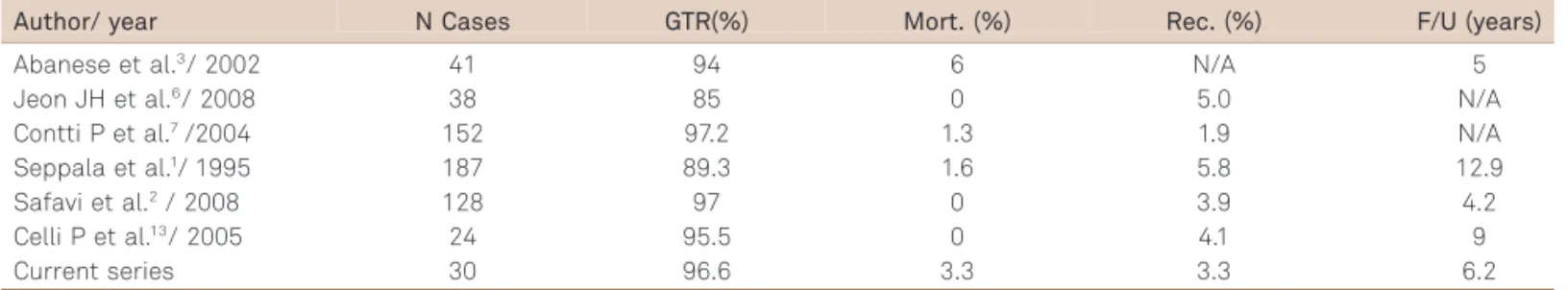
Table 2.Summary of the main surgical series on spinal nerve sheet tumors.
Author/ year N Cases GTR(%) Mort. (%) Rec. (%) F/U (years)
Abanese et al.3/ 2002 41 94 6 N/A 5
Jeon JH et al.6/ 2008 38 85 0 5.0 N/A
Contti P et al.7/2004 152 97.2 1.3 1.9 N/A
Seppala et al.1/ 1995 187 89.3 1.6 5.8 12.9
Safavi et al.2/ 2008 128 97 0 3.9 4.2
Celli P et al.13/ 2005 24 95.5 0 4.1 9
Current series 30 96.6 3.3 3.3 6.2
2. Safavi-Abbasi S, Senoglu M, Theodore N, et al. Microsurgical management of spinal schwannomas: evaluation of 128 cases. J Neurosurg Spine 2008;9:40-47.
3. Albanese V, Platania N. Spinal intradural extramedullary tumors. Personal experience. J Neurosurg Sci 2002;46:18-24.
4. Kim P, Ebersold MJ, Onofrio BM, Quast LM. Surgery of spinal nerve schwannoma. Risk of neurological deficit after resection of involved root. J Neurosurg 1989;71:810-814.
5. Seppälä MT, Haltia MJJ, Sankila RJ, Jääskeläinen JE, Heiskanen O. Long-term outcome after removal of spinal neurofibroma. J Neurosurg 1995;82:572-577.
6. Jeon JH, Hwang HS, Jeong JH, Park SH, Moon JG, Kim CH. Spinal Schwannoma; Analysis of 40 Cases. J Korean Neurosurg Soc 2008;43:135-138.
7. Conti P, Pansini G, Mouchaty H, Capuano C, Conti R. Spinal neurinomas: retrospective analysis and long-term outcome of 179 consecutively operated cases and review of the literature. Surg Neurol 2004;61:34-43.
8. Klekamp J, Samii M. Surgery of spinal nerve sheath tumors with special reference to neurofibromatosis. Neurosurg 1998;42:279-290.
9. Hasegawa M, Fujisawa H, Hayashi Y, Tachibana O, Kida S, Yamashita J. Surgical pathology of spinal schwannoma: has the nerve of its origin been preserved or already degenerated during tumor growth? Clin Neuropathol 2005;24:19-25.
10. Halliday AL, Sobel RA, Martuza RL. Benign spinal nerve sheath tumors: their occurrence sporadically and in neurofibromatosis types 1 and 2. J Neurosurg 1991;74:248-253.
11. Jinnai T, Koyama T. Clinical characteristics of spinal nerves sheath tumors: analysis of 149 cases. Neurosurg 2008;56:510-515.
12. Schultheiss R, Gullotta G. Resection of relevant nerve roots in surgery of spinal neurinomas without persisting neurological deficit. Acta Neurochir (Wien) 1993;122:91-96.
13. Celli P. Treatment of relevant nerve roots involving nerves sheath tumors: removal or preservation? Neurosurg 2002;51:684-692.
14. McCormick PC. Surgical Management of Dumbbell Tumors of the Cervical Spine. Neurosurg 1996;38:294-300
15. Thorat JD, Rajendra T, Thirugnanam A, Ng IH. Single-stage posterior midline approach for dumbbell tumors of the thoracic spine, with intraoperative CT guidance. Surg Neurol Int 2011;2:31-35.
16. Lot G, Bernard G. Cervical Neuromas with extradural components: surgical management in a series of 57 patients. Neurosurg 1997;41:813-822.
17. Falavigna A1, Righesso NO, Teles AR, Ruschel L, da Silva PG. Abordagem cirúrgica posterior e posterolateral para neurinomas cervicais em ampulheta da raiz de C2. Coluna 2010;9:157-166.